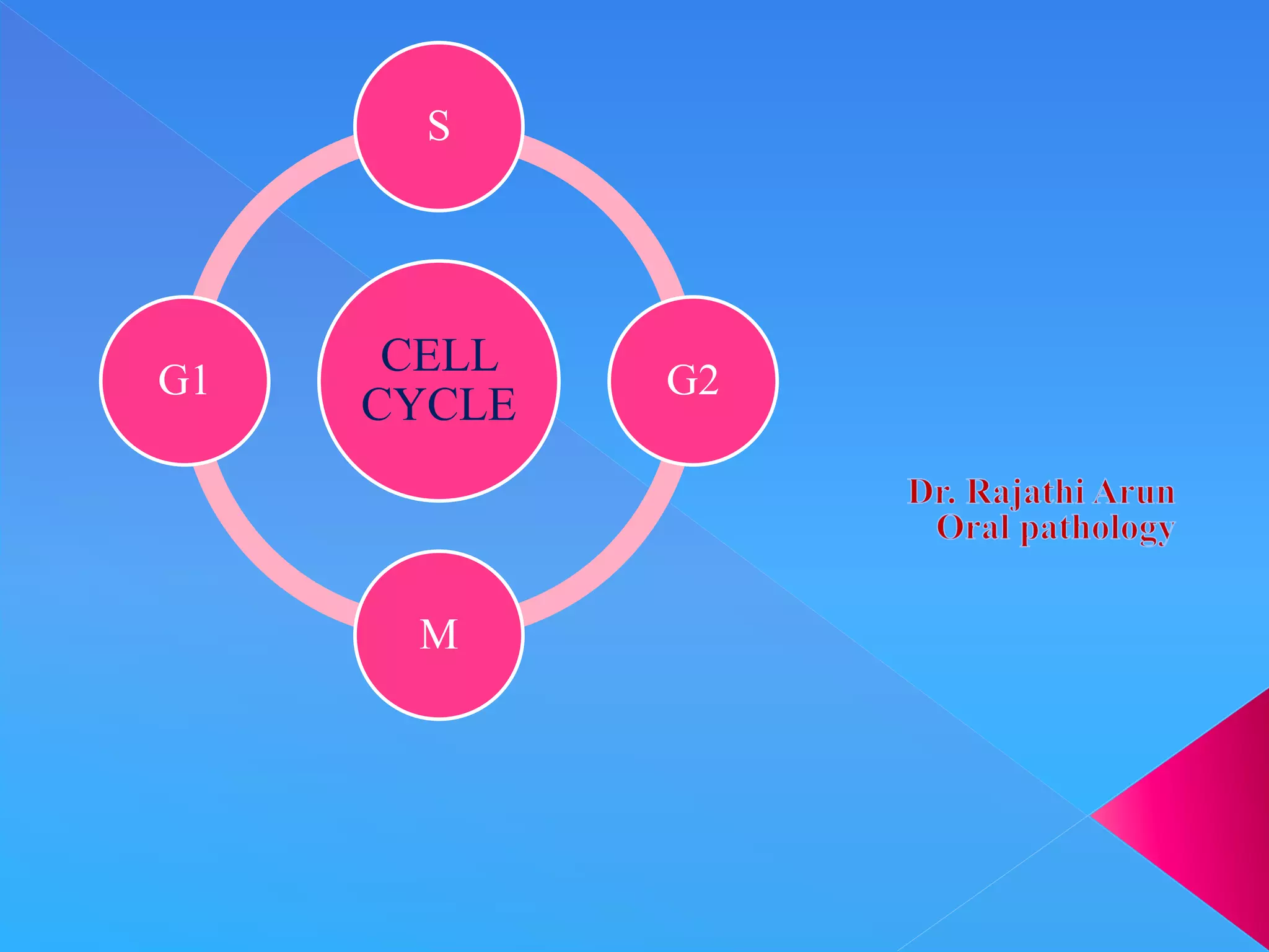
The document summarizes key aspects of the cell cycle. It describes the main phases (G1, S, G2, M), checkpoints that regulate progression, cyclins and CDKs that drive the cycle, and tissue types classified by their proliferative activity (continuously dividing, quiescent, nondividing). The cell cycle is highly regulated to ensure DNA replication fidelity and proper chromosome segregation through cyclin/CDK activity and checkpoint controls.
describe cell cycle and cell cycle control system for downloading the presentation , more presentations , infographics and blogs visit :
studyscienceblog.wordpress.com
Infer the significance of cell division.
Differentiate a DNA molecule, a chromosome, and a chromatid.
Characterize the phases of the cell cycle and their control points.
Describe the major events associated with stages of mitosis.
Explain the process of cytokinesis.
Learning Objectives
Describe the role of apoptosis in the life cycle of a cell.
Relate cancer as a result of the malfunction of the cell during the cell cycle.
Cell cycle and mitosis presentation (3)Ritu Sharma
The cell cycle is an ordered set of events, culminating in cell growth and division into two daughter cell.Cell division and tissue growth is a controlled and complex process. Cancer is a disease where regulation of the cell cycle goes messy and normal cell growth and behavior is lost.
Cell cycle and cell division are fundamental processes governing the growth, development, and reproduction of all living organisms. Understanding these processes is crucial in the field of biology as they play a pivotal role in shaping life at both the cellular and organismal levels.
For more information, visit-www.vavaclasses.com
cell division & physiology of cell division, types, binary fission, meiosis, mitosis, regulation of cell cycle, cell cycle checkpoints, what is cyclin-dependent kinases and its importance
WHAT IS CELL?
WHAT IS CELL DIVISION OR CELL CYCLE?
WHY DO CELL DIVIDE?
HISTORY
CELL CYCLE
INTERPHASE
M-PHASE
MOLECULAR EVENT DURING CELL CYCLE AND CELL REGULATION
TYPES OF CELL DIVISION
IMPORTANCE OF CELL DIVISION
ABNORMALTIES OF CELL CYCLE
REFRENCES
This presentation include the process of cell division. It hope it will helpful for all the medical students. Cell division is the series of events of equally dividing of one single mother cell into two identical daughter cell. Cell cycle and cell division terms are alternately used. Cell division is an important part of the all living processes.
At the time of cell division, RNA replication is a natural process.
The cell cycle, or cell-division cycle, is the series of events that take place in a cell that cause it to divide into two daughter cells.
These events include the duplication of its DNA (DNA replication) and some of its organelles, and subsequently the partitioning of its cytoplasm and other components into two daughter cells in a process called cell division.
There are two types of cell division
A) Mitosis and Binary fission – (Asexual reproduction) and B) Meiosis – (Sexual reproduction)
In prokaryotic cell, the cell division occurs via a process termed as Binary fission.
• In eukaryotic cell, the cell cycle can be divided in two periods i.e Interphase and Mitosis.
• During Interphase, the cell grows and DNA is replicated.
During Mitotic phase, the replicated DNA and cytoplasmic contents are separated, and cell divides.
The duration of cycle varies from hours to years. A typical human cell cycle has duration of 24 hours.
Some cells, such as skin cells, are constantly going through cell cycle, while other cells may divide rarely.
Some cells don’t grow and divide once they mature for ex. Neuron
Eukaryotic cell have a more complex cell cycle than prokaryotic cell.
The cell cycle, or cell-division cycle, is the series of events that take place in a cell leading to duplication of its DNA (DNA replication) and division of cytoplasm and organelles to produce two daughter cells.
Prix Galien International 2024 Forum ProgramLevi Shapiro
June 20, 2024, Prix Galien International and Jerusalem Ethics Forum in ROME. Detailed agenda including panels:
- ADVANCES IN CARDIOLOGY: A NEW PARADIGM IS COMING
- WOMEN’S HEALTH: FERTILITY PRESERVATION
- WHAT’S NEW IN THE TREATMENT OF INFECTIOUS,
ONCOLOGICAL AND INFLAMMATORY SKIN DISEASES?
- ARTIFICIAL INTELLIGENCE AND ETHICS
- GENE THERAPY
- BEYOND BORDERS: GLOBAL INITIATIVES FOR DEMOCRATIZING LIFE SCIENCE TECHNOLOGIES AND PROMOTING ACCESS TO HEALTHCARE
- ETHICAL CHALLENGES IN LIFE SCIENCES
- Prix Galien International Awards Ceremony
Tom Selleck Health: A Comprehensive Look at the Iconic Actor’s Wellness Journeygreendigital
Tom Selleck, an enduring figure in Hollywood. has captivated audiences for decades with his rugged charm, iconic moustache. and memorable roles in television and film. From his breakout role as Thomas Magnum in Magnum P.I. to his current portrayal of Frank Reagan in Blue Bloods. Selleck's career has spanned over 50 years. But beyond his professional achievements. fans have often been curious about Tom Selleck Health. especially as he has aged in the public eye.
Follow us on: Pinterest
Introduction
Many have been interested in Tom Selleck health. not only because of his enduring presence on screen but also because of the challenges. and lifestyle choices he has faced and made over the years. This article delves into the various aspects of Tom Selleck health. exploring his fitness regimen, diet, mental health. and the challenges he has encountered as he ages. We'll look at how he maintains his well-being. the health issues he has faced, and his approach to ageing .
Early Life and Career
Childhood and Athletic Beginnings
Tom Selleck was born on January 29, 1945, in Detroit, Michigan, and grew up in Sherman Oaks, California. From an early age, he was involved in sports, particularly basketball. which played a significant role in his physical development. His athletic pursuits continued into college. where he attended the University of Southern California (USC) on a basketball scholarship. This early involvement in sports laid a strong foundation for his physical health and disciplined lifestyle.
Transition to Acting
Selleck's transition from an athlete to an actor came with its physical demands. His first significant role in "Magnum P.I." required him to perform various stunts and maintain a fit appearance. This role, which he played from 1980 to 1988. necessitated a rigorous fitness routine to meet the show's demands. setting the stage for his long-term commitment to health and wellness.
Fitness Regimen
Workout Routine
Tom Selleck health and fitness regimen has evolved. adapting to his changing roles and age. During his "Magnum, P.I." days. Selleck's workouts were intense and focused on building and maintaining muscle mass. His routine included weightlifting, cardiovascular exercises. and specific training for the stunts he performed on the show.
Selleck adjusted his fitness routine as he aged to suit his body's needs. Today, his workouts focus on maintaining flexibility, strength, and cardiovascular health. He incorporates low-impact exercises such as swimming, walking, and light weightlifting. This balanced approach helps him stay fit without putting undue strain on his joints and muscles.
Importance of Flexibility and Mobility
In recent years, Selleck has emphasized the importance of flexibility and mobility in his fitness regimen. Understanding the natural decline in muscle mass and joint flexibility with age. he includes stretching and yoga in his routine. These practices help prevent injuries, improve posture, and maintain mobilit
Ethanol (CH3CH2OH), or beverage alcohol, is a two-carbon alcohol
that is rapidly distributed in the body and brain. Ethanol alters many
neurochemical systems and has rewarding and addictive properties. It
is the oldest recreational drug and likely contributes to more morbidity,
mortality, and public health costs than all illicit drugs combined. The
5th edition of the Diagnostic and Statistical Manual of Mental Disorders
(DSM-5) integrates alcohol abuse and alcohol dependence into a single
disorder called alcohol use disorder (AUD), with mild, moderate,
and severe subclassifications (American Psychiatric Association, 2013).
In the DSM-5, all types of substance abuse and dependence have been
combined into a single substance use disorder (SUD) on a continuum
from mild to severe. A diagnosis of AUD requires that at least two of
the 11 DSM-5 behaviors be present within a 12-month period (mild
AUD: 2–3 criteria; moderate AUD: 4–5 criteria; severe AUD: 6–11 criteria).
The four main behavioral effects of AUD are impaired control over
drinking, negative social consequences, risky use, and altered physiological
effects (tolerance, withdrawal). This chapter presents an overview
of the prevalence and harmful consequences of AUD in the U.S.,
the systemic nature of the disease, neurocircuitry and stages of AUD,
comorbidities, fetal alcohol spectrum disorders, genetic risk factors, and
pharmacotherapies for AUD.
micro teaching on communication m.sc nursing.pdfAnurag Sharma
Microteaching is a unique model of practice teaching. It is a viable instrument for the. desired change in the teaching behavior or the behavior potential which, in specified types of real. classroom situations, tends to facilitate the achievement of specified types of objectives.
Flu Vaccine Alert in Bangalore Karnatakaaddon Scans
As flu season approaches, health officials in Bangalore, Karnataka, are urging residents to get their flu vaccinations. The seasonal flu, while common, can lead to severe health complications, particularly for vulnerable populations such as young children, the elderly, and those with underlying health conditions.
Dr. Vidisha Kumari, a leading epidemiologist in Bangalore, emphasizes the importance of getting vaccinated. "The flu vaccine is our best defense against the influenza virus. It not only protects individuals but also helps prevent the spread of the virus in our communities," he says.
This year, the flu season is expected to coincide with a potential increase in other respiratory illnesses. The Karnataka Health Department has launched an awareness campaign highlighting the significance of flu vaccinations. They have set up multiple vaccination centers across Bangalore, making it convenient for residents to receive their shots.
To encourage widespread vaccination, the government is also collaborating with local schools, workplaces, and community centers to facilitate vaccination drives. Special attention is being given to ensuring that the vaccine is accessible to all, including marginalized communities who may have limited access to healthcare.
Residents are reminded that the flu vaccine is safe and effective. Common side effects are mild and may include soreness at the injection site, mild fever, or muscle aches. These side effects are generally short-lived and far less severe than the flu itself.
Healthcare providers are also stressing the importance of continuing COVID-19 precautions. Wearing masks, practicing good hand hygiene, and maintaining social distancing are still crucial, especially in crowded places.
Protect yourself and your loved ones by getting vaccinated. Together, we can help keep Bangalore healthy and safe this flu season. For more information on vaccination centers and schedules, residents can visit the Karnataka Health Department’s official website or follow their social media pages.
Stay informed, stay safe, and get your flu shot today!
The prostate is an exocrine gland of the male mammalian reproductive system
It is a walnut-sized gland that forms part of the male reproductive system and is located in front of the rectum and just below the urinary bladder
Function is to store and secrete a clear, slightly alkaline fluid that constitutes 10-30% of the volume of the seminal fluid that along with the spermatozoa, constitutes semen
A healthy human prostate measures (4cm-vertical, by 3cm-horizontal, 2cm ant-post ).
It surrounds the urethra just below the urinary bladder. It has anterior, median, posterior and two lateral lobes
It’s work is regulated by androgens which are responsible for male sex characteristics
Generalised disease of the prostate due to hormonal derangement which leads to non malignant enlargement of the gland (increase in the number of epithelial cells and stromal tissue)to cause compression of the urethra leading to symptoms (LUTS
Explore natural remedies for syphilis treatment in Singapore. Discover alternative therapies, herbal remedies, and lifestyle changes that may complement conventional treatments. Learn about holistic approaches to managing syphilis symptoms and supporting overall health.
TEST BANK for Operations Management, 14th Edition by William J. Stevenson, Ve...kevinkariuki227
TEST BANK for Operations Management, 14th Edition by William J. Stevenson, Verified Chapters 1 - 19, Complete Newest Version.pdf
ARTIFICIAL INTELLIGENCE IN HEALTHCARE.pdfAnujkumaranit
Artificial intelligence (AI) refers to the simulation of human intelligence processes by machines, especially computer systems. It encompasses tasks such as learning, reasoning, problem-solving, perception, and language understanding. AI technologies are revolutionizing various fields, from healthcare to finance, by enabling machines to perform tasks that typically require human intelligence.
2. The cell cycle, is the series of events that leads to
the duplication and division of a cell.
The cell cycle is driven by changing states of the
cytoplasm created by shifting balances of protein
phosphorylation and degradation machinery.
3. The orderly progression of cells through the
various phases of cell cycle is orchestrated by
Cyclins
Cyclin dependent kinases &
By their inhibitors.
4. The tissues of the body are divided into three
groups on the basis of their proliferative activity
In Continuously dividing tissues(labile tissues)
cells proliferate throughout life replacing those
that are destroyed.
Surface epithelia such as stratified surfaces of
skin, oral cavity, vagina, & cervix;
Lining mucosa of all the excretory ducts of the
glands of the body(ex.,salivary glands,
pancreas,biliary tract)
5. Columnar epithelium of GIT & uterus
Transitional epithelium of urinary tract, &
Cells of bone marrow & hematopoietic tissues.
In most of these tissues, mature cells are derived
from stem cells, which have an unlimited
capacity to proliferate & whose progeny may
undergo various streams of differentiation.
6. QUIESCENT OR STABLE TISSUES normally
have a low level of replication; however cells
from these tissues can undergo rapid division in
response to stimuli and are thus capable of
reconstituting the tissue of origin.
Considered to be in G0 stage of the cell cycle but
can be stimulated to enter G1 .
7. Parenchymal cells of liver, kidneys, & pancreas;
Mesenchymal cells, such as, fibroblasts &
smooth muscle;
Vascular endothelial cells; &
Resting leukocytes & other leukocytes.
8. Regenerative capacity of stable cells best
exemplied by the ability of liver to regenerate
after partial hepatectomy & after acute chemical
injury.
Fibroblasts, endothelial cells, smooth muscle
cells, chondrocytes, & osteocytes are quiescent in
adult mammals but proliferate widely,
constituting the connective tissue response to
inflammation.
9. NONDIVIDING (PERMANENT) TISSUES
contain cells that have left the cell cycle cannot
undergo mitotic division in postnatal life.
Neurons
Skeletal muscle cells &
Cardiac muscle cells.
10. If neurons in the central nervous system, are
destroyed, the tissue is generally replaced by the
proliferation of central nervous system
supportive elements, the glial cells.
Recent results demonstrate that neurogenesis
from stem cells may occur in adult brains.
11. Mature skeletal muscle cells do not divide ,
skeletal muscle does have some regenerative
capacity, through the differentiation of the
satellite cells that are attached to the endomysial
sheaths.
If the ends of severed muscle fibers are closely
juxtaposed, muscle regeneration in mammals
can be excellent, but this condition that can
rarely be attained under practical conditions.
12. Cardiac muscle has very limited, if any
regenerative capacity, and a large injury to the
heart muscle, as may occur in myocardial
infarction, is followed by scar formation.
13. Newly born cells are in the G1 phase of the cell
cycle.
These cells need to decide whether to commit
themselves to a round of proliferation or to
withdraw from the proliferation rat race and enter
a quiescent or differentiated state called G0 cells.
14. Cells that are considering proliferation must pass
two inspections
Restriction point – a biocemical control circuit
that determines whether internal & external
conditions are suitable for proliferation
Malfunctions cancer
Second quality control ( the G1 phase
checkpoint) verifies that the chromosomes are
intact before allowing the cell to replicate its
DNA.
15. Cells that decide to proliferate must replicate
their DNA in a timely and accurate manner.
S phase
During G2 phase cells conduct a final “cockpit
check” before embarking on the great adventure
of division.
16. Another key cell cycle checkpoint looks for
damaged or unreplicated DNA and restrains cells
from entering into mitosis before it is repaired.
This is also the last point in the cell cycle at
which the genome is scanned for damage so that
it can be repaired before division.
17. The goal of the cell cycle in most cases is to
produce two daughter cells that are accurate
copies of the parent .
The cell cycle integrates
a continuous growth cycle (the increase in cell
mass) with a
discontinuous division or chromosome cycle
(the replication and partitioning of the genome
into two daughter cells).
18. The chromosome cycle is driven by a sequence
of enzymatic cascades that produce a sequence of
discrete biochemical "states" of the cytoplasm.
Each state arises by destruction or inactivation of
key enzymatic activities characteristic of the
preceding state and expression or activation of a
new cohort of activities
19. convenient to divide the process into a series of
phases.
Recognition of cell-cycle phases began in 1882,
when Flemming named the process of nuclear
division mitosis (from the Greek mito, or
"thread") after the appearance of the condensed
chromosomes
20. It initially appeared that cells were active only
during mitosis, so the rest of the cell cycle was
called interphase (or resting stage).
Once DNA was recognized as the agent of
heredity in the 1940s, it was deduced that DNA
must be duplicated at some time during
interphase so that daughter cells can each receive
a full complement of genetic material.
21. A key experiment identified the relationship
between the timing of DNA synthesis and the
mitotic cycle and defined the four cell-cycle
phases.
22. M phase: Cell division, comprising mitosis, when
a fully grown cell segregates the replicated
chromosomes to opposite ends of a molecular
scaffold, termed the spindle, and cytokinesis,
when the cell cleaves between the separated
chromosomes to produce two daughter cells.
In general, each daughter cell receives a
complement of genetic material and organelles
identical to that of the parent cell
23. Interphase: The portion of the cell cycle when
cells grow and replicate their DNA. Interphase
has three sections.
The G1 (first gap) phase is the interval between
mitosis and the onset of DNA replication.
(Presynthetic phase)
The S (synthetic) phase is the time when DNA is
replicated.
24.
25. The G2 (second gap) phase is the interval
between the termination of DNA replication and
the onset of mitosis.(Premitotic phase)
In multicellular organisms, many differentiated
cells no longer actively divide.
These nondividing cells (which may
physiologically be extremely active) are in the G0
phase, a branch of the G1 phase
26. Each cell is born at the completion of the M
phase, which includes mitosis, the partitioning
of the chromosomes and other cellular
components, and cytokinesis, the division of the
cytoplasm.
All cycling cells have a M phase and a S phase.
However, some early embryos have minimal G1
and G2 phases.
27. The G1 phase is typically the longest and most
variable cell-cycle phase.
When cells are "born" at cytokinesis, they are
half the size they were before mitosis, and during
G1, they grow back toward an optimal size.
During this time, many genes involved in cell-
cycle progression are switched off so that the cell
cannot initiate a new round of proliferation. This
repressive system is termed the restriction point
28. If the supply of nutrients is poor or if cells
receive an antiproliferative stimulus such as a
signal to embark on terminal differentiation, they
delay their progress through the cell cycle in G1
or exit the cycle to enter G0.
However, if appropriate positive stimuli are
received, cells overcome the restriction point
block and trigger a program of gene expression
that commits them to a new cycle of DNA
replication and cell division.
29. Faulty restriction point control may result in cell
proliferation under inappropriate conditions.
Cancer cells often have defects in restriction
point control and continue to attempt to divide
even in the absence of appropriate environmental
signals.
30. Most cells of multicellular organisms
differentiate to carry out specialized functions
and no longer divide.
Such cells are considered to be in the G0 phase.
G0 cells are not dormant; indeed, they are often
actively engaged in protein synthesis and
secretion, and they may be highly motile.
31. The G0 phase is not necessarily permanent.
In some cases, G0 cells may be recruited to
reenter the cell cycle in response to a variety of
stimuli.
This process must be highly regulated, as the
uncontrolled proliferation of cells in a
multicellular organism can lead to cancer.
32. Chromosomes of higher eukaryotes are so large
that replication of the DNA must be initiated at
many different sites, termed origins of
replication.
In budding yeast, the approximately 400 origins
are spaced an average of 30,000 base pairs apart.
33. An average human chromosome contains about
150 × 106 base pairs of DNA, about 10 times the
size of the entire budding yeast genome, so many
more origins are required.
Each region of the chromosome that is replicated
from a single origin is referred to as a replicon.
Proliferating diploid cells must replicate their
DNA once and only once each cell cycle.
34. Each origin of replication is prepared for
replication by the formation of a prereplication
complex (a process that is referred to as
licensing) during G1.
As each origin "fires" during S phase, the
prereplication complex is dismantled and cannot
be reassembled until the next G1 phase.
This ensures that each origin fires only once per
cell cycle.
35. The cyclic nature of origin licensing is driven at
least in part by fluctuations in the activity of
cyclin-dependent kinases.
During replication, the duplicated DNA
molecules, called sister chromatids, become
linked to each other by a protein complex called
cohesin.
This pairing of sister chromatids is important for
their symmetrical segregation later in mitosis
36. In most cells of metazoans, G2 is a relatively
brief period during which key enzymatic
activities that will trigger the entry into mitosis
gradually accumulate and are converted to active
forms.
When their activities reach a critical threshold
level, the cell enters mitosis
37. In parallel, the chromatin and cytoskeleton are
prepared for the dramatic structural changes that
will occur during mitosis.
If unreplicated or damaged DNA is detected
during G2, a mechanism called a checkpoint
delays entry of the cell into mitosis.
38. During M phase (mitosis and the subsequent
cytokinesis), chromosomes and cytoplasm are
partitioned into two daughter cells.
Mitosis is normally divided into 5 discrete phases
Prophase
Prometaphase
Metaphase
Anaphase
Telophase
39. Prophase is defined by the onset of chromosome
condensation inside the intact nucleus and is
actually the final part of G2 phase.
The duplicated centrosomes (centrioles and
associated pericentriolar material ) separate and
form the two poles of the mitotic spindle.
40. Prometaphase begins when the nuclear envelope
breaks down (in higher eukaryotes) and
chromosomes begin to attach randomly to
microtubules emanating from the two poles of
the forming mitotic spindle.
Chromosomes may also nucleate some spindle
microtubules.
41. Once both kinetochores on a pair of sister
chromatids are attached to opposite spindle poles,
the chromosome slowly moves to a point midway
between the poles.
When all chromosomes are properly attached,
the cell is said to be in metaphase.
42. The exit from mitosis begins at anaphase with
the abrupt separation of the two sister
chromatids from one another.
The metaphase-anaphase transition is triggered
by the proteolytic degradation of molecules that
regulate sister chromatid cohesion.
During anaphase, the separated sister chromatids
move to the two spindle poles (anaphase A),
which themselves move apart (anaphase B).
43. As the chromatids approach the spindle poles, the
nuclear envelope reforms on the surface of the
chromatin.
At this point, the cell is said to be in telophase.
Finally, during telophase, a contractile ring of
actin and myosin assembles as a circumferential
belt in the cortex midway between spindle poles
and constricts the equator of the cell.
The separation of the two daughter cells from
one another is called cytokinesis
44. The cell cycle is highly regulated, and
checkpoints control transitions between cell-
cycle stages.
Checkpoints are biochemical circuits that detect
external or internal problems and send inhibitory
signals to the cell-cycle system.
Checkpoints: Biochemical circuits that regulate
cell-cycle transitions in response to the
physiological condition of the cell and the state
of its environment.
45. Checkpoints detect the
presence or absence of external signals telling the
cell to proliferate,
damage to the DNA, and
problems that arise during DNA replication and
chromosome segregation.
46. There are four major types of checkpoints.
RESTRICTION POINT.
DNA DAMAGE CHECKPOINT.
DNA REPLICATION CHECKPOINT.
SPINDLE ASSEMBLY CHECKPOINT.
47. The restriction point in the G1 phase is sensitive
to the physiological state of the cell and to its
interactions with the surrounding extracellular
matrix.
Cells that do not receive appropriate growth
stimuli from their environment do not progress
past this point in the G1 phase and may commit
suicide by apoptosis
48. DNA damage checkpoints operate in G1, S, and
G2 phases of the cell cycle. In general, these
checkpoints block cell-cycle progression, but
they can also trigger cell death by apoptosis.
49. The DNA replication checkpoint detects the
presence of unreplicated or stalled DNA
replication forks.
This checkpoint shares some components with
the DNA damage checkpoints but has the
additional feature that it specifically stabilizes
stalled replication forks so that they can be
repaired
50. During mitosis, the spindle assembly
checkpoint (also called the metaphase
checkpoint) delays the onset of chromosome
segregation until all chromosomes have attached
properly to the mitotic spindle.
51. The checkpoints in G1, S, and G2 use common
strategies to regulate cell-cycle progression.
DNA damage is detected by sensors.
These activate transducers, which are often
protein kinases but may also be transcriptional
activators.
53. The transducers act on effectors, which
ultimately block cell-cycle progression and may
also fulfill other functions.
Two key protein kinases, ataxia-telangiectasia
mutated (ATM) and ataxia-telangiectasia and
Rad9 related (ATR), lie at the head of the
pathway and may act as sensors of DNA damage.
54.
55. They activate two transducer kinases Chk1 and
Chk2 and also stabilize a transcription factor
called p53 that induces the expression of a cohort
of genes involved in halting cell-cycle
progression as well as genes that trigger cell
death by apoptosis.
In general, DNA damage checkpoints block cell-
cycle progression by inhibiting the cyclin-
dependent kinases by a variety of mechanisms.
56. Transitions between cell-cycle phases are
triggered by a network of protein kinases and
phosphatases that is linked to the discontinuous
events of the chromosome cycle by the periodic
accumulation, modification, and destruction of
several key components.
57. Genetic studies revealed that the yeast cell cycle
is a dependent pathway whereby events in the
cycle occur normally only after earlier processes
are completed.
The cell cycle can be modeled as a line of
dominoes, each domino corresponding to the
action of a gene product that is essential for cell-
cycle progression and the nth domino falling only
when knocked down by the (n - 1)th domino
58.
59. According to the model, mutations in genes that
are essential for cell-cycle progression cause an
entire culture of yeast to accumulate at a single
point in the cell cycle (the point at which the
defective gene product first becomes essential).
This is referred to as the arrest point.
Figure shows this by including a "mutant"
domino that does not fall over when struck by the
upstream domino.
60. Mutants that meet this criterion are called cell
division cycle mutants or CDC mutants. (Cdc
is used in fission yeast.)
Genetic screens for CDC mutants have identified
many important genes involved in cell-cycle
control.
61. Genetic analysis of the cell cycle in the fission
yeast Schizosaccharomyces pombe identified a
gene called cell division cycle-2+ (cdc2+) that is
essential for cell-cycle progression during both
the G1 → S and G2 → M transitions
The product of this gene, a protein kinase of
34,000 D originally called p34cdc2, is the
prototype for a family of protein kinases that is
crucial for cell-cycle progression in all
eukaryotes.
62. Humans have more than 10 distinct protein
kinases related to p34cdc2, although only a few are
involved in cell-cycle control.
To be active, these enzymes must each associate
with a regulatory subunit called a cyclin. Thus,
they have been termed cyclin-dependent kinases
63. p34cdc2, now termed Cdk1, seems to function
primarily in the regulation of the G2 → M
transition in animal cells.
A second family member, Cdk2, is involved in
regulation of the G1 → S and G2 → M
transitions,
Two other family members-Cdk4 and Cdk6-are
involved in passage of the restriction point.
64. Cdk7 is important for activation of other Cdks,
and also appears to participate in RNA
transcription and repair of damaged DNA.
Other Cdks participate in diverse processes
ranging from transcriptional regulation to
neuronal differentiation and may play as-yet-
undiscovered roles in cell-cycle regulation.
65. The defining feature of Cdks is that they require
binding of cyclins for catalytic activity.
Cyclins are a diverse group of proteins ranging in
size between 35 kD and 130 kD, all with a
similar core structure based on two symmetrical
domains of five α-helices.
66. Cyclins were discovered in rapidly dividing
invertebrate embryos as proteins that accumulate
gradually during interphase and are abruptly
destroyed during mitosis.
This process of cyclic accumulation and
destruction is the derivation of their name.
67. Subsequently, at least 16 different cyclins have
been identified in humans, although only a
handful are involved in cell-cycle control.
Of those that are, some function during G1 phase,
others during G2 phase, and still others during M
phase.
68. Cyclins are synthesized during specific phases of
the cell cycle, & their function is to activate
Cdks.
Cyclins D,E,A & B appear sequentially during
the cell cycle and bind to one or more Cdks.
69. Cdks have a bilobed structure with the active site
in a deep cleft between a small N-terminal and
larger C-terminal domain.
However, newly synthesized monomeric Cdks
differ from other kinases in that they appear to be
incompletely folded: a flexible loop (T loop)
blocks the mouth of the catalytic pocket.
70. In addition, misorientation of a short α-helix
causes a glutamic acid required for adenosine
triphosphate (ATP) hydrolysis to point away
from the catalytic cleft.
As a result, ATP bound by the monomeric kinase
is distorted and cannot transfer its α-phosphate to
protein substrates
71. Cdks are expressed consitutively during the cell
cycle but in an inactive form.
They are activated by phosphorylation after
binding to the family of proteins called cyclins.
72. At least four different mechanisms regulate Cdk
activity.
cyclin binding and phosphorylation of the T loop
stimulate enzyme activity.
On the other, phosphorylation of residues
adjacent to the ATP-binding site and binding of
inhibitory proteins inhibit Cdks.
73. Cyclin binding profoundly changes
Cdk structure, causing the retraction of th
of the T loop back from the mouth
of the catalytic pocket
74.
Cdk-cyclin complex has only partial
c catalytic activity.
Complete activation of most Cdks
requir requires the action of a kinase called
Cdk-a Cdk -activating kinase (CAK), which
phosphphosphorylates threonine160 in the T loop
of Cdk of Cdk2-cyclin A (this threonine gives
the loo the loop its name).
75.
76. At least two mechanisms slow or stop the cell
cycle by inactivating Cdks .
During G2 phase, the protein kinases Myt1 and
Wee1 hold Cdk1 in check by phosphorylating
threonine14 and tyrosine15 in the roof of the ATP-
binding site.
These phosphates interfere with ATP binding and
hydrolysis.
77.
78. Because threonine14 and tyrosine15 are accessible
to the regulatory kinases only following cyclin
binding, this phosphorylation of Cdks depends, at
least in part, on the availability of cyclins.
Three Cdc25 phosphatases reverse these
inhibitory phosphorylations.
79. Cdc25A is involved in the regulation of both the
G1 → S and G2 → M transitions and is essential
for life of the cell.
Ccd25B is dispensable for mitosis, but it is
essential for the production of gametes in
meiosis.
Cdc25C is a target of the G2 DNA damage
checkpoint that prevents cells from undergoing
mitosis with damaged DNA , but cells can
survive without it.
80. A second strategy for inactivating Cdks involves
the binding of small inhibitory subunits of the
cyclin-dependent kinase inhibitor (CKI) and
inhibitor of Cdk4 (INK4) families.
The activity of cyclin-CDK complexes is tightly
regulated by inhibitors, called CDK inhibitors.
Two main classes of CDK inhibitors:
Cip/Kip &
INK4/ARF families.
81. These inhibitors function as tumor supressors &
are frequently altered in tumors.
The Cip/Kip family(CKI) has three components,
p21, p27, & p57, which bind to inactivate the
complexes formed between cyclins and CDKs.
The human INK4a/ARF locus (a notation for
“inhibitor of kinase 4/alternative reading frame”)
encodes two proteins, p16INK4a & p14ARF,
which block the cell cycle and act as tumor
suppressors.
82. CKI molecules inactivate Cdk-cyclin A
complexes most efficiently.
The CKI p27Kip1 inactivates Cdk2-cyclin A
complexes in two ways.
One part of p27Kip1 associates with the cyclin
subunit, while another invades the N-terminal
domain of the Cdk, profoundly disrupting its
structure and competing with ATP for binding to
the active site.
83.
84. Members of the INK4 family preferentially
inactivate Cdk4 and Cdk6. They do this in two
ways .
First, interaction with monomeric Cdk opposite
the catalytic cleft distorts the orientation of the
N- and C-terminal lobes so that cyclin D does not
bind.
85.
86. INK4 family inhibitors also inhibit preformed
Cdk4/6-cyclin D complexes by binding the Cdk
and distorting the ATP-
binding site so that the
kinase uses ATP much
less efficiently.
87.
88. Cdk inhibitors are important for growth
regulation during the G1 and G0 phases of the cell
cycle .
They also play a critical role in the cell-cycle
arrest that occurs in response to DNA damage
and to anti-proliferative signals.
89. Mitosis is a state of the cytoplasm dominated by
high levels of active Cdk1-cyclin B-Cks.
Phosphorylation of key components by this
kinase leads to dramatic reorganization of the cell
and, ultimately, to separation of sister chromatids
on the mitotic spindle.
90. Once chromatids are separated, the cell must
return to a state with low levels of Cdk activity
so that nuclear envelope reassembly, spindle
disassembly, and cytokinesis can occur.
Thus, exit from mitosis requires Cdk
inactivation.
91. This occurs through the action of the ubiquitin-
directed proteolytic machinery that targets,
among other key proteins, A- and B-type cyclins
and a protein called securin, which regulates the
onset of sister chromatid separation at anaphase.
Destruction of cyclins inactivates the Cdk1 and
Cdk2 kinases, allowing various phosphatases to
reverse the action of Cdks and bring mitosis to a
close.
92. The proteasome is the second major cellular
compartment for proteolysis.
Proteasomes are multisubunit structures about
half the size of a ribosome that are located in
both the cytoplasm and nucleoplasm .
They are abundant, often accounting for up to 1%
of total cellular protein.
93.
94. Proteasomes contain an array of proteolytic
active sites arrayed on the interior wall of a
cylindrical chamber.
They degrade abnormal and misfolded proteins
as well as selected normal proteins down to the
level of small peptides .
95.
96. Proteasomes degrade key substrates in response
to signaling cascades or at key transitions of the
cell cycle.
One class of proteasomes processes intracellular
antigens for presentation by the immune system.
The proteasome has two major structural
components: the core and the cap.
The core, referred to as the 20S proteasome
(named according to its sedimentation
coefficient)
97. The proteasomes of eukaryotes and Archaea are
"capped" on one or both ends of the 20S barrel
with regulatory complexes to form the 26S
proteasome.
The type of regulatory complex varies depending
on the function of the proteasome.
The key to regulating degradation by
proteasomes is controlling access of molecules
into the central proteolytic chamber.
98. The best-characterized targeting mechanism
involves the reversible, covalent linkage of a
small protein, ubiquitin, onto the target protein.
Ubiquitin is a very abundant and highly
conserved protein of 76 residues.
The C-terminal four amino acids extend from
the compact globular structure, and its C-
terminus is linked to target proteins
99. Ubiquitination directs the selective degradation
of many different proteins:
abnormally folded proteins;
regulatory proteins, including some that control
cell cycle progression;
components of signal transduction systems; and
regulators of transcription.
100. Reversible ubiquitination is also involved in
other cellular functions, such as the
assembly of ribosomes, proteasomes, and other
multimeric complexes,
DNA repair, and
chromosomal structure.
101. Proteins with bound ubiquitin are directed to
their various fates by interaction with proteins
that contain ubiquitin-binding domains.
Low-affinity interactions of ubiquitin-binding
domains with ubiquitinated proteins allow the
system a great degree of dynamic flexibility.
Humans also have more than 80 deubiquitinating
enzymes that remove ubiquitin from target
proteins, thereby increasing the flexibility of
ubiquitin-based signaling pathways.
102. Ubiquitination of protein substrates proceeds
through a tightly regulated multistep pathway,
which has been elucidated through biochemical
purification of mammalian components and in
vitro reconstitution of partial reactions.
The overall scheme can be subdivided into three
stages
Activation of ubiquitin
Substrate recognition
Specific ubiquitinisation
103. Ubiquitin-mediated destruction of cyclins
involves the action of a series of enzymes .
E1 enzyme ubiquitin-activating enzyme
activates the small protein ubiquitinby forming a
thioester bond between the C-terminus of
ubiquitin and a cysteine on the enzyme..
Activated ubiquitin is then transferred to another
thioester bond on an E2 enzyme (ubiquitin-
conjugating enzyme).
104.
105. E2 may either transfer ubiquitin directly to the ε-
amino group of a lysine of a target protein or
combine with a third component (an E3 or
ubiquitin-protein ligase) to do so.
E3s are particularly important for imparting
substrate specificity.
Finally, the same enzymes build a chain of
ubiquitins by successive conjugations of the C-
terminus of a new ubiquitin to a lysine side chain
of the previous ubiquitin
106. The resulting polyubiquitinated proteins are
usually targets for destruction by the cylindrical
26S proteasome.
Tight regulation of ubiquitination pathways
ensures that only the appropriate target proteins
are recognized, ubiquitinated, and consequently
degraded.
107. The proteasome is a large multienzyme complex
that functions like a cytoplasmic garbage
disposal, grinding target proteins down to short
peptides and spitting out intact ubiquitin
monomers for reuse in further rounds of protein
degradation.
Its role was originally thought to be the removal
of damaged proteins from the cytoplasm;
however, it is now recognized as a central factor
in cell-cycle control.
108. The primary responsibility for substrate
selectivity lies with the E3 family of enzymes.
The key factor regulating proteolysis of cyclins is
a large (20S) complex with E3 activity consisting
of 12 to 13 subunits called the anaphase-
promoting complex/cyclosome (APC/C).
The APC/C is inactive during the S and G2
phases of the cell cycle.
109. Binding of protein "specificity factors" such as
Cdc20 and phosphorylation by Cdk1-cyclin B-
Cks1 activate the APC/C in early mitosis.
APC/CCdc20 is responsible for triggering the
metaphase-anaphase transition.
110. Later in mitosis, the APC/C binds a second
specificity factor, Cdh1.
Cdk phosphorylation blocks Cdh1 binding to
APC/C, so APC/CCdh1 forms only after cyclin
levels (and therefore Cdk activity) start to fall
late in mitosis.
111.
112. Further repression of Cdk activity and
destruction of Cdc20 by APC/CCdh1 are critical
both for mitotic exit and during G1 phase in
preparing chromatin for the initiation of DNA
replication
113. As cells pass from G1 into S phase, a newly
synthesized protein, Emi1, binds to APC/CCdh1
and inactivates it.
This allows the accumulation of cyclins during S
and G2.
Remarkably, APC/CCdh1 also has a role in
nondividing neurons, where it is involved with
regulating the activity of synapses.
114.
115. CELL BIOLOGY – THOMAS D. POLLARD –
SECOND EDITION.
PATHOLOGIC BASICS OF DISEASE –
ROBBINS – SEVENTH EDITION.