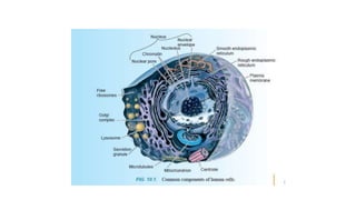
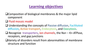
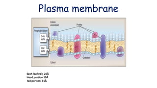
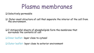
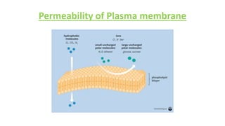
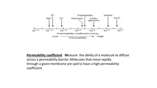
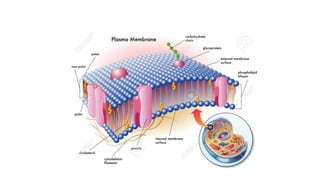
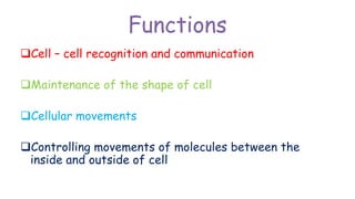
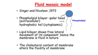
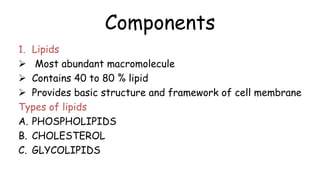
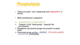
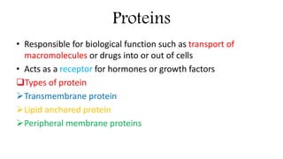
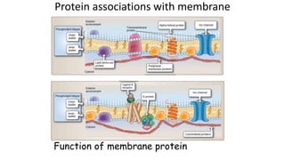
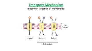
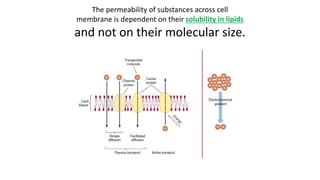
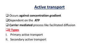
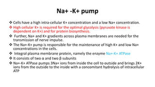
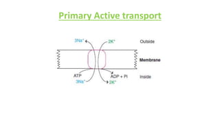
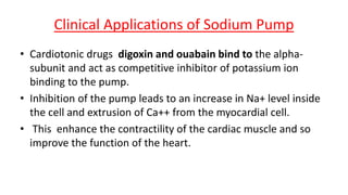
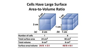
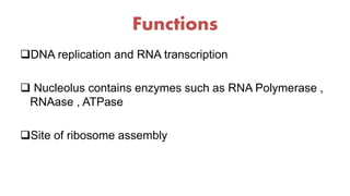
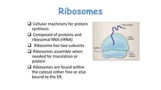
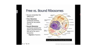
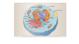
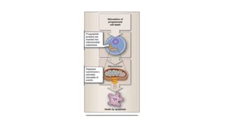
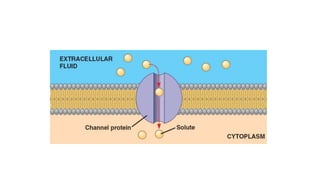
CELL
BIOLOGY
Rupesh Gupta
Department of Biochemistry
JNU IMSRC
Learning objectives
Composition of biological membranes & the major lipid
component
 Fluid mosaic model
 Understanding the concepts of Passive diffusion, Facilitated
diffusion, Active transport, Endocytosis, and Exocytosis.
 Recognize transporters, ion channels, the Na+ − K+-ATPase,
receptors, and gap junctions
Variety of disorders result from abnormalities of membrane
structure and function
Plasma membrane
Each leaflet is 25Å
Head portion 10Å
Tail portion 15Å
Plasma membranes
 Selectively permeable
 Outer-most structure of cell that separate the interior of the cell from
the environment.
 2 Antiparallel sheets of phospholipids form the membrane that
surrounds the contents of cell
 Inner leaflet- layer close to cytosol
 Outer leaflet- layer close to exterior environment
Permeability of Plasma membrane
Permeability coefficient Measure the ability of a molecule to diffuse
across a permeability barrier. Molecules that move rapidly
through a given membrane are said to have a high permeability
coefficient
Functions
Cell – cell recognition and communication
Maintenance of the shape of cell
Cellular movements
Controlling movements of molecules between the
inside and outside of cell
Fluid mosaic model
• Singer and Nicolson ,1972
• Phospholipid bilayer –polar head
(extracellular)
hydrophobic tail (cytoplasmic)
• Lipid bilayer shows free lateral
movement of its component ,hence the
membrane is fluid in nature
• The cholesterol content of membrane
alters the fluidity of membrane
Phospholipid
Components
1. Lipids
 Most abundant macromolecule
 Contains 40 to 80 % lipid
 Provides basic structure and framework of cell membrane
Types of lipids
A. PHOSPHOLIPIDS
B. CHOLESTEROL
C. GLYCOLIPIDS
Phospholipids
• These are polar, ionic compounds and Amphipathic in
nature
• Most predominant component
A. HYDROPHILIC COMPONENTS
 Present in the ‘’head group’’- towards the
environment
 Phosphate and alcohol groups are present on polar
head
 Functional group can be – SERINE , ETHANOLAMINE
, INOSITOL (CHOLINE)
B. HYDROPHOBIC COMPONENTS- Present in
the ‘’tail group’’ – extend inwards
Contains long chain of fatty acids
Can be Saturated & Unsaturated fatty acids
Helps in motions of PM such as flexion, rotation
and lateral movement
No flip – flop movements
CHOLESTEROL
• Major component of cell membrane
• An Amphipathic molecule
• Contains polar hydroxyl group and
hydrophobic steroid ring and
attached hydrocarbons
• Dispersed throughout the PM and
intercalating between
phospholipids
• Fits into spaces created by the kinks
of UFA leads to decrease in motion
of PM , causes stiffening and
strengthening of membrane
GLYCOLIPIDS
• Lipids attached to carbohydrate
• Found in PM in lower concentration
than phospholipids and cholesterol
• Oriented towards outside of cell
• Helps to form carbohydrate coat
which are involved in cell to cell
interactions
• Lipid rafts Specialized cholesterol-
enriched micro domains within cell
membranes are known as lipid rafts
Cholesterol transport
Endocytosis
Signal transduction
Proteins
• Responsible for biological function such as transport of
macromolecules or drugs into or out of cells
• Acts as a receptor for hormones or growth factors
Types of protein
Transmembrane protein
Lipid anchored protein
Peripheral membrane proteins
Protein associations with membrane
Function of membrane protein
Transport Mechanism
(Based on direction of movement)
The permeability of substances across cell
membrane is dependent on their solubility in lipids
and not on their molecular size.
Comparison of Transporters and Ion
Channels
Simple Diffusion
Driven by the concentration gradient
Occurs from higher to lower concentration
Rate of entry is proportional to the solubility of solute in the
hydrophobic core of the membrane
This does not require any energy
Very slow process
Ex- Solutes and gases enter into the cells passively
Facilitated Diffusion
Carrier mediated process
Important features
 The carrier mechanism could be saturated
Structurally similar solutes can competitively inhibit the entry
of the solutes.
Can operate bi-directionally.
does not require energy
rate of transport is more rapid than simple diffusion
• The carrier molecules can exist in two conformations, Ping and
Pong states.
• In the pong state, the active sites are exposed to the exterior
environment
• Solutes bind to the pong & conformational change occurs
• In the ping state, the active sites are facing the interior of the cell,
where the concentration of the solute is minimal.
• This will cause the release of the solute molecules and the protein
molecule reverts to the pong state.
• For example, glucose transport across membrane is by facilitated
diffusion involving a family of glucose transporters.
Active transport
 Occurs against concentration gradient
Dependent on the ATP
 Carrier mediated process like facilitated diffusion
2 Types
I. Primary active transport
II. Secondary active transport
Na+ -K+ pump
 Cells have a high intra-cellular K+ concentration and a low Na+ concentration.
 High cellular K+ is required for the optimal glycolysis (pyruvate kinase is
dependent on K+) and for protein biosynthesis.
 Further, Na+ and K+ gradients across plasma membranes are needed for the
transmission of nerve impulse.
 The Na+-K+ pump is responsible for the maintenance of high K+ and low Na+
concentrations in the cells.
 Integral plasma membrane protein, namely the enzyme Na+-K+ ATPase
 It consists of two α and two β subunits
 Na+-K+ ATPase pumps 3Na+ ions from inside the cell to outside and brings 2K+
ions from the outside to the inside with a concomitant hydrolysis of intracellular
ATP
Primary Active transport
Clinical Applications of Sodium Pump
• Cardiotonic drugs digoxin and ouabain bind to the alpha-
subunit and act as competitive inhibitor of potassium ion
binding to the pump.
• Inhibition of the pump leads to an increase in Na+ level inside
the cell and extrusion of Ca++ from the myocardial cell.
• This enhance the contractility of the cardiac muscle and so
improve the function of the heart.
Secondary active transport
(Ex- sodium dependent glucose transporter)
Phlorizin
Types of ATP-Driven Active
Transporters
I. P (in P-type) signifies phosphorylation (these proteins auto-phosphorylate).
II. F (in F-type) signifies energy coupling factors.
III. V (in V-type) signifies vacuolar.
IV. ABC signifies ATP-binding cassette transporter (all have two nucleotide-binding
domains and two transmembrane segments).
Proton pump in the stomach
This is an antiport transport system of gastric parietal cells
Enzyme H+–K+ ATPase to maintain highly acidic conditions in the
lumen of the stomach
Proton pump antiports two cytoplasmic protons (2H+) and two
extracellular potassium (2K+) ions for a molecule of ATP
hydrolysed.
The chloride ions secreted by Cl– channels combine with protons to
form gastric HCl.
Omeprazole is a drug used in the treatment of peptic ulcer.
Inhibits H+-K+ ATPase and results in reduced secretion of HCl.
Proton pump in the stomach
Channelopathies
Group of disorders that result from abnormalities in the
proteins forming the ion pores or channels.
Examples
I. Cystic fibrosis -chloride channel
II. Liddle's syndrome - sodium channel
III. Periodic paralysis - potassium channel
1. Cystic fibrosis (CF)
• This is the most common lethal genetic disease in Caucasians of Northern
European
• Prevalence = 1:3,000 births.
• This autosomal recessive disorder
• Cause= mutations to the gene for the CF transmembrane conductance regulator
(CFTR) protein that functions as a chloride channel on epithelium.
• Defective CFTR results in decreased secretion of chloride ion
• In the pancreas, the decreased hydration results in thickened secretions such
that pancreatic enzymes are not able to reach the intestine, leading to
pancreatic insufficiency.
• Treatment includes replacement of these enzymes and supplementation with
fat-soluble vitamins.
• CF also causes chronic lung infections with progressive pulmonary disease
CASE
A 3-year-old boy with salt deposition on skin
• A 3-year-old child was brought to the hospital OPD with complaints of
cough, difficulty in breathing, and indigestion. He had recurrent episodes
of infections of respiratory tract for which he was treated with antibiotics
• During hot weather, acute salt deposition occurred on child’s skin.
Moreover, a fond kiss left a salty taste in mouth, which he affectionately
referred to as the ‘salty kiss’.
• A first cousin of the child had somewhat similar signs and symptoms,
though of a milder degree.
• On examination, the child appeared weak and malnourished. Analysis of
the sample obtained by rectal biopsy showed thick mucus, that was
blocking various tubular structures of the glands.
Ionophores
Ex- valinomycin, a diffusion (shuttle) type ion transporter
It is obtained from Streptomyces.
• It permits transport of K across cell membranes.
• It has a polar interior that interacts with K, and a non-polar exterior which
enables it to interact with the membrane.
• The non-polar exterior is due to the hydrophobic side chains of the non-polar
amino acids that constitute this antibiotic.
• These side chains easily intercalate with the hydrophobic membrane matrix,
which enables the valinomycin molecule to shuttle back and forth across the
membrane.
• During the process, the K, held in the centre of the antibiotic, is carried across
the permeability barrier.
1. gramicidin, a pore type ion transporter.
2. Gramicidin A is an antibiotic isolated from Bacillus brevis.
3. It forms a tail-to-tail dimer, which makes a pore across the
membrane
4. The two helical monomers span the membrane.
5. Length of the helix is about 3 nm, which is consistent with the
hydrophobic region of a typical phospholipid bilayer.
6. The helical pore, having a diameter of 0.4 nm, can transport
several cations, such as H, Na, K, etc.
7. Ion transport by gramicidin is 1000 times faster than that by
valinomycin.
Ion Channels
Ion channels are transmembrane proteins that allow the
selective entry of various ions
Quick transporter of electrolytes such as Ca++, K+, Na+ and Cl--
 Channels generally remain closed, but in response to stimulus,
they open allowing rapid flux of ions down the gradient
Important for nerve impulse propagation, synaptic
transmission and secretion of biologically active substances
from the cells
Ligand Gated Channels
Ligand gated channels are opened by binding of effectors.
The binding of a ligand to a receptor site on the channel results
in the opening/closing of the channel
The ligand may be an extracellular signalling molecule or an
intracellular messenger
Ex- Acetyl choline receptor , Calcium channels
Voltage Gated Channels
• Voltage gated channels are opened by membrane depolarization
• The channel is usually closed in the ground state. .
• In voltage gated channels, the channels open or close in response to changes in
membrane potential.
• They pass from closed through open to inactivated state on depolarization.
• Once in the inactivated state, a channel cannot re-open until it has been
reprimed by repolarization of the membrane.
• Ex- sodium & Potassium channels, seen in nerve cells and are involved in the
conduction of nerve impulses.
• Ion channels allow passage of molecules in accordance with the concentration
gradient. Ion pumps can transport molecules against the gradient.
What limits the dimensions
of a cell?
The upper limit of cell size is set by the rate of diffusion of
solute molecules in aqueous systems.
Cells Have Large Surface
Area-to-Volume Ratio
Subcellular separation
Tween 20
Nucleus
Nucleus
Present in all eukaryotic cells except RBC
Chromosomes are present in nucleus
Nuclear envelop – outer most double
layered phospholipids membrane
Nuclear pores – permit trasfer of
materials between nucleus and cytosol
Nucleoplasm – interior of nucleus
,contains fluid in which chromosomes are
found
Nucleolus – prominent structure within
nucleus ,site of ribosome production
Functions
DNA replication and RNA transcription
 Nucleolus contains enzymes such as RNA Polymerase ,
RNAase , ATPase
Site of ribosome assembly
Ribosomes
 Cellular machinery for protein
synthesis
 Composed of proteins and
ribosomal RNA (rRNA)
 Ribosome has two subunits
 Ribosomes assemble when
needed for translation or
protein
 Ribosomes are found within
the cytosol either free or else
bound to the ER.
ENDOPLASMIC RETICULUM
 Network of membranous tubules within the
cell
ER is often observed to surround the nucleus
The outer layer of the nuclear envelope is
actually continous with the ER.
In muscle cells, this organelle is known as the
sarcoplasmic reticulum.
Types- SER & RER
Function of SER
contains enzymes for the synthesis of
Triacylglycerols and Phospholipids
Contains cytochrome P450 involved in
metabolism of drugs and toxic chemicals such as
ethanol
 Synthesis of steroid hormones.
Glycogen is stored in liver cells that are rich in
SER
Function of RER
 Involved in the synthesis of proteins.
Proteins produced on these ribosomes enter
the lumen of the RER, travel to the Golgi
complex in vesicles, and are subsequently
secreted from the cell
Post-translational modifications of these
proteins, such as glycosylation occur in the
RER
Golgi complex
 Consists of a curved stack of flattened vesicles in the
cytoplasm
 Divided into three compartments:
 cis Golgi network – convex and faces the nucleus
 medial Golgi network – concave
 trans Golgi network - faces the plasma membrane
Functions
 Involved in modifying proteins produced in the RER and in
sorting and distributing these proteins to the lysosomes,
secretory vesicles, or the plasma membrane.
 Participates in post-translational modification of proteins
such as sulfation, phosphorylation and proteolysis
• “What Is true of E. coli is
true of the elephant.”
JacquesMonod
MITOCHONDRIA
 Spherical, oval or rod-like bodies
Powerhouse of the cell, where energy released
from oxidation of food stuffs is trapped as
chemical energy in the form of ATP
• Surrounded by two membranes
 Outer membrane – contains pores made from
porins, permeable to molecules upto 1,000
g/mol.
 Inner membrane – Highly impermeable, forms
invaginations known as cristae containing the
electron-transport chain and ATP synthase.
FUNCTIONS
Energy production
 Electron transport chain, ATP generation, TCA cycle, beta oxidation of fatty
acids, ketone body production
Role as independent units within the eukaryotic
cells
 contain DNA (mtDNA) and ribosomes for the production of RNA and some
mitochondrial proteins
Function in cell survival
• When the process of programmed cell death or apoptosis is stimulated in
a cell, proapoptotic proteins insert into the mitochondrial membrane,
forming pores. A protein known as cytochrome c then leave the
intermembrane space of the mitochondria through the pores, entering the
cytosol. Cytochrome c in the cytosol stimulates a cascade of biochemical
events resulting in apoptotic death of the cell
Clinical significance
 Kearns-Sayre syndrome
 A single large deletion of mtDNA is responsible for the development of this
syndrome
 Paralysis of eye muscles and degeneration of the retina
 Leber hereditary optic neuropathy
 point mutation in mtDNA causes this disorder
 results in blindness in young men
 Pearson syndrome
 Deletions in mtDNA
 Bone marrow and pancreas dysfunctions
Peroxisomes
• Single membraned, cytoplasmic organelles, involved in oxidative
reactions using molecular oxygen.
• These reactions produce the toxic chemical hydrogen peroxide
(H2O2), which is degraded within the peroxisome by catalase
 Function
 Oxidation of very long-chain fatty acids to shorter chain fatty acids
 Conversion of cholesterol to bile acids
 Synthesis ether lipids called plasmalogens.
 Involved in the synthesis of myelin
Membrane disruption
Toxic product
Clinical significance
Zellweger syndrome
 caused by a defect in the transporting of peroxisomal enzymes
into the peroxisomes in liver, kidneys, and brain
 Affected individuals do not survive beyond 6 months of age
X-linked Adrenoleukodystrophy
 is characterized by the deterioration of myelin sheaths of
neurons
 failure of proper fatty acid metabolism
LYSOSOMES
Contains hydrolytic enzymes
Intracellular organelles of digestion ,enclosed by a
single membrane
 Formed from digestive vesicles called endosomes
which are involved in receptor-mediated
endocytosis.
Function
 Eliminating unwanted material
 Destruction of infectious bacteria and yeast
 Tissue remodeling
 Involution of tissues during development
A. Lysosomal Hydrolases
• Enzymes include nucleases, phosphatases,
glycosidases, esterases, and proteases
• cleave amide, ester, and other bonds through
the addition of water.
• highest activity near a pH of approximately 5.5
PHAGOCYTOSIS
Clinical significance
 Inclusion cell (I- cell) disease
 Rare condition in which lysosomes lack in enzymes
 Silicosis
 Results from inhalation of silica particles into the lungs which are
taken up by phagocytes.
 Lysosomal membrane ruptures, releasing the enzymes.
 stimulates fibroblast to proliferate and deposit collagen fibers,
resulting in fibrosis and decreased lungs elasticity
 Following cell death, the lysosomes rupture releasing the hydrolytic
enzymes which bring about postmortem autolysis
 GOUT
 LYSOSOMAL STORAGE DISEASE
• Yoshinori Ohsumi, 71,
received Nobel Prize for
uncovering “mechanisms
for autophagy”, a
fundamental process in
cells that scientists believe
can be harnessed to fight
cancer and dementia.
Cell biology

Cell biology

  • 1.
  • 3.
    Learning objectives Composition ofbiological membranes & the major lipid component  Fluid mosaic model  Understanding the concepts of Passive diffusion, Facilitated diffusion, Active transport, Endocytosis, and Exocytosis.  Recognize transporters, ion channels, the Na+ − K+-ATPase, receptors, and gap junctions Variety of disorders result from abnormalities of membrane structure and function
  • 4.
    Plasma membrane Each leafletis 25Å Head portion 10Å Tail portion 15Å
  • 5.
    Plasma membranes  Selectivelypermeable  Outer-most structure of cell that separate the interior of the cell from the environment.  2 Antiparallel sheets of phospholipids form the membrane that surrounds the contents of cell  Inner leaflet- layer close to cytosol  Outer leaflet- layer close to exterior environment
  • 6.
  • 7.
    Permeability coefficient Measurethe ability of a molecule to diffuse across a permeability barrier. Molecules that move rapidly through a given membrane are said to have a high permeability coefficient
  • 9.
    Functions Cell – cellrecognition and communication Maintenance of the shape of cell Cellular movements Controlling movements of molecules between the inside and outside of cell
  • 10.
    Fluid mosaic model •Singer and Nicolson ,1972 • Phospholipid bilayer –polar head (extracellular) hydrophobic tail (cytoplasmic) • Lipid bilayer shows free lateral movement of its component ,hence the membrane is fluid in nature • The cholesterol content of membrane alters the fluidity of membrane Phospholipid
  • 11.
    Components 1. Lipids  Mostabundant macromolecule  Contains 40 to 80 % lipid  Provides basic structure and framework of cell membrane Types of lipids A. PHOSPHOLIPIDS B. CHOLESTEROL C. GLYCOLIPIDS
  • 12.
    Phospholipids • These arepolar, ionic compounds and Amphipathic in nature • Most predominant component A. HYDROPHILIC COMPONENTS  Present in the ‘’head group’’- towards the environment  Phosphate and alcohol groups are present on polar head  Functional group can be – SERINE , ETHANOLAMINE , INOSITOL (CHOLINE)
  • 13.
    B. HYDROPHOBIC COMPONENTS-Present in the ‘’tail group’’ – extend inwards Contains long chain of fatty acids Can be Saturated & Unsaturated fatty acids Helps in motions of PM such as flexion, rotation and lateral movement No flip – flop movements
  • 15.
    CHOLESTEROL • Major componentof cell membrane • An Amphipathic molecule • Contains polar hydroxyl group and hydrophobic steroid ring and attached hydrocarbons • Dispersed throughout the PM and intercalating between phospholipids • Fits into spaces created by the kinks of UFA leads to decrease in motion of PM , causes stiffening and strengthening of membrane
  • 16.
    GLYCOLIPIDS • Lipids attachedto carbohydrate • Found in PM in lower concentration than phospholipids and cholesterol • Oriented towards outside of cell • Helps to form carbohydrate coat which are involved in cell to cell interactions • Lipid rafts Specialized cholesterol- enriched micro domains within cell membranes are known as lipid rafts Cholesterol transport Endocytosis Signal transduction
  • 17.
    Proteins • Responsible forbiological function such as transport of macromolecules or drugs into or out of cells • Acts as a receptor for hormones or growth factors Types of protein Transmembrane protein Lipid anchored protein Peripheral membrane proteins
  • 18.
    Protein associations withmembrane Function of membrane protein
  • 19.
    Transport Mechanism (Based ondirection of movement)
  • 20.
    The permeability ofsubstances across cell membrane is dependent on their solubility in lipids and not on their molecular size.
  • 21.
  • 22.
    Simple Diffusion Driven bythe concentration gradient Occurs from higher to lower concentration Rate of entry is proportional to the solubility of solute in the hydrophobic core of the membrane This does not require any energy Very slow process Ex- Solutes and gases enter into the cells passively
  • 23.
    Facilitated Diffusion Carrier mediatedprocess Important features  The carrier mechanism could be saturated Structurally similar solutes can competitively inhibit the entry of the solutes. Can operate bi-directionally. does not require energy rate of transport is more rapid than simple diffusion
  • 24.
    • The carriermolecules can exist in two conformations, Ping and Pong states. • In the pong state, the active sites are exposed to the exterior environment • Solutes bind to the pong & conformational change occurs • In the ping state, the active sites are facing the interior of the cell, where the concentration of the solute is minimal. • This will cause the release of the solute molecules and the protein molecule reverts to the pong state. • For example, glucose transport across membrane is by facilitated diffusion involving a family of glucose transporters.
  • 26.
    Active transport  Occursagainst concentration gradient Dependent on the ATP  Carrier mediated process like facilitated diffusion 2 Types I. Primary active transport II. Secondary active transport
  • 27.
    Na+ -K+ pump Cells have a high intra-cellular K+ concentration and a low Na+ concentration.  High cellular K+ is required for the optimal glycolysis (pyruvate kinase is dependent on K+) and for protein biosynthesis.  Further, Na+ and K+ gradients across plasma membranes are needed for the transmission of nerve impulse.  The Na+-K+ pump is responsible for the maintenance of high K+ and low Na+ concentrations in the cells.  Integral plasma membrane protein, namely the enzyme Na+-K+ ATPase  It consists of two α and two β subunits  Na+-K+ ATPase pumps 3Na+ ions from inside the cell to outside and brings 2K+ ions from the outside to the inside with a concomitant hydrolysis of intracellular ATP
  • 28.
  • 29.
    Clinical Applications ofSodium Pump • Cardiotonic drugs digoxin and ouabain bind to the alpha- subunit and act as competitive inhibitor of potassium ion binding to the pump. • Inhibition of the pump leads to an increase in Na+ level inside the cell and extrusion of Ca++ from the myocardial cell. • This enhance the contractility of the cardiac muscle and so improve the function of the heart.
  • 31.
    Secondary active transport (Ex-sodium dependent glucose transporter) Phlorizin
  • 32.
    Types of ATP-DrivenActive Transporters I. P (in P-type) signifies phosphorylation (these proteins auto-phosphorylate). II. F (in F-type) signifies energy coupling factors. III. V (in V-type) signifies vacuolar. IV. ABC signifies ATP-binding cassette transporter (all have two nucleotide-binding domains and two transmembrane segments).
  • 33.
    Proton pump inthe stomach This is an antiport transport system of gastric parietal cells Enzyme H+–K+ ATPase to maintain highly acidic conditions in the lumen of the stomach Proton pump antiports two cytoplasmic protons (2H+) and two extracellular potassium (2K+) ions for a molecule of ATP hydrolysed. The chloride ions secreted by Cl– channels combine with protons to form gastric HCl. Omeprazole is a drug used in the treatment of peptic ulcer. Inhibits H+-K+ ATPase and results in reduced secretion of HCl.
  • 34.
    Proton pump inthe stomach
  • 35.
    Channelopathies Group of disordersthat result from abnormalities in the proteins forming the ion pores or channels. Examples I. Cystic fibrosis -chloride channel II. Liddle's syndrome - sodium channel III. Periodic paralysis - potassium channel
  • 36.
    1. Cystic fibrosis(CF) • This is the most common lethal genetic disease in Caucasians of Northern European • Prevalence = 1:3,000 births. • This autosomal recessive disorder • Cause= mutations to the gene for the CF transmembrane conductance regulator (CFTR) protein that functions as a chloride channel on epithelium. • Defective CFTR results in decreased secretion of chloride ion • In the pancreas, the decreased hydration results in thickened secretions such that pancreatic enzymes are not able to reach the intestine, leading to pancreatic insufficiency. • Treatment includes replacement of these enzymes and supplementation with fat-soluble vitamins. • CF also causes chronic lung infections with progressive pulmonary disease
  • 37.
    CASE A 3-year-old boywith salt deposition on skin • A 3-year-old child was brought to the hospital OPD with complaints of cough, difficulty in breathing, and indigestion. He had recurrent episodes of infections of respiratory tract for which he was treated with antibiotics • During hot weather, acute salt deposition occurred on child’s skin. Moreover, a fond kiss left a salty taste in mouth, which he affectionately referred to as the ‘salty kiss’. • A first cousin of the child had somewhat similar signs and symptoms, though of a milder degree. • On examination, the child appeared weak and malnourished. Analysis of the sample obtained by rectal biopsy showed thick mucus, that was blocking various tubular structures of the glands.
  • 38.
    Ionophores Ex- valinomycin, adiffusion (shuttle) type ion transporter It is obtained from Streptomyces. • It permits transport of K across cell membranes. • It has a polar interior that interacts with K, and a non-polar exterior which enables it to interact with the membrane. • The non-polar exterior is due to the hydrophobic side chains of the non-polar amino acids that constitute this antibiotic. • These side chains easily intercalate with the hydrophobic membrane matrix, which enables the valinomycin molecule to shuttle back and forth across the membrane. • During the process, the K, held in the centre of the antibiotic, is carried across the permeability barrier.
  • 40.
    1. gramicidin, apore type ion transporter. 2. Gramicidin A is an antibiotic isolated from Bacillus brevis. 3. It forms a tail-to-tail dimer, which makes a pore across the membrane 4. The two helical monomers span the membrane. 5. Length of the helix is about 3 nm, which is consistent with the hydrophobic region of a typical phospholipid bilayer. 6. The helical pore, having a diameter of 0.4 nm, can transport several cations, such as H, Na, K, etc. 7. Ion transport by gramicidin is 1000 times faster than that by valinomycin.
  • 42.
    Ion Channels Ion channelsare transmembrane proteins that allow the selective entry of various ions Quick transporter of electrolytes such as Ca++, K+, Na+ and Cl--  Channels generally remain closed, but in response to stimulus, they open allowing rapid flux of ions down the gradient Important for nerve impulse propagation, synaptic transmission and secretion of biologically active substances from the cells
  • 43.
    Ligand Gated Channels Ligandgated channels are opened by binding of effectors. The binding of a ligand to a receptor site on the channel results in the opening/closing of the channel The ligand may be an extracellular signalling molecule or an intracellular messenger Ex- Acetyl choline receptor , Calcium channels
  • 44.
    Voltage Gated Channels •Voltage gated channels are opened by membrane depolarization • The channel is usually closed in the ground state. . • In voltage gated channels, the channels open or close in response to changes in membrane potential. • They pass from closed through open to inactivated state on depolarization. • Once in the inactivated state, a channel cannot re-open until it has been reprimed by repolarization of the membrane. • Ex- sodium & Potassium channels, seen in nerve cells and are involved in the conduction of nerve impulses. • Ion channels allow passage of molecules in accordance with the concentration gradient. Ion pumps can transport molecules against the gradient.
  • 46.
    What limits thedimensions of a cell? The upper limit of cell size is set by the rate of diffusion of solute molecules in aqueous systems.
  • 47.
    Cells Have LargeSurface Area-to-Volume Ratio
  • 48.
  • 49.
  • 50.
    Nucleus Present in alleukaryotic cells except RBC Chromosomes are present in nucleus Nuclear envelop – outer most double layered phospholipids membrane Nuclear pores – permit trasfer of materials between nucleus and cytosol Nucleoplasm – interior of nucleus ,contains fluid in which chromosomes are found Nucleolus – prominent structure within nucleus ,site of ribosome production
  • 51.
    Functions DNA replication andRNA transcription  Nucleolus contains enzymes such as RNA Polymerase , RNAase , ATPase Site of ribosome assembly
  • 53.
    Ribosomes  Cellular machineryfor protein synthesis  Composed of proteins and ribosomal RNA (rRNA)  Ribosome has two subunits  Ribosomes assemble when needed for translation or protein  Ribosomes are found within the cytosol either free or else bound to the ER.
  • 55.
  • 56.
    ENDOPLASMIC RETICULUM  Networkof membranous tubules within the cell ER is often observed to surround the nucleus The outer layer of the nuclear envelope is actually continous with the ER. In muscle cells, this organelle is known as the sarcoplasmic reticulum. Types- SER & RER
  • 57.
    Function of SER containsenzymes for the synthesis of Triacylglycerols and Phospholipids Contains cytochrome P450 involved in metabolism of drugs and toxic chemicals such as ethanol  Synthesis of steroid hormones. Glycogen is stored in liver cells that are rich in SER
  • 58.
    Function of RER Involved in the synthesis of proteins. Proteins produced on these ribosomes enter the lumen of the RER, travel to the Golgi complex in vesicles, and are subsequently secreted from the cell Post-translational modifications of these proteins, such as glycosylation occur in the RER
  • 59.
  • 60.
     Consists ofa curved stack of flattened vesicles in the cytoplasm  Divided into three compartments:  cis Golgi network – convex and faces the nucleus  medial Golgi network – concave  trans Golgi network - faces the plasma membrane Functions  Involved in modifying proteins produced in the RER and in sorting and distributing these proteins to the lysosomes, secretory vesicles, or the plasma membrane.  Participates in post-translational modification of proteins such as sulfation, phosphorylation and proteolysis
  • 62.
    • “What Istrue of E. coli is true of the elephant.” JacquesMonod
  • 64.
  • 65.
     Spherical, ovalor rod-like bodies Powerhouse of the cell, where energy released from oxidation of food stuffs is trapped as chemical energy in the form of ATP • Surrounded by two membranes  Outer membrane – contains pores made from porins, permeable to molecules upto 1,000 g/mol.  Inner membrane – Highly impermeable, forms invaginations known as cristae containing the electron-transport chain and ATP synthase.
  • 66.
    FUNCTIONS Energy production  Electrontransport chain, ATP generation, TCA cycle, beta oxidation of fatty acids, ketone body production Role as independent units within the eukaryotic cells  contain DNA (mtDNA) and ribosomes for the production of RNA and some mitochondrial proteins Function in cell survival • When the process of programmed cell death or apoptosis is stimulated in a cell, proapoptotic proteins insert into the mitochondrial membrane, forming pores. A protein known as cytochrome c then leave the intermembrane space of the mitochondria through the pores, entering the cytosol. Cytochrome c in the cytosol stimulates a cascade of biochemical events resulting in apoptotic death of the cell
  • 68.
    Clinical significance  Kearns-Sayresyndrome  A single large deletion of mtDNA is responsible for the development of this syndrome  Paralysis of eye muscles and degeneration of the retina  Leber hereditary optic neuropathy  point mutation in mtDNA causes this disorder  results in blindness in young men  Pearson syndrome  Deletions in mtDNA  Bone marrow and pancreas dysfunctions
  • 69.
    Peroxisomes • Single membraned,cytoplasmic organelles, involved in oxidative reactions using molecular oxygen. • These reactions produce the toxic chemical hydrogen peroxide (H2O2), which is degraded within the peroxisome by catalase  Function  Oxidation of very long-chain fatty acids to shorter chain fatty acids  Conversion of cholesterol to bile acids  Synthesis ether lipids called plasmalogens.  Involved in the synthesis of myelin
  • 70.
  • 72.
    Clinical significance Zellweger syndrome caused by a defect in the transporting of peroxisomal enzymes into the peroxisomes in liver, kidneys, and brain  Affected individuals do not survive beyond 6 months of age X-linked Adrenoleukodystrophy  is characterized by the deterioration of myelin sheaths of neurons  failure of proper fatty acid metabolism
  • 73.
  • 74.
    Intracellular organelles ofdigestion ,enclosed by a single membrane  Formed from digestive vesicles called endosomes which are involved in receptor-mediated endocytosis. Function  Eliminating unwanted material  Destruction of infectious bacteria and yeast  Tissue remodeling  Involution of tissues during development
  • 75.
    A. Lysosomal Hydrolases •Enzymes include nucleases, phosphatases, glycosidases, esterases, and proteases • cleave amide, ester, and other bonds through the addition of water. • highest activity near a pH of approximately 5.5
  • 77.
  • 79.
    Clinical significance  Inclusioncell (I- cell) disease  Rare condition in which lysosomes lack in enzymes  Silicosis  Results from inhalation of silica particles into the lungs which are taken up by phagocytes.  Lysosomal membrane ruptures, releasing the enzymes.  stimulates fibroblast to proliferate and deposit collagen fibers, resulting in fibrosis and decreased lungs elasticity  Following cell death, the lysosomes rupture releasing the hydrolytic enzymes which bring about postmortem autolysis  GOUT  LYSOSOMAL STORAGE DISEASE
  • 80.
    • Yoshinori Ohsumi,71, received Nobel Prize for uncovering “mechanisms for autophagy”, a fundamental process in cells that scientists believe can be harnessed to fight cancer and dementia.