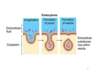
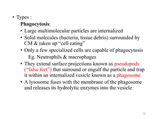
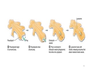
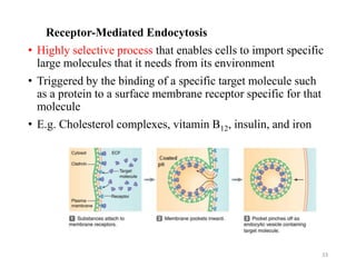
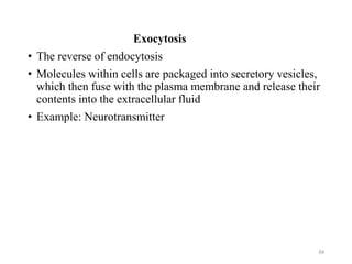
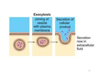
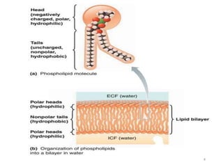
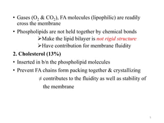
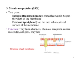
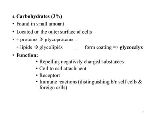
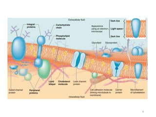
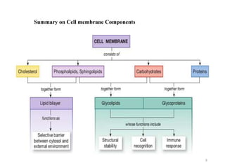
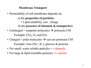
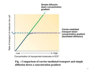
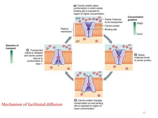
CHAPTER TWO
MEMBRANE PHYSIOLOGY
1
Cell Membrane
• A thin, pliable, elastic structure that envelops the cell
• Only 7.5 to 10 nanometers thick
• General function:
• Physical isolation
• Regulation of exchange with the environment
• Communication b/n the cell and its environment
• Structural support
2
• Major components:
1. Phospholipids (25%)
• Form the basic structure of cell membrane
• Amphipathic – have polar (water- soluble) & non polar
(water- insoluble) regions
• In aqueous solution:
a. Hydrophilic head: oriented towards the outer surface
(interact with water)
b. Hydrophobic tails: oriented themselves into the center
away from water
• They form double phospholipid layers = Lipid bilayer
• Lipid bilayer – creates a semi-permeable barrier
3
4
• Gases (O2 & CO2), FA molecules (lipophilic) are readily
cross the membrane
• Phospholipids are not held together by chemical bonds
Make the lipid bilayer is not rigid structure
Have contribution for membrane fluidity
2. Cholesterol (13%)
• Inserted in b/n the phospholipid molecules
• Prevent FA chains form packing together & crystallizing
≠ contributes to the fluidity as well as stability of
the membrane
5
3. Membrane proteins (55%)
• Two types:
Integral (transmembrane): embedded within & span
the width of the membrane
Extrinsic (peripheral): on the internal or external
surface of the membrane
• Function: They form channels, chemical receptors, carrier
molecules, antigens, enzymes
6
Structure of cell membrane
4. Carbohydrates (3%)
• Found in small amount
• Located on the outer surface of cells
• + proteins  glycoproteins
+ lipids  glycolipids form coating => glycocalyx
• Function:
• Repelling negatively charged substances
• Cell to cell attachment
• Receptors
• Immune reactions (distinguishing b/n self cells &
foreign cells)
7
8
Summary on Cell membrane Components
9
Membrane Transport
• Permeability of cell membrane depends on:
a) the properties of particles:
• Lipid solubility, size , charge
b) the presence of channels & transporters
• Uncharged + nonpolar molecules  permeate CM
Example: CO2, O2 and FAs
• Charged + polar molecules  can not permeate CM
Example: Ions (Na+, K+), glucose & proteins
• For small, water soluble particles => channels
• For large & lipid insoluble particles => carriers
10
Transport Across Cell Membranes
• Passive:- occur down conc. or electrochemical gradient
(“downhill”)
• Does not require metabolic energy
• Simple diffusion, facilitated diffusion & osmosis
• Active transport:- occur against conc. or electrochemical
gradient (“uphill”)
• Requires metabolic energy in the form of
ATP
• 1o active transport, 2o active transport &
• Vesicular transport: endocytosis & exocytosis
• NB: Facilitated diffusion and active transport are carrier-
mediated
11
Simple Diffusion
• Not carrier mediated
• Occur down conc. or electrochemical gradient (“downhill”)
• Does not require metabolic energy
• The rate at which a material diffuses through a membrane
(flux) is given by Fick's law of diffusion
12
13
Simple diffusion can occur across the CM through:
(a) (b)
Intermolecular spaces Membrane openings
(Channels)
Osmosis
• Net diffusion of water down its concentration gradient
through a selectively permeable membrane
• Water moves toward an area of higher solutes conc. or from
high osmotic potential to low osmotic potential
• Water molecules are strongly polar, but they are small.
• So, they can readily permeate the cell membrane
• However, this type of water movement across the membrane
is relatively slow
• Aquaporins, which are channels specific for the passage of
water (aqua means “water”), greatly increases membrane
permeability to water
14
Tonicity
• refers to the effect the conc. of non-penetrating solutes in a
solution has on cell volume
1. Isotonic solution
• Has the same conc. of non-penetrating solutes as normal
body cells do.
• No water enters or leaves the cell by osmosis
• Cell volume remains constant
2. Hypotonic solution
• A solution with a below-normal conc. of non-penetrating
solutes (and therefore a higher conc. of water)
• Water enters the cells by osmosis, causes them to swell,
rupture or lyse
15
3. Hypertonic solution
• a solution with an above-normal conc. of non-penetrating
solutes (and therefore a lower conc. of water)
• The cells shrink as they lose water by osmosis
• Cell decreases in volume with a crenated, or spiky, shape
16
17
Carrier-mediated transport
• A carrier protein spans the thickness of the plasma membrane
and can change its conformation (shape) so that specific
binding sites within the carrier are alternately exposed to the
ECF and the ICF.
Characteristics
1. Specificity:
• Carrier proteins bind only with select substances that fit into its
binding site
2. Saturation:
• Carrier binds substances until their maximum capacity. This
limit is known as the transport maximum (Tm)
• When the Tm reached, the carrier is saturated
18
Fig. : Comparison of carrier-mediated transport and simple
diffusion down a concentration gradient
19
3. Competition
• Different substances with similar chemical structures may be
able bind to the same carrier protein & the therefore compute
for transport across the membrane
• E.g. AAs glycine and alanine
20
Facilitated Diffusion
• Occurs down an electrochemical gradient(“downhill”), similar
to simple diffusion
• Does not require metabolic energy and therefore is passive.
• Is more rapid than simple diffusion
• Is carrier-mediated and therefore exhibits specificity,
saturation, and competition
• E.g.
a. Glucose transport by the glucose transporter (GLUT) across
intestinal epithelium
b. The transport of glucose into RBCs, muscles and adipose
tissue in the presence of insulin
21
22
Mechanism of facilitated diffusion
Active Transport
• Uses carrier proteins
• Carriers transport the sub. uphill against its conc. gradient
• Requires energy
• Two forms: 1o active transport, 2o active transport
1o active transport
• Occurs against an electrochemical gradient(“uphill”).
• Requires direct input of metabolic energy in the form of ATP
and therefore is active.
• Is carrier-mediated and therefore exhibits specificity,
saturation, and competition
23
• Examples:
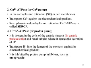
1. Na+/K+-ATPase (or Na+–K+ pump)
• In cell membranes
• Transports Na+ from ICF to ECF and K+ from ECF to ICF It
maintains low intracellular [Na+] and high intracellular [K+]
• Both Na+ and K+ are transported against their electrochemical
gradients
• The usual stoichiometry is 3 Na+/2 K+
• Produces net movement of positive charge out of the cell (an
electrogenic pump)
• This electrical potential is a basic requirement in nerve and
muscle fibers for transmitting electrical signals
• Specific inhibitors of Na+, K+-ATPase are the cardiac glycoside
drugs ouabain and digitalis
24
2. Ca2+-ATPase (or Ca2+pump)
• In the sarcoplasmic reticulum (SR) or cell membranes
• Transports Ca2+against an electrochemical gradient.
• Sarcoplasmic and endoplasmic reticulum Ca2+-ATPase is
called SERCA
3. H+/K+-ATPase (or proton pump)
• It is present in the cells of the gastric mucosa (in gastric
parietal cells) and renal tubules where it causes the secretion
of H+
• Transports H+ into the lumen of the stomach against its
electrochemical gradient
• It is inhibited by proton pump inhibitors, such as
omeprazole
25
Secondary Active Transport
• The transport of two or more solutes is coupled
• One of the solutes (usually Na+) is transported “downhill” and
provides energy for the “uphill” transport of the other solute(s)
• Metabolic energy is not provided directly, but indirectly from
the Na+ gradient that is maintained across cell membranes.
• Thus, inhibition of Na+ ,K+-ATPase will decrease transport of
Na+ out of the cell, decrease the transmembrane Na+ gradient,
and eventually inhibit secondary active transport
• If the solutes move in the same direction across the cell
membrane, it is called cotransport, or symport
26
• Examples:
• Na+– amino acids, Na+–glucose cotransport in the
small intestine
• Na+–K+–2Cl– cotransport in the renal thick ascending
limb
• If the solutes move in opposite directions across the cell
membranes, it is called counter -transport, exchange, or
antiport.
Examples: Na+–Ca2+exchange (in heart muscle cell)
Na+–H+ exchange (in renal tubules)
27
Vesicular Transport
• Allows the transport of macromolecules and multimolecular
particles between the ECF and ICF
• Requires energy expenditure by the cell
• Energy is needed to accomplish vesicle formation and vesicle
movement within the cell
• There are two mechanisms: endocytosis & exocytosis
Endocytosis
• Engulfing of materials by invaginating (folding inward) the
outer part of the membrane
28
29
• Types :
Phagocytosis:
• Large multimolecular particles are internalized
• Solid molecules (bacteria, tissue debris) surrounded by
CM & taken up “cell eating”
• Only a few specialized cells are capable of phagocytosis
Eg. Neutrophils & macrophages
• They extend surface projections known as pseudopods
(“false feet”) that surround or engulf the particle and trap
it within an internalized vesicle known as a phagosome
• A lysosome fuses with the membrane of the phagosome
and releases its hydrolytic enzymes into the vesicle
30
31
• Pinocytosis: droplets of ECF is taken up nonselectively “cell
drinking”
32
Receptor-Mediated Endocytosis
• Highly selective process that enables cells to import specific
large molecules that it needs from its environment
• Triggered by the binding of a specific target molecule such
as a protein to a surface membrane receptor specific for that
molecule
• E.g. Cholesterol complexes, vitamin B12, insulin, and iron
33
Exocytosis
• The reverse of endocytosis
• Molecules within cells are packaged into secretory vesicles,
which then fuse with the plasma membrane and release their
contents into the extracellular fluid
• Example: Neurotransmitter
34
35
The end
36

Chapter 2- Membrane physiology.power pointspptx

  • 1.
  • 2.
    Cell Membrane • Athin, pliable, elastic structure that envelops the cell • Only 7.5 to 10 nanometers thick • General function: • Physical isolation • Regulation of exchange with the environment • Communication b/n the cell and its environment • Structural support 2
  • 3.
    • Major components: 1.Phospholipids (25%) • Form the basic structure of cell membrane • Amphipathic – have polar (water- soluble) & non polar (water- insoluble) regions • In aqueous solution: a. Hydrophilic head: oriented towards the outer surface (interact with water) b. Hydrophobic tails: oriented themselves into the center away from water • They form double phospholipid layers = Lipid bilayer • Lipid bilayer – creates a semi-permeable barrier 3
  • 4.
  • 5.
    • Gases (O2& CO2), FA molecules (lipophilic) are readily cross the membrane • Phospholipids are not held together by chemical bonds Make the lipid bilayer is not rigid structure Have contribution for membrane fluidity 2. Cholesterol (13%) • Inserted in b/n the phospholipid molecules • Prevent FA chains form packing together & crystallizing ≠ contributes to the fluidity as well as stability of the membrane 5
  • 6.
    3. Membrane proteins(55%) • Two types: Integral (transmembrane): embedded within & span the width of the membrane Extrinsic (peripheral): on the internal or external surface of the membrane • Function: They form channels, chemical receptors, carrier molecules, antigens, enzymes 6 Structure of cell membrane
  • 7.
    4. Carbohydrates (3%) •Found in small amount • Located on the outer surface of cells • + proteins  glycoproteins + lipids  glycolipids form coating => glycocalyx • Function: • Repelling negatively charged substances • Cell to cell attachment • Receptors • Immune reactions (distinguishing b/n self cells & foreign cells) 7
  • 8.
  • 9.
    Summary on Cellmembrane Components 9
  • 10.
    Membrane Transport • Permeabilityof cell membrane depends on: a) the properties of particles: • Lipid solubility, size , charge b) the presence of channels & transporters • Uncharged + nonpolar molecules  permeate CM Example: CO2, O2 and FAs • Charged + polar molecules  can not permeate CM Example: Ions (Na+, K+), glucose & proteins • For small, water soluble particles => channels • For large & lipid insoluble particles => carriers 10
  • 11.
    Transport Across CellMembranes • Passive:- occur down conc. or electrochemical gradient (“downhill”) • Does not require metabolic energy • Simple diffusion, facilitated diffusion & osmosis • Active transport:- occur against conc. or electrochemical gradient (“uphill”) • Requires metabolic energy in the form of ATP • 1o active transport, 2o active transport & • Vesicular transport: endocytosis & exocytosis • NB: Facilitated diffusion and active transport are carrier- mediated 11
  • 12.
    Simple Diffusion • Notcarrier mediated • Occur down conc. or electrochemical gradient (“downhill”) • Does not require metabolic energy • The rate at which a material diffuses through a membrane (flux) is given by Fick's law of diffusion 12
  • 13.
    13 Simple diffusion canoccur across the CM through: (a) (b) Intermolecular spaces Membrane openings (Channels)
  • 14.
    Osmosis • Net diffusionof water down its concentration gradient through a selectively permeable membrane • Water moves toward an area of higher solutes conc. or from high osmotic potential to low osmotic potential • Water molecules are strongly polar, but they are small. • So, they can readily permeate the cell membrane • However, this type of water movement across the membrane is relatively slow • Aquaporins, which are channels specific for the passage of water (aqua means “water”), greatly increases membrane permeability to water 14
  • 15.
    Tonicity • refers tothe effect the conc. of non-penetrating solutes in a solution has on cell volume 1. Isotonic solution • Has the same conc. of non-penetrating solutes as normal body cells do. • No water enters or leaves the cell by osmosis • Cell volume remains constant 2. Hypotonic solution • A solution with a below-normal conc. of non-penetrating solutes (and therefore a higher conc. of water) • Water enters the cells by osmosis, causes them to swell, rupture or lyse 15
  • 16.
    3. Hypertonic solution •a solution with an above-normal conc. of non-penetrating solutes (and therefore a lower conc. of water) • The cells shrink as they lose water by osmosis • Cell decreases in volume with a crenated, or spiky, shape 16
  • 17.
  • 18.
    Carrier-mediated transport • Acarrier protein spans the thickness of the plasma membrane and can change its conformation (shape) so that specific binding sites within the carrier are alternately exposed to the ECF and the ICF. Characteristics 1. Specificity: • Carrier proteins bind only with select substances that fit into its binding site 2. Saturation: • Carrier binds substances until their maximum capacity. This limit is known as the transport maximum (Tm) • When the Tm reached, the carrier is saturated 18
  • 19.
    Fig. : Comparisonof carrier-mediated transport and simple diffusion down a concentration gradient 19
  • 20.
    3. Competition • Differentsubstances with similar chemical structures may be able bind to the same carrier protein & the therefore compute for transport across the membrane • E.g. AAs glycine and alanine 20
  • 21.
    Facilitated Diffusion • Occursdown an electrochemical gradient(“downhill”), similar to simple diffusion • Does not require metabolic energy and therefore is passive. • Is more rapid than simple diffusion • Is carrier-mediated and therefore exhibits specificity, saturation, and competition • E.g. a. Glucose transport by the glucose transporter (GLUT) across intestinal epithelium b. The transport of glucose into RBCs, muscles and adipose tissue in the presence of insulin 21
  • 22.
  • 23.
    Active Transport • Usescarrier proteins • Carriers transport the sub. uphill against its conc. gradient • Requires energy • Two forms: 1o active transport, 2o active transport 1o active transport • Occurs against an electrochemical gradient(“uphill”). • Requires direct input of metabolic energy in the form of ATP and therefore is active. • Is carrier-mediated and therefore exhibits specificity, saturation, and competition 23
  • 24.
    • Examples: 1. Na+/K+-ATPase(or Na+–K+ pump) • In cell membranes • Transports Na+ from ICF to ECF and K+ from ECF to ICF It maintains low intracellular [Na+] and high intracellular [K+] • Both Na+ and K+ are transported against their electrochemical gradients • The usual stoichiometry is 3 Na+/2 K+ • Produces net movement of positive charge out of the cell (an electrogenic pump) • This electrical potential is a basic requirement in nerve and muscle fibers for transmitting electrical signals • Specific inhibitors of Na+, K+-ATPase are the cardiac glycoside drugs ouabain and digitalis 24
  • 25.
    2. Ca2+-ATPase (orCa2+pump) • In the sarcoplasmic reticulum (SR) or cell membranes • Transports Ca2+against an electrochemical gradient. • Sarcoplasmic and endoplasmic reticulum Ca2+-ATPase is called SERCA 3. H+/K+-ATPase (or proton pump) • It is present in the cells of the gastric mucosa (in gastric parietal cells) and renal tubules where it causes the secretion of H+ • Transports H+ into the lumen of the stomach against its electrochemical gradient • It is inhibited by proton pump inhibitors, such as omeprazole 25
  • 26.
    Secondary Active Transport •The transport of two or more solutes is coupled • One of the solutes (usually Na+) is transported “downhill” and provides energy for the “uphill” transport of the other solute(s) • Metabolic energy is not provided directly, but indirectly from the Na+ gradient that is maintained across cell membranes. • Thus, inhibition of Na+ ,K+-ATPase will decrease transport of Na+ out of the cell, decrease the transmembrane Na+ gradient, and eventually inhibit secondary active transport • If the solutes move in the same direction across the cell membrane, it is called cotransport, or symport 26
  • 27.
    • Examples: • Na+–amino acids, Na+–glucose cotransport in the small intestine • Na+–K+–2Cl– cotransport in the renal thick ascending limb • If the solutes move in opposite directions across the cell membranes, it is called counter -transport, exchange, or antiport. Examples: Na+–Ca2+exchange (in heart muscle cell) Na+–H+ exchange (in renal tubules) 27
  • 28.
    Vesicular Transport • Allowsthe transport of macromolecules and multimolecular particles between the ECF and ICF • Requires energy expenditure by the cell • Energy is needed to accomplish vesicle formation and vesicle movement within the cell • There are two mechanisms: endocytosis & exocytosis Endocytosis • Engulfing of materials by invaginating (folding inward) the outer part of the membrane 28
  • 29.
  • 30.
    • Types : Phagocytosis: •Large multimolecular particles are internalized • Solid molecules (bacteria, tissue debris) surrounded by CM & taken up “cell eating” • Only a few specialized cells are capable of phagocytosis Eg. Neutrophils & macrophages • They extend surface projections known as pseudopods (“false feet”) that surround or engulf the particle and trap it within an internalized vesicle known as a phagosome • A lysosome fuses with the membrane of the phagosome and releases its hydrolytic enzymes into the vesicle 30
  • 31.
  • 32.
    • Pinocytosis: dropletsof ECF is taken up nonselectively “cell drinking” 32
  • 33.
    Receptor-Mediated Endocytosis • Highlyselective process that enables cells to import specific large molecules that it needs from its environment • Triggered by the binding of a specific target molecule such as a protein to a surface membrane receptor specific for that molecule • E.g. Cholesterol complexes, vitamin B12, insulin, and iron 33
  • 34.
    Exocytosis • The reverseof endocytosis • Molecules within cells are packaged into secretory vesicles, which then fuse with the plasma membrane and release their contents into the extracellular fluid • Example: Neurotransmitter 34
  • 35.
  • 36.