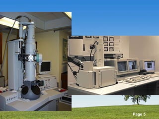
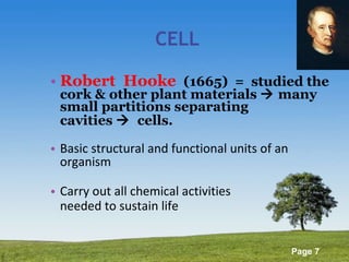
The Cell
Antoni  van  Leeuwenhoek (1632-1723), Dutch, maker of first  single  lens  microscope The  first  to  document  the  structure  of  RBC  & the  nature  of  the  circulatory  system protozoans  &  bacteria, life  cycles  of  many species  of  insects. MICROMETER  ( µ m) = 1/1000
Types  of  Microscopes 1.  COMPOUND/LIGHT MICROSCOPE Principle: sunlight (light  source)  2.  ELECTRON MICROSCOPE Principle:  beam of electrons  a.  Transmission (magnification: >/=1Mx) b.  Scanning – 3D image <250,000x Magnification = extent to which an image is enlarged
 
 
Image under Compound  microscope Scanning  EM Transmission  EM
CELL Robert  Hooke   (1665)  =  studied the cork & other plant materials    many small partitions separating cavities     cells. Basic structural and functional units of an organism Carry out all chemical activities  needed to sustain life
Cell Diversity Cells that connect body parts Cells that cover and line body organs Cells that move organs and body parts Cell that stores nutrients Cell that fights disease Cell that gathers information and controls body functions Cells of reproduction
3 Main Parts 1. PLASMA MEMBRANE 2. CYTOPLASM 3. NUCLEUS
1. Plasma Membrane syn:  plasmalemma, cell membrane - outer covering of the cell - surrounds each cell    separate its contents from external environment - regulates  what  enters  &  leaves  the  cell - allows communication
Plasma Membrane some, cholesterol and glycolipids LIPID BILAYER + integral and peripheral proteins SELECTIVE PERMEABILITY  - water & nonpolar (lipid-soluble) molecules e.g. FA, fat-soluble vit., steroids, O2, CO2  mainly made up of phospholipids and proteins (latter, mainly glycoproteins)
Plasma Membrane Impermeable to: ions, glucose, a.a. transport assisted by: ion channels, transporters Integral proteins act as: receptors, enzymes, cell identity markers
Plasma Membrane
rane
Definition of Terms Intracellular fluid (ICF)  – inside body cells i.e. fluid in cytoplasm, 2/3 of body fluid  Extracellular fluid (ECF) – fluid outside body cells Interstitial fluid  – the ECF between cells  e.g. plasma (in blood vessels), lymph (in lymphatic vessels)  ** materials dissolved in body fluids: gases, nutrients, ions, etc.
Definition of Terms Solute – any material dissolved in a fluid Solvent – fluid in w/c the solute is dissolved e.g. water Solution – homogenous mixture of 2 or more components (e.g. air, seawater, alcohol)
Definition of Terms CONCENTRATION  – the amount of a solute in a solution CONCENTRATION GRADIENT  – difference in concentration between 2 different areas  moving down vs. moving up
Substances move across cellular membranes by: PASSIVE PROCESSES  – substance moves down its concentration gradient using only its own energy of motion (kinetic energy) SIMPLE DIFFUSION, OSMOSIS ACTIVE PROCESSES   – cellular energy (e.g. ATP) is used to push the substance through the membrane  against its concentration gradient ACTIVE TRANSPORT, VESICLES
Passive Processes DIFFUSION  – a substance moves from one place to another due to the substance's kinetic energy  particles move from a region of higher to lower concentration  endpoint:  EQUILIBRIUM –   substance is evenly distributed throughout the solution and the concentration gradient disappears  may or may not involve a membrane
Diffusion
2 Types of Diffusion 1. SIMPLE DIFFUSION  lipid-soluble substances diffuse through the lipid bilayer e.g. O2, CO2, N, f.a., steroids, vit. ADEK, H2O, urea used in: exchange of gases, absorption of nutrients, release of wastes
Simple Diffusion
2 Types of Diffusion 1. SIMPLE DIFFUSION  Ion channels – allow a specific type of ion to move across the membrane through the channel's pore e.g. K, Cl, Na, Ca
 
2 Types of Diffusion 2. FACILITATED DIFFUSION  an integral membrane protein assists a specific substance across the membrane substance binds to a specific TRANSPORTER on one side of the membrane --> released on the other side after the transporter undergoes a change in shape movement along a conc. gradient, NO ATP needed e.g. glucose, fructose, galactose
 
Passive Processes 2.  OSMOSIS  = net movement of water through a selectively permeable membrane - water moves from an area of higher water to an area of lower water concentration - or, from an area of lower solute concentration to an area of higher solute concentration OSMOTIC PRESSURE – depends on the concentration of its solute particles  - the higher the solute conc., the higher the osmotic pressure
Osmosis
EFFECT  of  OSMOSIS  in  CELLS ISOTONIC   = any solution in w/c cells maintain their normal shape and volume, concentrations of solutes are same on both sides   =  tissue  fluids, blood  plasma, 0.9%  NSS, 5%  dextrose (glucose) HYPOTONIC   = lower concentration of solutes (higher conc. of H2O) than the cytosol inside the cell    = cell swells --> ruptures HYPERTONIC  = higher concentration of solutes (lower conc. of water) than the cytosol inside the cell   = cell shrinks
Effects of Osmosis
Passive Processes 3. FILTRATION  = process by w/c H2O and solutes are forced through a membrane (or capillary wall) by  fluid  or  hydrostatic pressure  (pressure gradient) e.g.  filtering capacity of the kidney (e.g. in urine formation)
Active Processes 1.  ACTIVE TRANSPORT   - cellular energy is used to transport substances across the membrane against a concentration gradient (from an area of low to an area of high concentration), needs ATP  - splitting of ATP changes the shape of a transporter protein (PUMP) --> moves a substance across the membrane against its conc. gradient - Na, K, H, Ca, I, Cl
Active Transport
Active Processes 2.  TRANSPORT IN VESICLES  - VESICLE   –  small round sac formed by budding off from an existing membrane - transport substances, take in and release substances  - requires energy (ATP)
2 Types of Transport in Vesicles 1. ENDOCYTOSIS –   materials move into a cell in a vesicle formed from the plasma membrane   - substances are surrounded by a piece of the plasma membrane w/c buds off inside the cell to form a vesicle containing the ingested substances.
1. PHAGOCYTOSIS –   large solid particles (e.g. bacteria, viruses, aged or dead cells), are taken in by the cell  - fuses w/ a lysosome --> break down of material - e.g. WBCs, macrophages 2 Types of Endocytosis
2. BULK-PHASE ENDOCYTOSIS (PINOCYTOSIS) –   cells take up tiny droplets of ECF  - fuses with a lysosome --> enzymatic breakdown of engulfed solutes 2 Types of Endocytosis
2 Types of Transport in Vesicles 2. EXOCYTOSIS   – results in secretion (release of materials from a cell) a. SECRETORY CELLS – release digestive enzymes, hormones,  mucus, etc. b. NERVE CELLS – during release of neurotransmitters  * membrane-enclosed secretory vesicles form inside the cell, fuse w/ the cell membrane, and release contents into the ECF
Exocytosis
Table. 3.2
Specializations of the Plasma Membrane Microvilli Membrane junctions
Membrane Junctions 1. TIGHT JUNCTIONS  = formed from fusion of adjacent cell membrane --> impermeable or leak-proof sheets = keep digestive juices & harmful substances from damaging the organs or getting into the bloodstream 2. DESMOSOMES  = anchoring junctions (button-like thickenings),  prevent cells under mechanical stress from being pulled apart e.g.  skin 3. GAP JUNCTIONS  = allows communication e.g.  heart, nervous system
2. Cytoplasm consists of all cellular contents bet. the cell membrane and nucleus Includes:   CYTOSOL   (ICF)   – fluid portion of the cytoplasm, 55% of total cell volume, 75-90% H2O, site of chemical reactions ORGANELLES   – specialized structures inside cells w/ specific functions
1. Cytoskeleton network of 3 different types of protein filaments 1. MICROFILAMENTS  – thinnest, concentrated at the periphery --> strength and shape - provides mechanical support and generates movement - anchor cytoskeleton to integral proteins - support for microvilli - intercellular attachment
1. Cytoskeleton 2. INTERMEDIATE FILAMENTS   – found in parts of cells subject to tension (stretching), hold organelles in place, intercellular attachment  3. MICROTUBULES   – long, hollow tubes, determines cell shape, movement of organelles w/in the cell, migration of chromosomes during cell division, movement of cilia and flagella
 
found near the nucleus Includes: 1. Centrioles (paired) – composed of microtubules 2. Pericentriolar material – composed of tubulins, organizing centers for growth of the mitotic spindle (role in cell division) 2 . Centrosome
3. Cilia and Flagella CILIA  – short, hairlike projections extending from the surface of the cell propel fluids across surfaces of cells FLAGELLA  - move an entire cell
4. Ribosomes Tiny, round, dark bodies Actual site of protein production high rRNA content Free ribosomes attached to RER
5. Endoplasmic Reticulum (ER) network of folded membranes  2 Types Rough ER   –  extends from the nuclear envelope, studded w/ ribosomes, synthesis of secretory proteins and membrane molecules Smooth ER  –  extends from the RER --> network of membranous tubules, lacks ribosomes, f.a. & steroids (e.g. estrogen, testosterone) are produced,  detoxification (e.g. alc, pesticides, carcinogens)
5. Endoplasmic Reticulum (ER)
6. Golgi Complex Stack of flattened membranous sacs Modify and package proteins secretory vesicles carries proteins and phospholipids to become part of cell membrane incorporated in lysosomes
 
7. Lysosomes membrane-encosed vesicles, >/= 60 digestive enzymes digestion, recycling autophagy, autolysis
 
8. Peroxisomes Sacs containing oxidase enzymes Use oxygen to detoxify harmful or poisonous substances (e.g. alcohol, formaldehyde) Disarm “free radicals”  FR -> H2O2->H2O liver and kidney cells
9. Proteasomes continuous destruction of unneeded , damaged or faulty proteins  contain proteases
10. Mitochondria powerhouse of the cell (site of ATP production) increased in: muscles, liver, kidneys
3. Nucleus most prominent feature of a cell NUCLEAR ENVELOPE – separates the nucleus from the cytoplasm NUCLEAR PORES – control movement of substances NUCLEOLI – sites of assembly of ribosomes
3. Nucleus GENES  – hereditary units direct cellular activities  arranged along chromosomes  46 chromosomes (23/parent) in a nondividing cell, chromosomes appear as diffuse granular mass -->   CHROMATIN GENOME  – total genetic information carried in a cell or organism


Protein Synthesis DNA found in genes gives instructions for making proteins 1. Transcription – DNA is copied --> RNA 2. Translation – information in RNA (attached to a ribosome) is translated into a sequence of a.a. --> protein molecule
Transcription occurs in the nucleus genetic info in DNA  base triplets  is copied into complementary sequence of  CODONS  in a strand of RNA (helped by RNA polymerase) PROMOTER  – sequence of nucleotides in DNA where RNA polymerase attaches to  TERMINATOR  – sequence of nucleotides in DNA where transcription ends
3 Kinds of RNA 1.  Messenger RNA  (mRNA )  – directs synthesis of a protein 2.  Ribosomal RNA  (rRNA)  – joins w/ ribosomal proteins to make ribosomes 3.  Transfer RNA (tRNA)  – binds to an a.a. and holds it in place on a ribosome until it becomes part of a protein during translation
Base pairing
Translation mRNA attaches to ribosomes and directs protein synthesis by converting sequence of nucleotides (CODON) into a specific sequence of a.a. --> PROTEIN  tRNA contains the triplet of nucleotides called ANTICODON Protein synthesis ends when ribosome reaches a STOP CODON 15 a.a./second
 
Somatic Cell Division process by which damaged, diseased or worn out cells are replaced process by which cells reproduce themselves
2 Types of Cell Division 1. REPRODUCTIVE CELL DIVISION (MEIOSIS) - the process that produces gametes (sperm & oocytes) 2. SOMATIC CELL DIVISION - division of all body cells (except gametes) --> two identical cells
Prerequisite of somatic cell division: DNA Replication  DNA – building blocks  “nucleotides” Deoxyribose sugar Phosphate group  Nitrogen-containing base
duplication of the DNA sequences that make up the genes and chromosomes --> daughter cells w/ same genes and same number of chromosomes  Prerequisite of somatic cell division: DNA Replication
DNA Replication
Cell Cycle sequence of changes that a cell undergoes from the time it forms until it duplicates its contents and divides into two cells 2  MAJOR PERIODS 1.  INTERPHASE – cell is not dividing 2.  MITOTIC PHASE – cell is dividing
DNA replication occurs manufactures organelles and cytosolic components increased metabolic activity cell is growing Interphase
Mitotic Phase consists of: MITOSIS  – nuclear division CYTOKINESIS  – cytoplasmic division into 2 cells
PROPHASE   =  chromatin coil and shorten   chromosomes,  bar-like bodies Chromosome = 2 strands of  chromatids , held together by a buttonlike body,  centromere Mitosis
Mitosis STAGES : METAPHASE  – chromosomes cluster and align  at the center (metaphase plate)
Mitosis STAGES: ANAPHASE   –  movement of chromosomes toward    opposite ends of the cell
Mitosis STAGES: TELOPHASE   –  chromosomes uncoil and become  chromatin again, nuclear envelope forms around  each chromatin mass
Cytokinesis division of a cell's cytoplasm and organelles formation of cleavage furrow that extends around the center of the cell endpoint: 2 new and separate cells
 
END QUIZ NEXT MEETING!

Cell

  • 1.
  • 2.
    Antoni van Leeuwenhoek (1632-1723), Dutch, maker of first single lens microscope The first to document the structure of RBC & the nature of the circulatory system protozoans & bacteria, life cycles of many species of insects. MICROMETER ( µ m) = 1/1000
  • 3.
    Types of Microscopes 1. COMPOUND/LIGHT MICROSCOPE Principle: sunlight (light source) 2. ELECTRON MICROSCOPE Principle: beam of electrons a. Transmission (magnification: >/=1Mx) b. Scanning – 3D image <250,000x Magnification = extent to which an image is enlarged
  • 4.
  • 5.
  • 6.
    Image under Compound microscope Scanning EM Transmission EM
  • 7.
    CELL Robert Hooke (1665) = studied the cork & other plant materials  many small partitions separating cavities  cells. Basic structural and functional units of an organism Carry out all chemical activities needed to sustain life
  • 8.
    Cell Diversity Cellsthat connect body parts Cells that cover and line body organs Cells that move organs and body parts Cell that stores nutrients Cell that fights disease Cell that gathers information and controls body functions Cells of reproduction
  • 9.
    3 Main Parts1. PLASMA MEMBRANE 2. CYTOPLASM 3. NUCLEUS
  • 10.
    1. Plasma Membranesyn: plasmalemma, cell membrane - outer covering of the cell - surrounds each cell  separate its contents from external environment - regulates what enters & leaves the cell - allows communication
  • 11.
    Plasma Membrane some,cholesterol and glycolipids LIPID BILAYER + integral and peripheral proteins SELECTIVE PERMEABILITY - water & nonpolar (lipid-soluble) molecules e.g. FA, fat-soluble vit., steroids, O2, CO2 mainly made up of phospholipids and proteins (latter, mainly glycoproteins)
  • 12.
    Plasma Membrane Impermeableto: ions, glucose, a.a. transport assisted by: ion channels, transporters Integral proteins act as: receptors, enzymes, cell identity markers
  • 13.
  • 14.
  • 15.
    Definition of TermsIntracellular fluid (ICF) – inside body cells i.e. fluid in cytoplasm, 2/3 of body fluid Extracellular fluid (ECF) – fluid outside body cells Interstitial fluid – the ECF between cells e.g. plasma (in blood vessels), lymph (in lymphatic vessels) ** materials dissolved in body fluids: gases, nutrients, ions, etc.
  • 16.
    Definition of TermsSolute – any material dissolved in a fluid Solvent – fluid in w/c the solute is dissolved e.g. water Solution – homogenous mixture of 2 or more components (e.g. air, seawater, alcohol)
  • 17.
    Definition of TermsCONCENTRATION – the amount of a solute in a solution CONCENTRATION GRADIENT – difference in concentration between 2 different areas moving down vs. moving up
  • 18.
    Substances move acrosscellular membranes by: PASSIVE PROCESSES – substance moves down its concentration gradient using only its own energy of motion (kinetic energy) SIMPLE DIFFUSION, OSMOSIS ACTIVE PROCESSES – cellular energy (e.g. ATP) is used to push the substance through the membrane against its concentration gradient ACTIVE TRANSPORT, VESICLES
  • 19.
    Passive Processes DIFFUSION – a substance moves from one place to another due to the substance's kinetic energy particles move from a region of higher to lower concentration endpoint: EQUILIBRIUM – substance is evenly distributed throughout the solution and the concentration gradient disappears may or may not involve a membrane
  • 20.
  • 21.
    2 Types ofDiffusion 1. SIMPLE DIFFUSION lipid-soluble substances diffuse through the lipid bilayer e.g. O2, CO2, N, f.a., steroids, vit. ADEK, H2O, urea used in: exchange of gases, absorption of nutrients, release of wastes
  • 22.
  • 23.
    2 Types ofDiffusion 1. SIMPLE DIFFUSION Ion channels – allow a specific type of ion to move across the membrane through the channel's pore e.g. K, Cl, Na, Ca
  • 24.
  • 25.
    2 Types ofDiffusion 2. FACILITATED DIFFUSION an integral membrane protein assists a specific substance across the membrane substance binds to a specific TRANSPORTER on one side of the membrane --> released on the other side after the transporter undergoes a change in shape movement along a conc. gradient, NO ATP needed e.g. glucose, fructose, galactose
  • 26.
  • 27.
    Passive Processes 2. OSMOSIS = net movement of water through a selectively permeable membrane - water moves from an area of higher water to an area of lower water concentration - or, from an area of lower solute concentration to an area of higher solute concentration OSMOTIC PRESSURE – depends on the concentration of its solute particles - the higher the solute conc., the higher the osmotic pressure
  • 28.
  • 29.
    EFFECT of OSMOSIS in CELLS ISOTONIC = any solution in w/c cells maintain their normal shape and volume, concentrations of solutes are same on both sides = tissue fluids, blood plasma, 0.9% NSS, 5% dextrose (glucose) HYPOTONIC = lower concentration of solutes (higher conc. of H2O) than the cytosol inside the cell = cell swells --> ruptures HYPERTONIC = higher concentration of solutes (lower conc. of water) than the cytosol inside the cell = cell shrinks
  • 30.
  • 31.
    Passive Processes 3.FILTRATION = process by w/c H2O and solutes are forced through a membrane (or capillary wall) by fluid or hydrostatic pressure (pressure gradient) e.g. filtering capacity of the kidney (e.g. in urine formation)
  • 32.
    Active Processes 1. ACTIVE TRANSPORT - cellular energy is used to transport substances across the membrane against a concentration gradient (from an area of low to an area of high concentration), needs ATP - splitting of ATP changes the shape of a transporter protein (PUMP) --> moves a substance across the membrane against its conc. gradient - Na, K, H, Ca, I, Cl
  • 33.
  • 34.
    Active Processes 2. TRANSPORT IN VESICLES - VESICLE – small round sac formed by budding off from an existing membrane - transport substances, take in and release substances - requires energy (ATP)
  • 35.
    2 Types ofTransport in Vesicles 1. ENDOCYTOSIS – materials move into a cell in a vesicle formed from the plasma membrane - substances are surrounded by a piece of the plasma membrane w/c buds off inside the cell to form a vesicle containing the ingested substances.
  • 36.
    1. PHAGOCYTOSIS – large solid particles (e.g. bacteria, viruses, aged or dead cells), are taken in by the cell - fuses w/ a lysosome --> break down of material - e.g. WBCs, macrophages 2 Types of Endocytosis
  • 37.
    2. BULK-PHASE ENDOCYTOSIS(PINOCYTOSIS) – cells take up tiny droplets of ECF - fuses with a lysosome --> enzymatic breakdown of engulfed solutes 2 Types of Endocytosis
  • 38.
    2 Types ofTransport in Vesicles 2. EXOCYTOSIS – results in secretion (release of materials from a cell) a. SECRETORY CELLS – release digestive enzymes, hormones, mucus, etc. b. NERVE CELLS – during release of neurotransmitters * membrane-enclosed secretory vesicles form inside the cell, fuse w/ the cell membrane, and release contents into the ECF
  • 39.
  • 40.
  • 41.
    Specializations of thePlasma Membrane Microvilli Membrane junctions
  • 42.
    Membrane Junctions 1.TIGHT JUNCTIONS = formed from fusion of adjacent cell membrane --> impermeable or leak-proof sheets = keep digestive juices & harmful substances from damaging the organs or getting into the bloodstream 2. DESMOSOMES = anchoring junctions (button-like thickenings), prevent cells under mechanical stress from being pulled apart e.g. skin 3. GAP JUNCTIONS = allows communication e.g. heart, nervous system
  • 43.
    2. Cytoplasm consistsof all cellular contents bet. the cell membrane and nucleus Includes: CYTOSOL (ICF) – fluid portion of the cytoplasm, 55% of total cell volume, 75-90% H2O, site of chemical reactions ORGANELLES – specialized structures inside cells w/ specific functions
  • 44.
    1. Cytoskeleton networkof 3 different types of protein filaments 1. MICROFILAMENTS – thinnest, concentrated at the periphery --> strength and shape - provides mechanical support and generates movement - anchor cytoskeleton to integral proteins - support for microvilli - intercellular attachment
  • 45.
    1. Cytoskeleton 2.INTERMEDIATE FILAMENTS – found in parts of cells subject to tension (stretching), hold organelles in place, intercellular attachment 3. MICROTUBULES – long, hollow tubes, determines cell shape, movement of organelles w/in the cell, migration of chromosomes during cell division, movement of cilia and flagella
  • 46.
  • 47.
    found near thenucleus Includes: 1. Centrioles (paired) – composed of microtubules 2. Pericentriolar material – composed of tubulins, organizing centers for growth of the mitotic spindle (role in cell division) 2 . Centrosome
  • 48.
    3. Cilia andFlagella CILIA – short, hairlike projections extending from the surface of the cell propel fluids across surfaces of cells FLAGELLA - move an entire cell
  • 49.
    4. Ribosomes Tiny,round, dark bodies Actual site of protein production high rRNA content Free ribosomes attached to RER
  • 50.
    5. Endoplasmic Reticulum(ER) network of folded membranes 2 Types Rough ER – extends from the nuclear envelope, studded w/ ribosomes, synthesis of secretory proteins and membrane molecules Smooth ER – extends from the RER --> network of membranous tubules, lacks ribosomes, f.a. & steroids (e.g. estrogen, testosterone) are produced, detoxification (e.g. alc, pesticides, carcinogens)
  • 51.
  • 52.
    6. Golgi ComplexStack of flattened membranous sacs Modify and package proteins secretory vesicles carries proteins and phospholipids to become part of cell membrane incorporated in lysosomes
  • 53.
  • 54.
    7. Lysosomes membrane-encosedvesicles, >/= 60 digestive enzymes digestion, recycling autophagy, autolysis
  • 55.
  • 56.
    8. Peroxisomes Sacscontaining oxidase enzymes Use oxygen to detoxify harmful or poisonous substances (e.g. alcohol, formaldehyde) Disarm “free radicals” FR -> H2O2->H2O liver and kidney cells
  • 57.
    9. Proteasomes continuousdestruction of unneeded , damaged or faulty proteins contain proteases
  • 58.
    10. Mitochondria powerhouseof the cell (site of ATP production) increased in: muscles, liver, kidneys
  • 59.
    3. Nucleus mostprominent feature of a cell NUCLEAR ENVELOPE – separates the nucleus from the cytoplasm NUCLEAR PORES – control movement of substances NUCLEOLI – sites of assembly of ribosomes
  • 60.
    3. Nucleus GENES – hereditary units direct cellular activities arranged along chromosomes 46 chromosomes (23/parent) in a nondividing cell, chromosomes appear as diffuse granular mass --> CHROMATIN GENOME – total genetic information carried in a cell or organism
  • 61.
  • 62.
  • 63.
  • 64.
    Protein Synthesis DNAfound in genes gives instructions for making proteins 1. Transcription – DNA is copied --> RNA 2. Translation – information in RNA (attached to a ribosome) is translated into a sequence of a.a. --> protein molecule
  • 65.
    Transcription occurs inthe nucleus genetic info in DNA base triplets is copied into complementary sequence of CODONS in a strand of RNA (helped by RNA polymerase) PROMOTER – sequence of nucleotides in DNA where RNA polymerase attaches to TERMINATOR – sequence of nucleotides in DNA where transcription ends
  • 66.
    3 Kinds ofRNA 1. Messenger RNA (mRNA ) – directs synthesis of a protein 2. Ribosomal RNA (rRNA) – joins w/ ribosomal proteins to make ribosomes 3. Transfer RNA (tRNA) – binds to an a.a. and holds it in place on a ribosome until it becomes part of a protein during translation
  • 67.
  • 68.
    Translation mRNA attachesto ribosomes and directs protein synthesis by converting sequence of nucleotides (CODON) into a specific sequence of a.a. --> PROTEIN tRNA contains the triplet of nucleotides called ANTICODON Protein synthesis ends when ribosome reaches a STOP CODON 15 a.a./second
  • 69.
  • 70.
    Somatic Cell Divisionprocess by which damaged, diseased or worn out cells are replaced process by which cells reproduce themselves
  • 71.
    2 Types ofCell Division 1. REPRODUCTIVE CELL DIVISION (MEIOSIS) - the process that produces gametes (sperm & oocytes) 2. SOMATIC CELL DIVISION - division of all body cells (except gametes) --> two identical cells
  • 72.
    Prerequisite of somaticcell division: DNA Replication DNA – building blocks “nucleotides” Deoxyribose sugar Phosphate group Nitrogen-containing base
  • 73.
    duplication of theDNA sequences that make up the genes and chromosomes --> daughter cells w/ same genes and same number of chromosomes Prerequisite of somatic cell division: DNA Replication
  • 74.
  • 75.
    Cell Cycle sequenceof changes that a cell undergoes from the time it forms until it duplicates its contents and divides into two cells 2 MAJOR PERIODS 1. INTERPHASE – cell is not dividing 2. MITOTIC PHASE – cell is dividing
  • 76.
    DNA replication occursmanufactures organelles and cytosolic components increased metabolic activity cell is growing Interphase
  • 77.
    Mitotic Phase consistsof: MITOSIS – nuclear division CYTOKINESIS – cytoplasmic division into 2 cells
  • 78.
    PROPHASE = chromatin coil and shorten  chromosomes, bar-like bodies Chromosome = 2 strands of chromatids , held together by a buttonlike body, centromere Mitosis
  • 79.
    Mitosis STAGES :METAPHASE – chromosomes cluster and align at the center (metaphase plate)
  • 80.
    Mitosis STAGES: ANAPHASE – movement of chromosomes toward opposite ends of the cell
  • 81.
    Mitosis STAGES: TELOPHASE – chromosomes uncoil and become chromatin again, nuclear envelope forms around each chromatin mass
  • 82.
    Cytokinesis division ofa cell's cytoplasm and organelles formation of cleavage furrow that extends around the center of the cell endpoint: 2 new and separate cells
  • 83.
  • 84.
    END QUIZ NEXTMEETING!