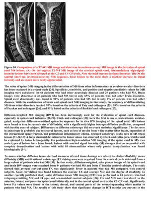
Downloaded 121 times

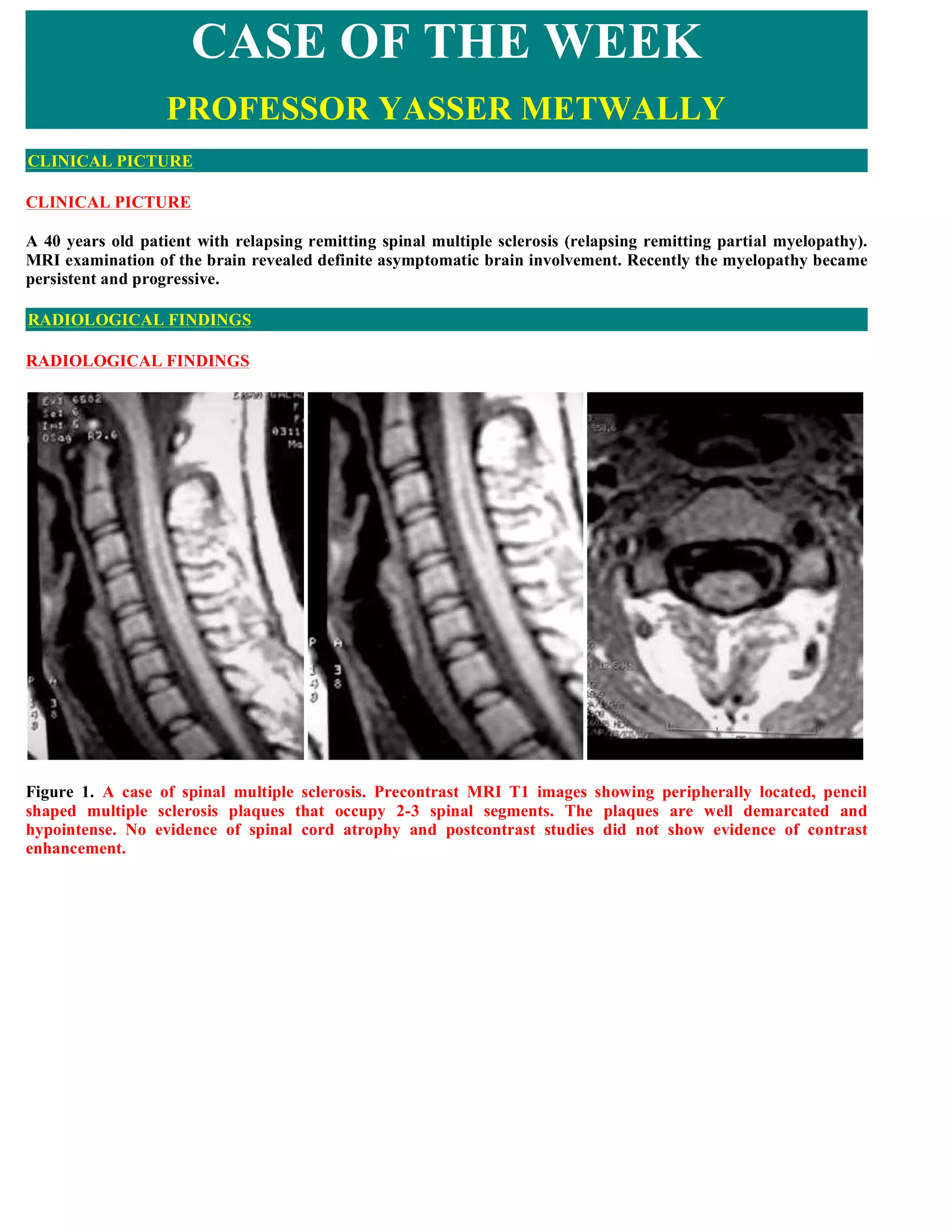
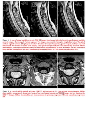
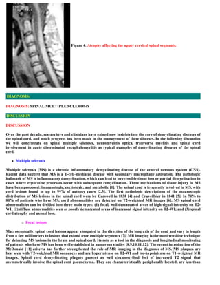
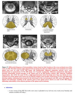


This document discusses a case of spinal multiple sclerosis in a 40-year-old patient. MRI images show well-defined pencil-shaped lesions occupying 2-3 spinal segments that are hypointense on T1-weighted images and hyperintense on T2-weighted images. Diffuse abnormalities are also seen as poorly demarcated hyperintense regions on T2-weighted images. The diagnosis is spinal multiple sclerosis. The document then discusses features of spinal MS lesions seen on MRI such as focal lesions, diffuse abnormalities, and spinal cord atrophy, and compares features of MS to other conditions like neuromyelitis optica.


