Short case...Cerebral sinus thrombosis with congestive encephalopathy
•
3 likes•517 views

A 40-year-old female patient presented with headache, seizures, and altered consciousness. She was diagnosed with venous congestive encephalopathy secondary to venous sinus thrombosis based on MRI findings. The MRI showed dilation and enhancement of the dural sinuses and cortical veins, subcortical abnormalities, and a thrombosed superior sagittal sinus. The case publication was edited by Professor Yasser Metwally and provides MRI images illustrating the findings.

Recommended
Recommended
More Related Content
Similar to Short case...Cerebral sinus thrombosis with congestive encephalopathy
Similar to Short case...Cerebral sinus thrombosis with congestive encephalopathy (20)
More from Professor Yasser Metwally
More from Professor Yasser Metwally (20)
Recently uploaded
Recently uploaded (20)
Short case...Cerebral sinus thrombosis with congestive encephalopathy
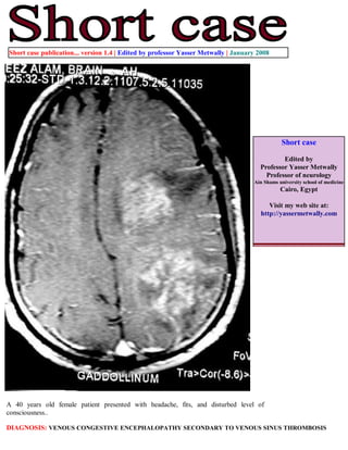
- 1. Short case publication... version 1.4 | Edited by professor Yasser Metwally | January 2008 Short case Edited by Professor Yasser Metwally Professor of neurology Ain Shams university school of medicine Cairo, Egypt Visit my web site at: http://yassermetwally.com A 40 years old female patient presented with headache, fits, and disturbed level of consciousness.. DIAGNOSIS: VENOUS CONGESTIVE ENCEPHALOPATHY SECONDARY TO VENOUS SINUS THROMBOSIS
- 2. Figure 1. Cerebral congestive encephalopathy. MRI T1 postcontrast study showing dural sinus and cortical venous dilations and enhancement due to widespread dural sinus & cerebral venous thrombosis. The enhanced cortical and transcerebral veins are seen forming the hyperdense cord signs which are seen radiating to the dilated and thrombosed dural sinuses. Also notice parenchymal subcortical hypointensities and patchy, irregular and linear enhancement with mass effect which could be due to edema, infarction or ischemia, forming what is called cerebral congestive encephalopathy. Figure 2. Venous congestive encephalopathy due to venous hypertension secondary to venous thrombosis. MRI T2 images showing subcortical white matter hyperintensities with mass effect, mixed with linear and patchy hypointensity and signal void structures. Changes are due to edema, petechial hemorrhages and dilated veins.
- 3. Figure 3. Cerebral congestive encephalopathy. MRI T1 postcontrast study showing dural sinus and cortical venous dilations and enhancement due to widespread dural sinus & cerebral venous thrombosis. The enhanced cortical and transcerebral veins are seen forming the hyperdense cord signs which are seen radiating to the dilated and thrombosed dural sinuses. Also notice parenchymal subcortical hypointensities and patchy, irregular and linear enhancement with mass effect which could be due to edema, infarction or ischemia, forming what is called cerebral congestive encephalopathy. Figure 4. MRI T1 postcontrast study showing dural sinus and cortical venous dilations and enhancement due to widespread dural sinus & cerebral venous thrombosis. The enhanced cortical and transcerebral veins are seen forming the hyperdense cord signs which are seen radiating to the dilated and thrombosed dural sinuses. Also notice parenchymal subcortical hypointensities and patchy, irregular and linear enhancement which could be due to edema, infarction or ischemia, forming what is called cerebral congestive encephalopathy.
- 4. Figure 5. MRI T1 postcontrast study showing enhancement and dilation of the thrombosed superior sagittal sinus with central hypointense filling defects which could be due to the intraluminal thrombi. Dilated enhanced cortical veins are seen pouring in the thrombosed sinus, subcortical parenchymal hypointensity could be due to edema or infarction Addendum A new version of this software is uploaded in my web site every week (every Saturday and remains available till Friday.) To download the current version follow the link quot;http://pdf.yassermetwally.com/short.pdfquot;. You can download the long case version of this short case during the same week from: http://pdf.yassermetwally.com/case.pdf or visit web site: http://pdf.yassermetwally.com To download the software version of the publication (crow.exe) follow the link: http://neurology.yassermetwally.com/crow.zip At the end of each year, all the publications are compiled on a single CD-ROM, please contact the author to know more details. Screen resolution is better set at 1024*768 pixel screen area for optimum display References 1. Metwally, MYM: Textbook of neurimaging, A CD-ROM publication, (Metwally, MYM editor) WEB-CD agency for electronic publishing, version 9.1a January 2008