SlideShare a Scribd company logo
1 of 10
Download to read offline
CASE OF THE WEEK
                   PROFESSOR YASSER METWALLY
CLINICAL PICTURE

CLINICAL PICTURE

A 22 years old male patient presented with quadriplegia with a high cervical sensory level of acute onset. The
neurological disability occurred 10 days following an attack of flu.

RADIOLOGICAL FINDINGS

RADIOLOGICAL FINDINGS


Figure 1. A case of transverse myelitis. A, precontrast MRI T1 image and B,C,D postcontrast MRI T1 images. showing
mild dilatation of C2,C3,C4,C5 cervical spinal segments with central intramedullary, multisegmental hypointensity
and with peripheral contrast enhancement.
Figure 2. A case of transverse myelitis. MRI T2 images showing multisegmental (about five spinal segments from C2-
C6) intramedullary hyperintensity with mild cord dilatation.


Figure 3. A case of transverse myelitis. Cross-sectional MRI T2 images showing mild dilatation of the spinal cord with
central hyperintensities occupying more than 2/3 of the cross-sectional area of the spinal cord with absence of the
central dot sign.
Figure 4. MRI T1 precontrast (A,B,C,D) and postcontrast (E,F,G) and MRI
                                      T2 image (H) showing a case of acute idiopathic transverse myelitis, notice
                                      cord swelling in the cervico dorsal region with patchy irregular and peripheral
                                      contrast enhancement. Also notice the central T2 hyperintensity. Peripheral
                                      contrast enhancement is outside and peripheral to the central T2
                                      hyperintensity.


                                                      Figure 5. MRI T2 showing a case of acute idiopathic transverse
                                                      myelitis. Notice cord swelling and the multisegmental, central
                                                      increased cord signal intensity at the cervicodorsal region


Table 1. The MRI picture characteristic of idiopathic transverse myelitis

        1. A centrally located multisegmental (3 to 8 spinal segments) MRI T2 hyperintensity that occupies more
           than two thirds of the cross-sectional area of the cord is characteristic of transverse myelitis. The MRI T2
           hyperintensity commonly shows a slow regression with clinical improvement. The central spinal cord
           MRI T2 hyperintensity represents evenly distributed central cord edema. MRI T1 Hypointensity might
           be present in the same spinal segments that show T2 hyperintensity although to a lesser extent. The MRI
           T2 hyperintensity is central, bilateral, more or less symmetrical and multisegmental.

        2. MRI T2 central isointensity, or dot (within and in the core of the MRI T2 hyperintensity) might be
           present and is believed to represent central gray matter squeezed by the uniform, evenly distributed
           edematous changes of the cord. (central dot sign). It might not be of any clinical significance.

        3. Contrast enhancement is commonly focal or peripheral and maximal at or near the segmental MRI T2
           hyperintensity. In idiopathic transverse myelitis enhancement is peripheral to the centrally located area
           of high T2 signal intensity rather than in the very same area. The prevalence of cord enhancement is
           significantly higher in patients with cord expansion.
4. Spinal cord expansion might or might not be present and when present is usually multisegmental and
            better appreciated on the sagittal MRI T1 images. Spinal cord expansion tapers smoothly to the normal
            cord, and is of lesser extent than the high T2 signal abnormality.

         5. Multiple sclerosis plaques (and subsequent T2 hyperintensity) are located peripherally, are less than 2
            vertebral segments in length, and occupies less than half the cross-sectional area of the cord. In contrast
            to transverse myelitis, enhancement in MS occurs in the same location of high-signal-intensity lesions
            seen on T2-weighted images.


Table 2. Differences between idiopathic transverse myelitis and spinal multiple sclerosis

                                         Number
                               T2           of
     Disease entity                                      Contrast element                         Pathology
                         hyperintensity segments
                                        involved
Idiopathic transverse    Central,       4-8      In       transverse        myelitis   Nonspecific necrosis that affects
myelitis                 multisegmental          enhancement is peripheral to the      gray    and     white     matter
                                                 centrally located area of high T2     indiscriminately and destroys
                                                 signal intensity rather than in       axons and cell bodies as well as
                                                 the very same area.                   myelin.
Spinal multiple          Peripheral     1-2      In contrast to transverse             White matter demyelination
sclerosis                                        myelitis, enhancement in MS           only.
                                                 occurs in the same location of
                                                 high-signal-intensity lesions seen
                                                 on T2-weighted images.

DIAGNOSIS:

DIAGNOSIS: ACUTE IDIOPATHIC TRANSVERSE MYELITIS

DISCUSSION

DISCUSSION

The first cases of acute transverse myelitis (ATM) were described in 1882 by Bastian [1]. In 1922 and 1923, 200 cases
of so-called “post-vaccination encephalomyelitis” were reported in Holland and England. It was in 1948 that the term
ATM was used in reporting a case of severe myelopathy after pneumonia [2].

Transverse myelitis is a clinical syndrome characterized by bilateral motor, sensory, and autonomic disturbances [3].
About 50% of patients have paraparesis; 80% to 94% have numbness, paresthesias, and band-like dysesthesias; and
all have bladder dysfunction [3]. The histopathologic features of TM include perivascular monocytic and lymphocytic
infiltration, demyelination, and axonal injury [4]. TM may exist as part of a multifocal CNS disease; as a multi-
systemic disease; or as an isolated, idiopathic entity. The immunopathogenesis of disease-associated TM is varied and
includes vasculitis neurosarcoidosis, MS, and lupus. Several reports of TM after vaccination have been published [5,6].
Recently, the term “parainfectious TM” has been introduced for TM cases with antecedent respiratory,
gastrointestinal, or systemic illness [4]. A variety of immune stimuli (eg, molecular mimicry, superantigen-mediated
immune activation) may trigger the immune system to injure the nervous system [4]. In a retrospective study of 288
patients who had TM, 45 (15.6%) met the criteria for idiopathic TM [7]. According to the published series,
approximately one third of patients recover with little or no sequelae, one third are left with a moderate degree of
permanent disability, and one third develop severe disability [8]. In 2002, the Transverse Myelitis Consortium
Working Group proposed criteria for idiopathic ATM, with incorporation of CSF testing and MR imaging findings
[8].

The criteria include (1) bilateral sensory, motor, or autonomic spinal cord dysfunction; (2) defined sensory level and
bilateral signs and symptoms; (3) proof of inflammation within the spinal cord by MR imaging or CSF examination;
(4) symptoms from onset to reach maximal deficit between few hours and 21 days; and (5) exclusion of extra-axial
compressive etiology [8]. The thoracic spine is most commonly involved, and middle-aged adults are usually affected.
MR imaging findings include focal, centrally located increased signal on T2-weighted MR images, usually occupying
more than two thirds of the cross-sectional area of the cord (Fig. 6) [9]. This was observed in 88% of patients in a
series of 17 patients who had idiopathic TM [10]. Usually, the signal abnormality extends more than three to four
vertebral segments in length. Cord expansion may or may not be present; it was found in 47% in published series [9].
Enhancement is usually absent; when enhancement was present, two patterns have been described: moderate patchy
enhancement or diffuse abnormal enhancement (Fig. 7, Fig. 8) [7,10,11,12]. Enhancement was found in only 38% of
cases of idiopathic TM in one series and in 47% and 53% in the two other series [7,9]. About 40% of TM cases display
a normal MR imaging study [13]. MS is the most important differential diagnosis of TM. Signal abnormality located
peripherally in the spinal cord that is less than two vertebral segments in length and occupying less than half the cross-
sectional area of the cord favors a diagnosis of MS rather than TM [9].


                                                             Figure 6. ATM. (A) Sagittal T2-weighted MR image
                                                             showing high-signal-intensity abnormality in the spinal
                                                             cord lesion extending over several segments of the upper
                                                             thoracic spine. (B) A focal, centrally located increased
                                                             signal occupying more than two thirds of the cross-
                                                             sectional area of the cord is demonstrated on the axial T2-
                                                             weighted MR image. (C) On a sagittal, diffusion-weighted
                                                             MR image performed using navigated interleaved
                                                             multishot echo planar imaging (5-mm slice thickness, b
                                                             max = 700 s/mm2), high signal indicates increased
                                                             diffusion in the area of increased signal on T2-WI. (D)
                                                             High signal was observed on the apparent diffusion
                                                             coefficient map, suggesting a T2 shine-through effect
                                                             rather than restricted diffusion in spinal cord areas
                                                             affected by myelitis.
Figure 7. Idiopathic ATM. (A) Sagittal T2-weighted MR image of the thoracic spine showing signal abnormality
extending from T7 to the L2 vertebral segment. (B) The lesion is isointense to the spinal cord on sagittal T-weighted
MR image. (C) Sagittal image showing focal enhancement in the cord.


                                                                                   Figure 8. A case of acute transverse
                                                                                   myelitis in a patient who presented
                                                                                   with sensory level. (A, B) T2-
                                                                                   weighted (A) and sagittal short-tau
                                                                                   inversion-recovery (B) MR images
                                                                                   show             high-signal-intensity
                                                                                   abnormality in the cervical spinal
                                                                                   cord extending from the C3 to the T1
                                                                                   level with cord swelling. (C) Sagittal
                                                                                   gadolinium-enhanced, T1-weighted
                                                                                   MR image showing moderate patchy
                                                                                   enhancement.


There is growing evidence that the length of the lesion is likely important from a pathogenic and a prognostic
standpoint. Patients who have acute partial transverse myelitis have signal abnormalities extending less than two
segments on MR imaging, and patients who have complete longitudinally extensive transverse myelitis have
abnormalities that extend to multiple segments (see Fig. 8). Patients in the first group are at higher risk for developing
MS compared with those in the second group, where the risk is low [14].

DTI was recently used to characterize inflammatory processes of the spinal cord [15]. In cases of inflammatory
myelitis, decreased FA values have been found in the region of a T2-weighted lesion and increased FA values in the
lesion's boundaries. This pattern is different from that seen in invasive tumors, in which FA is low in peripheral
regions of edema.
Novel biomarkers, such as cytokine interleukin-6 and collapsin response-mediator protein–5 are potentially useful
prognostic indicators and markers of disease severity. The “idiopathic” form of ATM is rarely seen [16].


SUMMARY


SUMMARY

Over the past decade, researchers and clinicians have gained new insights into the core of demyelinating diseases of the
spinal cord, and much progress has been made in the management of these diseases. Although we are starting to
uncover some of the structural and physiologic substrates of demyelination of the CNS, we are far from understanding
what causes many of these demyelinating disorders and how to prevent their progression. With further development of
new techniques, such as DTI and more potent MR units, spinal cord diseases may be distinguished from each other,
and effective therapeutic strategies may be initiated before any cord damage occurs (Fig. 9).

In particular MRI is very helpful in differentiation between Spinal multiple sclerosis and transverse myelitis In the
series reported by Choi et al, [18] the centrally located MRI T2 high signal intensity occupied more than two thirds of
the cross-sectional area of the cord in transverse myelitis. In multiple sclerosis, plaques are usually located
peripherally and occupy less than half the cross-sectional area of the cord. The central isointensity, or dot (commonly
seen in transverse myelitis), represents central gray matter squeezed by the uniform, evenly distributed oedematous
changes of the cord. Choi and colleagues [18] have demonstrated the role of contrast media in differentiating
transverse myelitis from multiple sclerosis. In transverse myelitis, enhancement is in the periphery of a centrally
located area of high T2 weighted images. In multiple sclerosis, the lesions show enhancement in the central zone of
peripherally located high signal intensity on T2 weighted images. [14]

In conclusion, certain MRI characteristics help in differentiating acute transverse myelitis from spinal form of
multiple sclerosis. These include: 1) centrally located high intensity signal extending over 3 to 4 segments and
occupying more than two thirds of the cord cross-sectional area and 2) peripheral contrast enhancement of high
intensity signal.
Figure 15. Differential diagnoses of intramedullary lesions based on their location at the cross-sectional area of the
cord. (A) MS: Dorsally located wedge-shaped lesion involving less then two thirds of the cross-sectional area of the
spinal cord seen on axial T2-Wi MR image. (B) Poliomyelitis: Bilateral enhancing anterior nerve roots demonstrated
on postcontrast T1-Wi MR image. (C) Vacuolar myelopathy: Bilateral, symmetrical, high-signal-intensity abnormality
located dorsally in the spinal cord in an HIV-positive patient. DD: Subacute combined degeneration. (D) ATM: On
axial T2-Wi, a high-signal-intensity lesion involving more than two thirds of cross-sectional area of the spinal cord is
observed. (E) Herpes-simplex-virus myelitis: Postcontrast T1-Wi axial MR image showing nodular enhancing lesion
located in the lateral part of the cervical spinal cord. DD: active MS plaque. (F) Spinal cord infarction: Swelling of the
anterior parts of the spinal cord is shown on axial T2-Wi MR images, indicating vulnerability of the anterior portions
of the spinal cord to ischemia.


      Addendum

            A new version of this PDF file (with a new case) is uploaded in my web site every week (every Saturday and
             remains available till Friday.)
   To download the current version follow the link quot;http://pdf.yassermetwally.com/case.pdfquot;.
            You can also download the current version from my web site at quot;http://yassermetwally.comquot;.
            To download the software version of the publication (crow.exe) follow the link:
             http://neurology.yassermetwally.com/crow.zip
            The case is also presented as a short case in PDF format, to download the short case follow the link:
             http://pdf.yassermetwally.com/short.pdf
            At the end of each year, all the publications are compiled on a single CD-ROM, please contact the author to know
             more details.
            Screen resolution is better set at 1024*768 pixel screen area for optimum display.
            For an archive of the previously reported cases go to www.yassermetwally.net, then under pages in the right
             panel, scroll down and click on the text entry quot;downloadable case records in PDF formatquot;


REFERENCES

References

[1]Bastian HC. Special diseases of the spinal cord. In: Quain R, editor. A dictionary of medicine: including general
pathology, general therapeutics, hygiene, and the diseases peculiar to women and children. London: Longmans,
Green; 1882. p. 1479–83.

[2]Suchett-Kaye AL. Acute transverse myelitis complicating pneumonia. Lancet. 1948;255:417.

[3] [53]Krishnan C, Kaplin AI, Pardo CA, et al.. Demyelinating disorders: update on transverse myelitis. Curr Neurol
Neurosci Rep. 2006;6(3):236–243.

[54]Kerr DA, Ayetey H. Immunopathogenesis of acute transverse myelitis. Curr Opin Neurol. 2002;15:339–347.

[5]Larner AJ, Farmer SF. Myelopathy following influenza vaccination in inflammatory CNS disorder treated with
chronic immunosuppression. Eur J Neurol. 2000;7:731–733.

[6]Bakshi R, Mazziotta JC. Acute transverse myelitis after influenza vaccination: magnetic resonance imaging
findings. J Neuroimaging. 1996;6(4):248–250.

[7]De Seze J, Lanctin C, Lebrun C, et al.. Idiopathic acute transverse myelitis: application of the recent diagnostic
criteria. Neurology. 2005;65:1950–1953. CrossRef

[8]Transverse Myelitis Consortium Working Group (TMCWG) . Proposed diagnostic criteria and nosology of acute
transverse myelitis. Neurology. 2002;59:499–505.

[9]Choi KH, Lee KS, Chung SO, et al.. Idiopathic transverse myelitis: MR characteristics. AJNR Am J Neuroradiol.
1996;17:1151–1160.

[10] [60]Kim KK. Idiopathic reccurent transverse myelitis. Arch Neurol. 2003;60:1290–1294.

[11]Brinar VV, Habek M, Brinar M, et al.. The differential diagnosis of acute transverse myelitis. Clin Neurol
Neurosurg. 2006;108:278–283.

[12]Holtas S, Basibüyük N, Fredriksson K. MRI in acute transverse myelopathy. Neuroradiology. 1993;35:221–226.

[13]Scotti G, Gerevini S. Diagnosis and differential diagnosis of acute transverse myelopathy: the role of
neuroradiological investigations and review of the literature. Neurol Sci. 2001;22(2):S69–S73. CrossRef

[14] Pittock SJ, Lucchinetti CF. Inflammatory transverse myelitis: evolving concepts. Current Opin Neurol.
2006;19:362–368.

[15]Renoux J, Facon D, Fillard P, et al.. MR diffusion tensor imaging and fiber tracking in inflammatory diseases of
the spinal cord. AJNR Am J Neuroradiol. 2006;27:1947–1951.
[16]Cree BA, Wingerchuk DM. Acute transverse myelitis: is the “idiopathic” form vanishing?. Neurology. 2005;65
(12):1857–1858. CrossRef

[17] Metwally, MYM: Textbook of neurimaging, A CD-ROM publication, (Metwally, MYM editor) WEB-CD agency
for electronic publication, version 9.1a January 2008

[18] Choi KH, Lee KS, Chung SO, et al.. Idiopathic transverse myelitis: MR characteristics. AJNR Am J Neuroradiol.
1996;17:1151–1160.


More Related Content

What's hot

Presentation1.pptx,radiological imaging of cord myelopathy.
Presentation1.pptx,radiological imaging of cord myelopathy.Presentation1.pptx,radiological imaging of cord myelopathy.
Presentation1.pptx,radiological imaging of cord myelopathy.Abdellah Nazeer
 
Mri in white matter diseases
Mri in white matter diseasesMri in white matter diseases
Mri in white matter diseasesSindhu Gowdar
 
Presentation1.pptx radio;ogical imaging of benign and malignant soft tissue t...
Presentation1.pptx radio;ogical imaging of benign and malignant soft tissue t...Presentation1.pptx radio;ogical imaging of benign and malignant soft tissue t...
Presentation1.pptx radio;ogical imaging of benign and malignant soft tissue t...Abdellah Nazeer
 
Shah alam sir om (2)
Shah alam sir om (2)Shah alam sir om (2)
Shah alam sir om (2)wasek_bd
 
Recent advances in inflammatory muscle diseases
Recent advances in inflammatory muscle diseasesRecent advances in inflammatory muscle diseases
Recent advances in inflammatory muscle diseasesNeurologyKota
 
Aipgmee2011part1
Aipgmee2011part1Aipgmee2011part1
Aipgmee2011part1isha9
 
Occipital Neuralgia
Occipital NeuralgiaOccipital Neuralgia
Occipital NeuralgiaAde Wijaya
 
Idiopathic Inflammatory Myopathies
Idiopathic Inflammatory MyopathiesIdiopathic Inflammatory Myopathies
Idiopathic Inflammatory MyopathiesAde Wijaya
 
Pyogenic spine infections
Pyogenic spine infectionsPyogenic spine infections
Pyogenic spine infectionsRobert Oluoch
 
Epidural abcess a case presentation
Epidural abcess a case presentationEpidural abcess a case presentation
Epidural abcess a case presentationRamy Mostafa
 
SPINAL EPIDURAL, AND SUBDURAL - INTRAMEDULLAR ABSCESSES
SPINAL EPIDURAL,  AND SUBDURAL - INTRAMEDULLAR ABSCESSESSPINAL EPIDURAL,  AND SUBDURAL - INTRAMEDULLAR ABSCESSES
SPINAL EPIDURAL, AND SUBDURAL - INTRAMEDULLAR ABSCESSESAlexander Bardis
 
Which patients of epilepsy require early neurosurgical referral
Which patients of epilepsy require early neurosurgical referral Which patients of epilepsy require early neurosurgical referral
Which patients of epilepsy require early neurosurgical referral Dr. Arghya Deb
 
Oncologic conditions of brain and spinal cord
Oncologic conditions of brain and spinal cordOncologic conditions of brain and spinal cord
Oncologic conditions of brain and spinal cordBobby Abraham
 
Recent advances in idiopathic inflammatory myopathies by Dr. Naman Madaan
Recent advances in idiopathic inflammatory myopathies by Dr. Naman MadaanRecent advances in idiopathic inflammatory myopathies by Dr. Naman Madaan
Recent advances in idiopathic inflammatory myopathies by Dr. Naman MadaanNaman Madaan
 
Spinal Tumors: approach and management
Spinal Tumors: approach and managementSpinal Tumors: approach and management
Spinal Tumors: approach and managementAmit Agrawal
 
Pyogenic spondylitis cme chitwan 2019
Pyogenic spondylitis cme chitwan 2019Pyogenic spondylitis cme chitwan 2019
Pyogenic spondylitis cme chitwan 2019SureshPandey32
 
Polymyositis Dermatomyositis
Polymyositis DermatomyositisPolymyositis Dermatomyositis
Polymyositis DermatomyositisZia ul Haq Nawaz
 

What's hot (20)

Intracranial lesions mimicking neoplasms
Intracranial lesions mimicking neoplasmsIntracranial lesions mimicking neoplasms
Intracranial lesions mimicking neoplasms
 
Presentation1.pptx,radiological imaging of cord myelopathy.
Presentation1.pptx,radiological imaging of cord myelopathy.Presentation1.pptx,radiological imaging of cord myelopathy.
Presentation1.pptx,radiological imaging of cord myelopathy.
 
Mri in white matter diseases
Mri in white matter diseasesMri in white matter diseases
Mri in white matter diseases
 
Presentation1.pptx radio;ogical imaging of benign and malignant soft tissue t...
Presentation1.pptx radio;ogical imaging of benign and malignant soft tissue t...Presentation1.pptx radio;ogical imaging of benign and malignant soft tissue t...
Presentation1.pptx radio;ogical imaging of benign and malignant soft tissue t...
 
Shah alam sir om (2)
Shah alam sir om (2)Shah alam sir om (2)
Shah alam sir om (2)
 
Recent advances in inflammatory muscle diseases
Recent advances in inflammatory muscle diseasesRecent advances in inflammatory muscle diseases
Recent advances in inflammatory muscle diseases
 
Aipgmee2011part1
Aipgmee2011part1Aipgmee2011part1
Aipgmee2011part1
 
Occipital Neuralgia
Occipital NeuralgiaOccipital Neuralgia
Occipital Neuralgia
 
Case record...Postinfectious cerebellitis
Case record...Postinfectious cerebellitisCase record...Postinfectious cerebellitis
Case record...Postinfectious cerebellitis
 
Idiopathic Inflammatory Myopathies
Idiopathic Inflammatory MyopathiesIdiopathic Inflammatory Myopathies
Idiopathic Inflammatory Myopathies
 
Pyogenic spine infections
Pyogenic spine infectionsPyogenic spine infections
Pyogenic spine infections
 
Epidural abcess a case presentation
Epidural abcess a case presentationEpidural abcess a case presentation
Epidural abcess a case presentation
 
SPINAL EPIDURAL, AND SUBDURAL - INTRAMEDULLAR ABSCESSES
SPINAL EPIDURAL,  AND SUBDURAL - INTRAMEDULLAR ABSCESSESSPINAL EPIDURAL,  AND SUBDURAL - INTRAMEDULLAR ABSCESSES
SPINAL EPIDURAL, AND SUBDURAL - INTRAMEDULLAR ABSCESSES
 
Which patients of epilepsy require early neurosurgical referral
Which patients of epilepsy require early neurosurgical referral Which patients of epilepsy require early neurosurgical referral
Which patients of epilepsy require early neurosurgical referral
 
Oncologic conditions of brain and spinal cord
Oncologic conditions of brain and spinal cordOncologic conditions of brain and spinal cord
Oncologic conditions of brain and spinal cord
 
Recent advances in idiopathic inflammatory myopathies by Dr. Naman Madaan
Recent advances in idiopathic inflammatory myopathies by Dr. Naman MadaanRecent advances in idiopathic inflammatory myopathies by Dr. Naman Madaan
Recent advances in idiopathic inflammatory myopathies by Dr. Naman Madaan
 
Spinal Tumors: approach and management
Spinal Tumors: approach and managementSpinal Tumors: approach and management
Spinal Tumors: approach and management
 
Pyogenic spondylitis cme chitwan 2019
Pyogenic spondylitis cme chitwan 2019Pyogenic spondylitis cme chitwan 2019
Pyogenic spondylitis cme chitwan 2019
 
Polymyositis Dermatomyositis
Polymyositis DermatomyositisPolymyositis Dermatomyositis
Polymyositis Dermatomyositis
 
Inflammatory disease of muscle
Inflammatory disease of muscleInflammatory disease of muscle
Inflammatory disease of muscle
 

Similar to Case of Quadriplegia After Flu

Case record...Idiopathic postinfectious transverse myelitis
Case record...Idiopathic postinfectious transverse myelitisCase record...Idiopathic postinfectious transverse myelitis
Case record...Idiopathic postinfectious transverse myelitisProfessor Yasser Metwally
 
Short case...Acute postinfectious transverse myelitis
Short case...Acute postinfectious transverse myelitisShort case...Acute postinfectious transverse myelitis
Short case...Acute postinfectious transverse myelitisProfessor Yasser Metwally
 
Case record...Acute postinfectious transverse myelitis (Transverse myelitis s...
Case record...Acute postinfectious transverse myelitis (Transverse myelitis s...Case record...Acute postinfectious transverse myelitis (Transverse myelitis s...
Case record...Acute postinfectious transverse myelitis (Transverse myelitis s...Professor Yasser Metwally
 
MRI of Spine and very easy details of sp
MRI of Spine and very easy details of spMRI of Spine and very easy details of sp
MRI of Spine and very easy details of spssuserc66686
 
Short case...Postinfectious transverse myelitis
Short case...Postinfectious transverse myelitisShort case...Postinfectious transverse myelitis
Short case...Postinfectious transverse myelitisProfessor Yasser Metwally
 
Short case...Postinfectious transverse myelitis
Short case...Postinfectious transverse myelitisShort case...Postinfectious transverse myelitis
Short case...Postinfectious transverse myelitisProfessor Yasser Metwally
 
Case record... Acute disseminated encephalomyelitis
Case record... Acute disseminated encephalomyelitisCase record... Acute disseminated encephalomyelitis
Case record... Acute disseminated encephalomyelitisProfessor Yasser Metwally
 
Radiology of demyelinating diseases
Radiology of demyelinating diseases Radiology of demyelinating diseases
Radiology of demyelinating diseases NeurologyKota
 
Orbital pathologies.pptx 1
Orbital pathologies.pptx 1Orbital pathologies.pptx 1
Orbital pathologies.pptx 1Anish Choudhary
 
Orbital pathologies.pptx (part 1)
Orbital pathologies.pptx (part 1)Orbital pathologies.pptx (part 1)
Orbital pathologies.pptx (part 1)Dr. Mohit Goel
 
Diagnostic Imaging of Degenerative & White Matter Diseases
Diagnostic Imaging of Degenerative & White Matter DiseasesDiagnostic Imaging of Degenerative & White Matter Diseases
Diagnostic Imaging of Degenerative & White Matter DiseasesMohamed M.A. Zaitoun
 
Atlas of peripheral nerve ultrasound
Atlas of peripheral nerve ultrasoundAtlas of peripheral nerve ultrasound
Atlas of peripheral nerve ultrasoundSpringer
 
radiologyofdemyelinatingdiseasesmbs-170105191815.pptx
radiologyofdemyelinatingdiseasesmbs-170105191815.pptxradiologyofdemyelinatingdiseasesmbs-170105191815.pptx
radiologyofdemyelinatingdiseasesmbs-170105191815.pptxShubham661884
 

Similar to Case of Quadriplegia After Flu (20)

Case record...Idiopathic postinfectious transverse myelitis
Case record...Idiopathic postinfectious transverse myelitisCase record...Idiopathic postinfectious transverse myelitis
Case record...Idiopathic postinfectious transverse myelitis
 
Short case...Acute postinfectious transverse myelitis
Short case...Acute postinfectious transverse myelitisShort case...Acute postinfectious transverse myelitis
Short case...Acute postinfectious transverse myelitis
 
Case record...Neuromyelitis optica
Case record...Neuromyelitis opticaCase record...Neuromyelitis optica
Case record...Neuromyelitis optica
 
Case record...Acute postinfectious transverse myelitis (Transverse myelitis s...
Case record...Acute postinfectious transverse myelitis (Transverse myelitis s...Case record...Acute postinfectious transverse myelitis (Transverse myelitis s...
Case record...Acute postinfectious transverse myelitis (Transverse myelitis s...
 
Case record...Spinal multiple sclerosis
Case record...Spinal multiple sclerosisCase record...Spinal multiple sclerosis
Case record...Spinal multiple sclerosis
 
MRI of Spine and very easy details of sp
MRI of Spine and very easy details of spMRI of Spine and very easy details of sp
MRI of Spine and very easy details of sp
 
Short case...Postinfectious transverse myelitis
Short case...Postinfectious transverse myelitisShort case...Postinfectious transverse myelitis
Short case...Postinfectious transverse myelitis
 
Short case...Intramedullary cavernoma
Short case...Intramedullary cavernomaShort case...Intramedullary cavernoma
Short case...Intramedullary cavernoma
 
Short case...Postinfectious transverse myelitis
Short case...Postinfectious transverse myelitisShort case...Postinfectious transverse myelitis
Short case...Postinfectious transverse myelitis
 
Short case...Spinal multiple sclerosis
Short case...Spinal multiple sclerosisShort case...Spinal multiple sclerosis
Short case...Spinal multiple sclerosis
 
Case record... Acute disseminated encephalomyelitis
Case record... Acute disseminated encephalomyelitisCase record... Acute disseminated encephalomyelitis
Case record... Acute disseminated encephalomyelitis
 
CT - Ring Enhancing Lesion
CT - Ring Enhancing LesionCT - Ring Enhancing Lesion
CT - Ring Enhancing Lesion
 
Radiology of demyelinating diseases
Radiology of demyelinating diseases Radiology of demyelinating diseases
Radiology of demyelinating diseases
 
Orbital pathologies.pptx 1
Orbital pathologies.pptx 1Orbital pathologies.pptx 1
Orbital pathologies.pptx 1
 
Orbital pathologies.pptx (part 1)
Orbital pathologies.pptx (part 1)Orbital pathologies.pptx (part 1)
Orbital pathologies.pptx (part 1)
 
Diagnostic Imaging of Degenerative & White Matter Diseases
Diagnostic Imaging of Degenerative & White Matter DiseasesDiagnostic Imaging of Degenerative & White Matter Diseases
Diagnostic Imaging of Degenerative & White Matter Diseases
 
Atlas of peripheral nerve ultrasound
Atlas of peripheral nerve ultrasoundAtlas of peripheral nerve ultrasound
Atlas of peripheral nerve ultrasound
 
Case record...Spinal multiple sclerosis
Case record...Spinal multiple sclerosisCase record...Spinal multiple sclerosis
Case record...Spinal multiple sclerosis
 
radiologyofdemyelinatingdiseasesmbs-170105191815.pptx
radiologyofdemyelinatingdiseasesmbs-170105191815.pptxradiologyofdemyelinatingdiseasesmbs-170105191815.pptx
radiologyofdemyelinatingdiseasesmbs-170105191815.pptx
 
Case record...Friedreich ataxia
Case record...Friedreich ataxiaCase record...Friedreich ataxia
Case record...Friedreich ataxia
 

More from Professor Yasser Metwally

The Snake, the Scorpion, the turtle in Egypt
The Snake, the Scorpion, the turtle in EgyptThe Snake, the Scorpion, the turtle in Egypt
The Snake, the Scorpion, the turtle in EgyptProfessor Yasser Metwally
 
Radiological pathology of epileptic disorders
Radiological pathology of epileptic disordersRadiological pathology of epileptic disorders
Radiological pathology of epileptic disordersProfessor Yasser Metwally
 
Radiological pathology of cerebrovascular disorders
Radiological pathology of cerebrovascular disordersRadiological pathology of cerebrovascular disorders
Radiological pathology of cerebrovascular disordersProfessor Yasser Metwally
 
Radiological pathology of spontaneous cerebral hemorrhage
Radiological pathology of spontaneous cerebral hemorrhageRadiological pathology of spontaneous cerebral hemorrhage
Radiological pathology of spontaneous cerebral hemorrhageProfessor Yasser Metwally
 
Radiological pathology of cerebral amyloid angiography
Radiological pathology of cerebral amyloid angiographyRadiological pathology of cerebral amyloid angiography
Radiological pathology of cerebral amyloid angiographyProfessor Yasser Metwally
 
Radiological pathology of cerebral microbleeds
Radiological pathology of cerebral microbleedsRadiological pathology of cerebral microbleeds
Radiological pathology of cerebral microbleedsProfessor Yasser Metwally
 
Issues in radiological pathology: Radiological pathology of watershed infarct...
Issues in radiological pathology: Radiological pathology of watershed infarct...Issues in radiological pathology: Radiological pathology of watershed infarct...
Issues in radiological pathology: Radiological pathology of watershed infarct...Professor Yasser Metwally
 
Radiological pathology of cortical laminar necrosis
Radiological pathology of cortical laminar necrosisRadiological pathology of cortical laminar necrosis
Radiological pathology of cortical laminar necrosisProfessor Yasser Metwally
 

More from Professor Yasser Metwally (20)

The Egyptian Zoo in Cairo 2015
The Egyptian Zoo in Cairo 2015The Egyptian Zoo in Cairo 2015
The Egyptian Zoo in Cairo 2015
 
End of the great nile river in Ras Elbar
End of the great nile river in Ras ElbarEnd of the great nile river in Ras Elbar
End of the great nile river in Ras Elbar
 
The Lion and The tiger in Egypt
The Lion and The tiger in EgyptThe Lion and The tiger in Egypt
The Lion and The tiger in Egypt
 
The monkeys in Egypt
The monkeys in EgyptThe monkeys in Egypt
The monkeys in Egypt
 
The Snake, the Scorpion, the turtle in Egypt
The Snake, the Scorpion, the turtle in EgyptThe Snake, the Scorpion, the turtle in Egypt
The Snake, the Scorpion, the turtle in Egypt
 
The Egyptian Parrot
The Egyptian ParrotThe Egyptian Parrot
The Egyptian Parrot
 
The Egyptian Deer
The Egyptian DeerThe Egyptian Deer
The Egyptian Deer
 
The Egyptian Pelican
The Egyptian PelicanThe Egyptian Pelican
The Egyptian Pelican
 
The Flamingo bird in Egypt
The Flamingo bird in EgyptThe Flamingo bird in Egypt
The Flamingo bird in Egypt
 
Egyptian Cats
Egyptian CatsEgyptian Cats
Egyptian Cats
 
Radiological pathology of epileptic disorders
Radiological pathology of epileptic disordersRadiological pathology of epileptic disorders
Radiological pathology of epileptic disorders
 
Radiological pathology of cerebrovascular disorders
Radiological pathology of cerebrovascular disordersRadiological pathology of cerebrovascular disorders
Radiological pathology of cerebrovascular disorders
 
Radiological pathology of spontaneous cerebral hemorrhage
Radiological pathology of spontaneous cerebral hemorrhageRadiological pathology of spontaneous cerebral hemorrhage
Radiological pathology of spontaneous cerebral hemorrhage
 
Radiological pathology of cerebral amyloid angiography
Radiological pathology of cerebral amyloid angiographyRadiological pathology of cerebral amyloid angiography
Radiological pathology of cerebral amyloid angiography
 
Radiological pathology of cerebral microbleeds
Radiological pathology of cerebral microbleedsRadiological pathology of cerebral microbleeds
Radiological pathology of cerebral microbleeds
 
The Egyptian Zoo in Cairo
The Egyptian Zoo in CairoThe Egyptian Zoo in Cairo
The Egyptian Zoo in Cairo
 
Progressive multifocal leukoencephalopathy
Progressive multifocal leukoencephalopathyProgressive multifocal leukoencephalopathy
Progressive multifocal leukoencephalopathy
 
Progressive multifocal leukoencephalopathy
Progressive multifocal leukoencephalopathyProgressive multifocal leukoencephalopathy
Progressive multifocal leukoencephalopathy
 
Issues in radiological pathology: Radiological pathology of watershed infarct...
Issues in radiological pathology: Radiological pathology of watershed infarct...Issues in radiological pathology: Radiological pathology of watershed infarct...
Issues in radiological pathology: Radiological pathology of watershed infarct...
 
Radiological pathology of cortical laminar necrosis
Radiological pathology of cortical laminar necrosisRadiological pathology of cortical laminar necrosis
Radiological pathology of cortical laminar necrosis
 

Recently uploaded

Introduction to ArtificiaI Intelligence in Higher Education
Introduction to ArtificiaI Intelligence in Higher EducationIntroduction to ArtificiaI Intelligence in Higher Education
Introduction to ArtificiaI Intelligence in Higher Educationpboyjonauth
 
Incoming and Outgoing Shipments in 1 STEP Using Odoo 17
Incoming and Outgoing Shipments in 1 STEP Using Odoo 17Incoming and Outgoing Shipments in 1 STEP Using Odoo 17
Incoming and Outgoing Shipments in 1 STEP Using Odoo 17Celine George
 
Kisan Call Centre - To harness potential of ICT in Agriculture by answer farm...
Kisan Call Centre - To harness potential of ICT in Agriculture by answer farm...Kisan Call Centre - To harness potential of ICT in Agriculture by answer farm...
Kisan Call Centre - To harness potential of ICT in Agriculture by answer farm...Krashi Coaching
 
How to Make a Pirate ship Primary Education.pptx
How to Make a Pirate ship Primary Education.pptxHow to Make a Pirate ship Primary Education.pptx
How to Make a Pirate ship Primary Education.pptxmanuelaromero2013
 
Introduction to AI in Higher Education_draft.pptx
Introduction to AI in Higher Education_draft.pptxIntroduction to AI in Higher Education_draft.pptx
Introduction to AI in Higher Education_draft.pptxpboyjonauth
 
Employee wellbeing at the workplace.pptx
Employee wellbeing at the workplace.pptxEmployee wellbeing at the workplace.pptx
Employee wellbeing at the workplace.pptxNirmalaLoungPoorunde1
 
Enzyme, Pharmaceutical Aids, Miscellaneous Last Part of Chapter no 5th.pdf
Enzyme, Pharmaceutical Aids, Miscellaneous Last Part of Chapter no 5th.pdfEnzyme, Pharmaceutical Aids, Miscellaneous Last Part of Chapter no 5th.pdf
Enzyme, Pharmaceutical Aids, Miscellaneous Last Part of Chapter no 5th.pdfSumit Tiwari
 
ECONOMIC CONTEXT - LONG FORM TV DRAMA - PPT
ECONOMIC CONTEXT - LONG FORM TV DRAMA - PPTECONOMIC CONTEXT - LONG FORM TV DRAMA - PPT
ECONOMIC CONTEXT - LONG FORM TV DRAMA - PPTiammrhaywood
 
Paris 2024 Olympic Geographies - an activity
Paris 2024 Olympic Geographies - an activityParis 2024 Olympic Geographies - an activity
Paris 2024 Olympic Geographies - an activityGeoBlogs
 
How to Configure Email Server in Odoo 17
How to Configure Email Server in Odoo 17How to Configure Email Server in Odoo 17
How to Configure Email Server in Odoo 17Celine George
 
call girls in Kamla Market (DELHI) 🔝 >༒9953330565🔝 genuine Escort Service 🔝✔️✔️
call girls in Kamla Market (DELHI) 🔝 >༒9953330565🔝 genuine Escort Service 🔝✔️✔️call girls in Kamla Market (DELHI) 🔝 >༒9953330565🔝 genuine Escort Service 🔝✔️✔️
call girls in Kamla Market (DELHI) 🔝 >༒9953330565🔝 genuine Escort Service 🔝✔️✔️9953056974 Low Rate Call Girls In Saket, Delhi NCR
 
Crayon Activity Handout For the Crayon A
Crayon Activity Handout For the Crayon ACrayon Activity Handout For the Crayon A
Crayon Activity Handout For the Crayon AUnboundStockton
 
Science 7 - LAND and SEA BREEZE and its Characteristics
Science 7 - LAND and SEA BREEZE and its CharacteristicsScience 7 - LAND and SEA BREEZE and its Characteristics
Science 7 - LAND and SEA BREEZE and its CharacteristicsKarinaGenton
 
Presentation by Andreas Schleicher Tackling the School Absenteeism Crisis 30 ...
Presentation by Andreas Schleicher Tackling the School Absenteeism Crisis 30 ...Presentation by Andreas Schleicher Tackling the School Absenteeism Crisis 30 ...
Presentation by Andreas Schleicher Tackling the School Absenteeism Crisis 30 ...EduSkills OECD
 
Interactive Powerpoint_How to Master effective communication
Interactive Powerpoint_How to Master effective communicationInteractive Powerpoint_How to Master effective communication
Interactive Powerpoint_How to Master effective communicationnomboosow
 
CARE OF CHILD IN INCUBATOR..........pptx
CARE OF CHILD IN INCUBATOR..........pptxCARE OF CHILD IN INCUBATOR..........pptx
CARE OF CHILD IN INCUBATOR..........pptxGaneshChakor2
 
Separation of Lanthanides/ Lanthanides and Actinides
Separation of Lanthanides/ Lanthanides and ActinidesSeparation of Lanthanides/ Lanthanides and Actinides
Separation of Lanthanides/ Lanthanides and ActinidesFatimaKhan178732
 
Call Girls in Dwarka Mor Delhi Contact Us 9654467111
Call Girls in Dwarka Mor Delhi Contact Us 9654467111Call Girls in Dwarka Mor Delhi Contact Us 9654467111
Call Girls in Dwarka Mor Delhi Contact Us 9654467111Sapana Sha
 

Recently uploaded (20)

Introduction to ArtificiaI Intelligence in Higher Education
Introduction to ArtificiaI Intelligence in Higher EducationIntroduction to ArtificiaI Intelligence in Higher Education
Introduction to ArtificiaI Intelligence in Higher Education
 
Código Creativo y Arte de Software | Unidad 1
Código Creativo y Arte de Software | Unidad 1Código Creativo y Arte de Software | Unidad 1
Código Creativo y Arte de Software | Unidad 1
 
Incoming and Outgoing Shipments in 1 STEP Using Odoo 17
Incoming and Outgoing Shipments in 1 STEP Using Odoo 17Incoming and Outgoing Shipments in 1 STEP Using Odoo 17
Incoming and Outgoing Shipments in 1 STEP Using Odoo 17
 
Kisan Call Centre - To harness potential of ICT in Agriculture by answer farm...
Kisan Call Centre - To harness potential of ICT in Agriculture by answer farm...Kisan Call Centre - To harness potential of ICT in Agriculture by answer farm...
Kisan Call Centre - To harness potential of ICT in Agriculture by answer farm...
 
9953330565 Low Rate Call Girls In Rohini Delhi NCR
9953330565 Low Rate Call Girls In Rohini  Delhi NCR9953330565 Low Rate Call Girls In Rohini  Delhi NCR
9953330565 Low Rate Call Girls In Rohini Delhi NCR
 
How to Make a Pirate ship Primary Education.pptx
How to Make a Pirate ship Primary Education.pptxHow to Make a Pirate ship Primary Education.pptx
How to Make a Pirate ship Primary Education.pptx
 
Introduction to AI in Higher Education_draft.pptx
Introduction to AI in Higher Education_draft.pptxIntroduction to AI in Higher Education_draft.pptx
Introduction to AI in Higher Education_draft.pptx
 
Employee wellbeing at the workplace.pptx
Employee wellbeing at the workplace.pptxEmployee wellbeing at the workplace.pptx
Employee wellbeing at the workplace.pptx
 
Enzyme, Pharmaceutical Aids, Miscellaneous Last Part of Chapter no 5th.pdf
Enzyme, Pharmaceutical Aids, Miscellaneous Last Part of Chapter no 5th.pdfEnzyme, Pharmaceutical Aids, Miscellaneous Last Part of Chapter no 5th.pdf
Enzyme, Pharmaceutical Aids, Miscellaneous Last Part of Chapter no 5th.pdf
 
ECONOMIC CONTEXT - LONG FORM TV DRAMA - PPT
ECONOMIC CONTEXT - LONG FORM TV DRAMA - PPTECONOMIC CONTEXT - LONG FORM TV DRAMA - PPT
ECONOMIC CONTEXT - LONG FORM TV DRAMA - PPT
 
Paris 2024 Olympic Geographies - an activity
Paris 2024 Olympic Geographies - an activityParis 2024 Olympic Geographies - an activity
Paris 2024 Olympic Geographies - an activity
 
How to Configure Email Server in Odoo 17
How to Configure Email Server in Odoo 17How to Configure Email Server in Odoo 17
How to Configure Email Server in Odoo 17
 
call girls in Kamla Market (DELHI) 🔝 >༒9953330565🔝 genuine Escort Service 🔝✔️✔️
call girls in Kamla Market (DELHI) 🔝 >༒9953330565🔝 genuine Escort Service 🔝✔️✔️call girls in Kamla Market (DELHI) 🔝 >༒9953330565🔝 genuine Escort Service 🔝✔️✔️
call girls in Kamla Market (DELHI) 🔝 >༒9953330565🔝 genuine Escort Service 🔝✔️✔️
 
Crayon Activity Handout For the Crayon A
Crayon Activity Handout For the Crayon ACrayon Activity Handout For the Crayon A
Crayon Activity Handout For the Crayon A
 
Science 7 - LAND and SEA BREEZE and its Characteristics
Science 7 - LAND and SEA BREEZE and its CharacteristicsScience 7 - LAND and SEA BREEZE and its Characteristics
Science 7 - LAND and SEA BREEZE and its Characteristics
 
Presentation by Andreas Schleicher Tackling the School Absenteeism Crisis 30 ...
Presentation by Andreas Schleicher Tackling the School Absenteeism Crisis 30 ...Presentation by Andreas Schleicher Tackling the School Absenteeism Crisis 30 ...
Presentation by Andreas Schleicher Tackling the School Absenteeism Crisis 30 ...
 
Interactive Powerpoint_How to Master effective communication
Interactive Powerpoint_How to Master effective communicationInteractive Powerpoint_How to Master effective communication
Interactive Powerpoint_How to Master effective communication
 
CARE OF CHILD IN INCUBATOR..........pptx
CARE OF CHILD IN INCUBATOR..........pptxCARE OF CHILD IN INCUBATOR..........pptx
CARE OF CHILD IN INCUBATOR..........pptx
 
Separation of Lanthanides/ Lanthanides and Actinides
Separation of Lanthanides/ Lanthanides and ActinidesSeparation of Lanthanides/ Lanthanides and Actinides
Separation of Lanthanides/ Lanthanides and Actinides
 
Call Girls in Dwarka Mor Delhi Contact Us 9654467111
Call Girls in Dwarka Mor Delhi Contact Us 9654467111Call Girls in Dwarka Mor Delhi Contact Us 9654467111
Call Girls in Dwarka Mor Delhi Contact Us 9654467111
 

Case of Quadriplegia After Flu

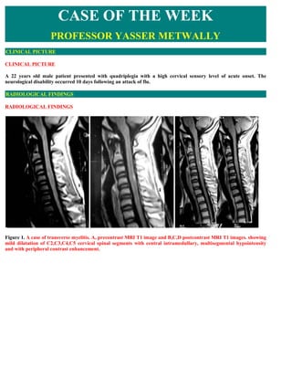
  • 1. CASE OF THE WEEK PROFESSOR YASSER METWALLY CLINICAL PICTURE CLINICAL PICTURE A 22 years old male patient presented with quadriplegia with a high cervical sensory level of acute onset. The neurological disability occurred 10 days following an attack of flu. RADIOLOGICAL FINDINGS RADIOLOGICAL FINDINGS Figure 1. A case of transverse myelitis. A, precontrast MRI T1 image and B,C,D postcontrast MRI T1 images. showing mild dilatation of C2,C3,C4,C5 cervical spinal segments with central intramedullary, multisegmental hypointensity and with peripheral contrast enhancement.
  • 2. Figure 2. A case of transverse myelitis. MRI T2 images showing multisegmental (about five spinal segments from C2- C6) intramedullary hyperintensity with mild cord dilatation. Figure 3. A case of transverse myelitis. Cross-sectional MRI T2 images showing mild dilatation of the spinal cord with central hyperintensities occupying more than 2/3 of the cross-sectional area of the spinal cord with absence of the central dot sign.
  • 3. Figure 4. MRI T1 precontrast (A,B,C,D) and postcontrast (E,F,G) and MRI T2 image (H) showing a case of acute idiopathic transverse myelitis, notice cord swelling in the cervico dorsal region with patchy irregular and peripheral contrast enhancement. Also notice the central T2 hyperintensity. Peripheral contrast enhancement is outside and peripheral to the central T2 hyperintensity. Figure 5. MRI T2 showing a case of acute idiopathic transverse myelitis. Notice cord swelling and the multisegmental, central increased cord signal intensity at the cervicodorsal region Table 1. The MRI picture characteristic of idiopathic transverse myelitis 1. A centrally located multisegmental (3 to 8 spinal segments) MRI T2 hyperintensity that occupies more than two thirds of the cross-sectional area of the cord is characteristic of transverse myelitis. The MRI T2 hyperintensity commonly shows a slow regression with clinical improvement. The central spinal cord MRI T2 hyperintensity represents evenly distributed central cord edema. MRI T1 Hypointensity might be present in the same spinal segments that show T2 hyperintensity although to a lesser extent. The MRI T2 hyperintensity is central, bilateral, more or less symmetrical and multisegmental. 2. MRI T2 central isointensity, or dot (within and in the core of the MRI T2 hyperintensity) might be present and is believed to represent central gray matter squeezed by the uniform, evenly distributed edematous changes of the cord. (central dot sign). It might not be of any clinical significance. 3. Contrast enhancement is commonly focal or peripheral and maximal at or near the segmental MRI T2 hyperintensity. In idiopathic transverse myelitis enhancement is peripheral to the centrally located area of high T2 signal intensity rather than in the very same area. The prevalence of cord enhancement is significantly higher in patients with cord expansion.
  • 4. 4. Spinal cord expansion might or might not be present and when present is usually multisegmental and better appreciated on the sagittal MRI T1 images. Spinal cord expansion tapers smoothly to the normal cord, and is of lesser extent than the high T2 signal abnormality. 5. Multiple sclerosis plaques (and subsequent T2 hyperintensity) are located peripherally, are less than 2 vertebral segments in length, and occupies less than half the cross-sectional area of the cord. In contrast to transverse myelitis, enhancement in MS occurs in the same location of high-signal-intensity lesions seen on T2-weighted images. Table 2. Differences between idiopathic transverse myelitis and spinal multiple sclerosis Number T2 of Disease entity Contrast element Pathology hyperintensity segments involved Idiopathic transverse Central, 4-8 In transverse myelitis Nonspecific necrosis that affects myelitis multisegmental enhancement is peripheral to the gray and white matter centrally located area of high T2 indiscriminately and destroys signal intensity rather than in axons and cell bodies as well as the very same area. myelin. Spinal multiple Peripheral 1-2 In contrast to transverse White matter demyelination sclerosis myelitis, enhancement in MS only. occurs in the same location of high-signal-intensity lesions seen on T2-weighted images. DIAGNOSIS: DIAGNOSIS: ACUTE IDIOPATHIC TRANSVERSE MYELITIS DISCUSSION DISCUSSION The first cases of acute transverse myelitis (ATM) were described in 1882 by Bastian [1]. In 1922 and 1923, 200 cases of so-called “post-vaccination encephalomyelitis” were reported in Holland and England. It was in 1948 that the term ATM was used in reporting a case of severe myelopathy after pneumonia [2]. Transverse myelitis is a clinical syndrome characterized by bilateral motor, sensory, and autonomic disturbances [3]. About 50% of patients have paraparesis; 80% to 94% have numbness, paresthesias, and band-like dysesthesias; and all have bladder dysfunction [3]. The histopathologic features of TM include perivascular monocytic and lymphocytic infiltration, demyelination, and axonal injury [4]. TM may exist as part of a multifocal CNS disease; as a multi- systemic disease; or as an isolated, idiopathic entity. The immunopathogenesis of disease-associated TM is varied and includes vasculitis neurosarcoidosis, MS, and lupus. Several reports of TM after vaccination have been published [5,6]. Recently, the term “parainfectious TM” has been introduced for TM cases with antecedent respiratory, gastrointestinal, or systemic illness [4]. A variety of immune stimuli (eg, molecular mimicry, superantigen-mediated immune activation) may trigger the immune system to injure the nervous system [4]. In a retrospective study of 288 patients who had TM, 45 (15.6%) met the criteria for idiopathic TM [7]. According to the published series, approximately one third of patients recover with little or no sequelae, one third are left with a moderate degree of permanent disability, and one third develop severe disability [8]. In 2002, the Transverse Myelitis Consortium Working Group proposed criteria for idiopathic ATM, with incorporation of CSF testing and MR imaging findings [8]. The criteria include (1) bilateral sensory, motor, or autonomic spinal cord dysfunction; (2) defined sensory level and bilateral signs and symptoms; (3) proof of inflammation within the spinal cord by MR imaging or CSF examination; (4) symptoms from onset to reach maximal deficit between few hours and 21 days; and (5) exclusion of extra-axial
  • 5. compressive etiology [8]. The thoracic spine is most commonly involved, and middle-aged adults are usually affected. MR imaging findings include focal, centrally located increased signal on T2-weighted MR images, usually occupying more than two thirds of the cross-sectional area of the cord (Fig. 6) [9]. This was observed in 88% of patients in a series of 17 patients who had idiopathic TM [10]. Usually, the signal abnormality extends more than three to four vertebral segments in length. Cord expansion may or may not be present; it was found in 47% in published series [9]. Enhancement is usually absent; when enhancement was present, two patterns have been described: moderate patchy enhancement or diffuse abnormal enhancement (Fig. 7, Fig. 8) [7,10,11,12]. Enhancement was found in only 38% of cases of idiopathic TM in one series and in 47% and 53% in the two other series [7,9]. About 40% of TM cases display a normal MR imaging study [13]. MS is the most important differential diagnosis of TM. Signal abnormality located peripherally in the spinal cord that is less than two vertebral segments in length and occupying less than half the cross- sectional area of the cord favors a diagnosis of MS rather than TM [9]. Figure 6. ATM. (A) Sagittal T2-weighted MR image showing high-signal-intensity abnormality in the spinal cord lesion extending over several segments of the upper thoracic spine. (B) A focal, centrally located increased signal occupying more than two thirds of the cross- sectional area of the cord is demonstrated on the axial T2- weighted MR image. (C) On a sagittal, diffusion-weighted MR image performed using navigated interleaved multishot echo planar imaging (5-mm slice thickness, b max = 700 s/mm2), high signal indicates increased diffusion in the area of increased signal on T2-WI. (D) High signal was observed on the apparent diffusion coefficient map, suggesting a T2 shine-through effect rather than restricted diffusion in spinal cord areas affected by myelitis.
  • 6. Figure 7. Idiopathic ATM. (A) Sagittal T2-weighted MR image of the thoracic spine showing signal abnormality extending from T7 to the L2 vertebral segment. (B) The lesion is isointense to the spinal cord on sagittal T-weighted MR image. (C) Sagittal image showing focal enhancement in the cord. Figure 8. A case of acute transverse myelitis in a patient who presented with sensory level. (A, B) T2- weighted (A) and sagittal short-tau inversion-recovery (B) MR images show high-signal-intensity abnormality in the cervical spinal cord extending from the C3 to the T1 level with cord swelling. (C) Sagittal gadolinium-enhanced, T1-weighted MR image showing moderate patchy enhancement. There is growing evidence that the length of the lesion is likely important from a pathogenic and a prognostic standpoint. Patients who have acute partial transverse myelitis have signal abnormalities extending less than two segments on MR imaging, and patients who have complete longitudinally extensive transverse myelitis have abnormalities that extend to multiple segments (see Fig. 8). Patients in the first group are at higher risk for developing MS compared with those in the second group, where the risk is low [14]. DTI was recently used to characterize inflammatory processes of the spinal cord [15]. In cases of inflammatory myelitis, decreased FA values have been found in the region of a T2-weighted lesion and increased FA values in the lesion's boundaries. This pattern is different from that seen in invasive tumors, in which FA is low in peripheral regions of edema.
  • 7. Novel biomarkers, such as cytokine interleukin-6 and collapsin response-mediator protein–5 are potentially useful prognostic indicators and markers of disease severity. The “idiopathic” form of ATM is rarely seen [16]. SUMMARY SUMMARY Over the past decade, researchers and clinicians have gained new insights into the core of demyelinating diseases of the spinal cord, and much progress has been made in the management of these diseases. Although we are starting to uncover some of the structural and physiologic substrates of demyelination of the CNS, we are far from understanding what causes many of these demyelinating disorders and how to prevent their progression. With further development of new techniques, such as DTI and more potent MR units, spinal cord diseases may be distinguished from each other, and effective therapeutic strategies may be initiated before any cord damage occurs (Fig. 9). In particular MRI is very helpful in differentiation between Spinal multiple sclerosis and transverse myelitis In the series reported by Choi et al, [18] the centrally located MRI T2 high signal intensity occupied more than two thirds of the cross-sectional area of the cord in transverse myelitis. In multiple sclerosis, plaques are usually located peripherally and occupy less than half the cross-sectional area of the cord. The central isointensity, or dot (commonly seen in transverse myelitis), represents central gray matter squeezed by the uniform, evenly distributed oedematous changes of the cord. Choi and colleagues [18] have demonstrated the role of contrast media in differentiating transverse myelitis from multiple sclerosis. In transverse myelitis, enhancement is in the periphery of a centrally located area of high T2 weighted images. In multiple sclerosis, the lesions show enhancement in the central zone of peripherally located high signal intensity on T2 weighted images. [14] In conclusion, certain MRI characteristics help in differentiating acute transverse myelitis from spinal form of multiple sclerosis. These include: 1) centrally located high intensity signal extending over 3 to 4 segments and occupying more than two thirds of the cord cross-sectional area and 2) peripheral contrast enhancement of high intensity signal.
  • 8. Figure 15. Differential diagnoses of intramedullary lesions based on their location at the cross-sectional area of the cord. (A) MS: Dorsally located wedge-shaped lesion involving less then two thirds of the cross-sectional area of the spinal cord seen on axial T2-Wi MR image. (B) Poliomyelitis: Bilateral enhancing anterior nerve roots demonstrated on postcontrast T1-Wi MR image. (C) Vacuolar myelopathy: Bilateral, symmetrical, high-signal-intensity abnormality located dorsally in the spinal cord in an HIV-positive patient. DD: Subacute combined degeneration. (D) ATM: On axial T2-Wi, a high-signal-intensity lesion involving more than two thirds of cross-sectional area of the spinal cord is observed. (E) Herpes-simplex-virus myelitis: Postcontrast T1-Wi axial MR image showing nodular enhancing lesion located in the lateral part of the cervical spinal cord. DD: active MS plaque. (F) Spinal cord infarction: Swelling of the anterior parts of the spinal cord is shown on axial T2-Wi MR images, indicating vulnerability of the anterior portions of the spinal cord to ischemia.  Addendum  A new version of this PDF file (with a new case) is uploaded in my web site every week (every Saturday and remains available till Friday.)
  • 9. To download the current version follow the link quot;http://pdf.yassermetwally.com/case.pdfquot;.  You can also download the current version from my web site at quot;http://yassermetwally.comquot;.  To download the software version of the publication (crow.exe) follow the link: http://neurology.yassermetwally.com/crow.zip  The case is also presented as a short case in PDF format, to download the short case follow the link: http://pdf.yassermetwally.com/short.pdf  At the end of each year, all the publications are compiled on a single CD-ROM, please contact the author to know more details.  Screen resolution is better set at 1024*768 pixel screen area for optimum display.  For an archive of the previously reported cases go to www.yassermetwally.net, then under pages in the right panel, scroll down and click on the text entry quot;downloadable case records in PDF formatquot; REFERENCES References [1]Bastian HC. Special diseases of the spinal cord. In: Quain R, editor. A dictionary of medicine: including general pathology, general therapeutics, hygiene, and the diseases peculiar to women and children. London: Longmans, Green; 1882. p. 1479–83. [2]Suchett-Kaye AL. Acute transverse myelitis complicating pneumonia. Lancet. 1948;255:417. [3] [53]Krishnan C, Kaplin AI, Pardo CA, et al.. Demyelinating disorders: update on transverse myelitis. Curr Neurol Neurosci Rep. 2006;6(3):236–243. [54]Kerr DA, Ayetey H. Immunopathogenesis of acute transverse myelitis. Curr Opin Neurol. 2002;15:339–347. [5]Larner AJ, Farmer SF. Myelopathy following influenza vaccination in inflammatory CNS disorder treated with chronic immunosuppression. Eur J Neurol. 2000;7:731–733. [6]Bakshi R, Mazziotta JC. Acute transverse myelitis after influenza vaccination: magnetic resonance imaging findings. J Neuroimaging. 1996;6(4):248–250. [7]De Seze J, Lanctin C, Lebrun C, et al.. Idiopathic acute transverse myelitis: application of the recent diagnostic criteria. Neurology. 2005;65:1950–1953. CrossRef [8]Transverse Myelitis Consortium Working Group (TMCWG) . Proposed diagnostic criteria and nosology of acute transverse myelitis. Neurology. 2002;59:499–505. [9]Choi KH, Lee KS, Chung SO, et al.. Idiopathic transverse myelitis: MR characteristics. AJNR Am J Neuroradiol. 1996;17:1151–1160. [10] [60]Kim KK. Idiopathic reccurent transverse myelitis. Arch Neurol. 2003;60:1290–1294. [11]Brinar VV, Habek M, Brinar M, et al.. The differential diagnosis of acute transverse myelitis. Clin Neurol Neurosurg. 2006;108:278–283. [12]Holtas S, Basibüyük N, Fredriksson K. MRI in acute transverse myelopathy. Neuroradiology. 1993;35:221–226. [13]Scotti G, Gerevini S. Diagnosis and differential diagnosis of acute transverse myelopathy: the role of neuroradiological investigations and review of the literature. Neurol Sci. 2001;22(2):S69–S73. CrossRef [14] Pittock SJ, Lucchinetti CF. Inflammatory transverse myelitis: evolving concepts. Current Opin Neurol. 2006;19:362–368. [15]Renoux J, Facon D, Fillard P, et al.. MR diffusion tensor imaging and fiber tracking in inflammatory diseases of the spinal cord. AJNR Am J Neuroradiol. 2006;27:1947–1951.
  • 10. [16]Cree BA, Wingerchuk DM. Acute transverse myelitis: is the “idiopathic” form vanishing?. Neurology. 2005;65 (12):1857–1858. CrossRef [17] Metwally, MYM: Textbook of neurimaging, A CD-ROM publication, (Metwally, MYM editor) WEB-CD agency for electronic publication, version 9.1a January 2008 [18] Choi KH, Lee KS, Chung SO, et al.. Idiopathic transverse myelitis: MR characteristics. AJNR Am J Neuroradiol. 1996;17:1151–1160.