Case record..Multiple sclerosis presented clinically with bilateral trigeminal neuralgia and a high cervicomedullary active demyelinating plaque
•
2 likes•5,436 views

Case record...Multiple sclerosis presented clinically with bilateral trigeminal neuralgia and a high cervicomedullary active demyelinating plaque

Recommended
More Related Content
What's hot
What's hot (20)
Similar to Case record..Multiple sclerosis presented clinically with bilateral trigeminal neuralgia and a high cervicomedullary active demyelinating plaque
Similar to Case record..Multiple sclerosis presented clinically with bilateral trigeminal neuralgia and a high cervicomedullary active demyelinating plaque (20)
More from Professor Yasser Metwally
More from Professor Yasser Metwally (20)
Recently uploaded
Recently uploaded (20)
Case record..Multiple sclerosis presented clinically with bilateral trigeminal neuralgia and a high cervicomedullary active demyelinating plaque
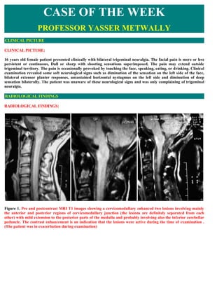
- 1. CASE OF THE WEEK PROFESSOR YASSER METWALLY CLINICAL PICTURE CLINICAL PICTURE: 16 years old female patient presented clinically with bilateral trigeminal neuralgia. The facial pain is more or less persistent or continuous, Dull or sharp with shooting sensations superimposed. The pain may extend outside trigeminal territory. The pain is occasionally provoked by touching the face, speaking, eating, or drinking. Clinical examination revealed some soft neurological signs such as diminution of the sensation on the left side of the face, bilateral extensor planter responses, unsustained horizontal nystagmus on the left side and diminution of deep sensation bilaterally. The patient was unaware of these neurological signs and was only complaining of trigeminal neuralgia. RADIOLOGICAL FINDINGS RADIOLOGICAL FINDINGS: Figure 1. Pre and postcontrast MRI T1 images showing a cervicomedullary enhanced two lesions involving mainly the anterior and posterior regions of cervicomedullary junction (the lesions are definitely separated from each other) with mild extension to the posterior parts of the medulla and probably involving also the inferior cerebellar peduncle. The contrast enhancement is an indication that the lesions were active during the time of examination . (The patient was in exacerbation during examination)
- 2. Figure 2. Pre and postcontrast MRI T1 images showing a cervicomedullary enhanced two lesions involving mainly the anterior and posterior regions of cervicomedullary junction (the lesions are definitely separated from each other) with mild extension to the posterior parts of the medulla and probably involving also the inferior cerebellar peduncle. The contrast enhancement is an indication that the lesions were active during the time of examination . (The patient was in exacerbation during examination)
- 3. Figure 3. MRI T2 image (A) and postcontrast MRI T1 image (B). The cervicomedullary pathology appear hyperintense on the MRI T2 image and is composed almost of two lesions (an oval lesion anteriorly and a linear one posteriorly), the two lesions ate separated from each other by an isointense line of cleavage on the MRI T2 image. Only parts of the lesions as demonstrated on the MRI T2 image (A) are enhanced on the postcontrast MRI T1 image (B). Other parts of the lesions are not enhanced. The multiplicity of the lesions provides evidence for dissemination in place. However MRI can also provide evidence for quot; Dissemination in timequot;. When some lesions, or some parts of the same lesion - as seen on the T2 images- enhance on the T1 postcontrast images and others do not enhance, this simply mean that some lesions are old quot;burnt outquot; plaques (those which do not enhance) and other lesions are new and active plaques (those which enhance), and this provides evidence of quot; Dissemination in timequot;. Ring enhancement also provides evidence for quot; Dissemination in timequot;. Contrast enhancement (as demonstrated on the postcontrast T1 image) occurred at the periphery of the lesions demonstrated on the MRI T2 image (A) and this is the picture of reactivation of old plaques which takes place at the periphery of old plaques rather than at the center.
- 4. Figure 4. MRI T2 images. The cervicomedullary pathology appear hyperintense on the MRI T2 images and is composed almost of two lesions (an oval lesion anteriorly and a linear one posteriorly), the two lesions ate separated from each other by an isointense line of cleavage on the MRI T2 images. The lesions are causing slight enlargement of the cervicomedullary junction. Figure 5. MRI FLAIR image showing the typical picture of multiple sclerosis. MRI examination of the brain and the spinal cord demonstrated dissemination in both time and place. The clinical diagnosis of multiple sclerosis was made. The clinical presentation of multiple sclerosis in this case was in the form of bilateral trigeminal neuralgia which is quite rare in multiple sclerosis as a presenting symptom of the disease. The demonstrated MS plaques in the cervicomedullary junction were active because of the following 1. The lesions showed partial contrast enhancement (parts of the lesions are enhanced (new or recently activated plaques) and parts are not enhanced (old or burnt- out plaques). Contrast enhancement occurs due to break
- 5. down of blood brain barrier. 2. The MRI T2 hyperintensity are partially due to the existence of vasogenic edema which occurred due to breakdown of blood brain barrier. Breakdown of the blood brain barrier is a radiological sign of plaque reactivation. The slight enlargement of the cervicomedullary junction demonstrated in this case is secondary to the existing vasogenic edema. Examination of the pons did not provide evidence for MS plaques at the root entry zone of the fifth nerve. The cervicomedullary lesions definitely involved the sensory nucleus of the trigeminal nerve in this patient, however, the contribution of lesions of the trigeminal nuclei to the clinical picture of trigeminal neuralgia in multiple sclerosis is still not clear. Although vascular compression of the fifth nerve may be present and has been suggested as the cause of TN even in multiple sclerosis patients presented with trigeminal neuralgia, we did not identify suspicious vessels along the fifth nerve in this patient. Although characteristic signal void makes even tiny arteries visible on high-resolution images, additional MRA acquisitions may be used to confirm negative conventional study results. Nevertheless, with the use of high-flow MRA, the chance remains that venous compression of the fifth nerve will be missed. Peripheral cranial nerve dysfunction caused by demyelination of fascicular brainstem fibers as demonstrated by MRI has been noted in sixth, seventh, and eighth nerve dysfunction. Therefore, peripheral cranial nerve deficits in patients with MS should give rise to the suspicion of a brainstem lesion. We conclude that in MS, trigeminal neuralgia is usually caused by demyelinating lesions affecting pontine trigeminal pathways, and like i said before the contribution of lesions of the trigeminal nuclei to the clinical picture of trigeminal neuralgia in multiple sclerosis is still not clear. Box 1. Criteria that characterize trigeminal neuralgia in multiple sclerosis patients and differentiate it from idiopathic trigeminal neuralgia 1. The young age of the patient (below 50 years of age) 2. The bilateral presentation of trigeminal neuralgia 3. The existence of soft neurological signs the patient is probably not aware of 4. The existence of sensory loss in the face 5. The pain is more continuous and might extend outside the trigeminal area 6. The pain shows no remission and has a continuous slow progression DIAGNOSIS: DIAGNOSIS: A CASE OF MULTIPLE SCLEROSIS PRESENTED CLINICALLY WITH BILATERAL TRIGEMINAL NEURALGIA AND A CERVICO-MEDULLARY ACTIVE DEMYELINATING PLAQUES DISCUSSION DISCUSSION: This case report illustrates that trigeminal neuralgia occasionally is associated with a serious systemic disease. Appropriate diagnostic tests are indicated for all patients with trigeminal neuralgia, since early diagnosis and treatment of an underlying systemic disease will have a significant impact on the prognosis.
- 6. Epidemiology of trigeminal neuralgia Trigeminal neuralgia has an incidence of 4.5 per 100,000 population. The disorder is more prevalent among females (F:M sex ratio of 1.74:1) and affects primarily older people; the average age of onset is between the fifth and seventh decades. The majority of cases are unilateral; approximately 4 percent of cases are bilateral. TN most commonly involves either the maxillary or mandibular division of the trigeminal nerve alone, while the ophthalmic division rarely is affected alone. Involvement of more than one division of the trigeminal nerve is not uncommon. Trigeminal neuralgia is characterized by spontaneous remissions that may last months or even years. Nevertheless, with time, pain attacks become more frequent, while remissions occur less often. Definition of trigeminal neuralgia Both the International Association for the Study of Pain (IASP) and International Headache Society (IHS) have suggested their own diagnostic criteria for trigeminal neuralgia. [72] These are remarkably similar and highlight the sudden, explosive nature of the pain (Table 1). In further descriptions of the condition, both classifications allude to vascular compression, MS and tumours as known aetiological causes. The IASP classification makes a distinction between trigeminal neuralgia (including MS) and secondary neuralgias (caused by structural lesions and injuries, but not including MS), while IHS separates idiopathic trigeminal neuralgia from the ‘symptomatic form’ depending on the presence of a structural lesion; it is not quite clear if vascular compression qualifies as such. Neither approach includes reference to variant forms of trigeminal neuralgia, which satisfy the diagnostic criteria but display additional features as well. [71] Table 1 Definition of trigeminal neuralgia provided by the International Association for the Study of Pain, IASP72 IASP definition IHS definition Sudden, usually unilateral, Painful unilateral affliction of the face, characterized by brief electric shock like severe brief stabbing recurrent pain limited to the distribution of one or more divisions of the trigeminal nerve. pains in the distribution of one or Pain is commonly evoked by trivial stimuli including washing, shaving, smoking, more branches of the Vth cranial talking, and brushing the teeth, but may also occur spontaneously. The pain is nerve abrupt in onset and termination and may remit for varying periods Diagnosis Table 2 demonstrates differences between typical trigeminal neuralgia, trigeminal neuralgia with atypical features and Trigeminal neuropathy. A typical form of trigeminal neuralgia may, in prolonged cases, develop atypical signs. By contrast, many cases of trigeminal neuralgia start with pain lacking the typical characteristics of trigeminal neuralgia, but respond well to carbamazepine, and later developing all the hallmark signs of trigeminal neuralgia (‘pre-trigeminal neuralgia’). [71] Table 2 Comparison of definitions of trigeminal neuralgia. *Key items [71] Trigeminal Typical trigeminal Atypical trigeminal neuralgia (IASP neuralgia neuralgia and IHS definition) Trigeminal neuropathy Site* Unilateral Unilateral Unilateral Uni- or bilateral Sharp, shooting, Sharp shooting, stabbing, Dull or sharp, smarting,
- 7. Quality of Sharp, stabbing, stabbing, lingering lingering aftersensations, steady pain with shooting pain* burning, superficial aftersensations burning, smarting sensations superimposed Duration of Any duration, usually pain* Brief A few seconds at most Several seconds hours Duration of Seconds to 2 Continuous with pain- paroxysms minutes Seconds to minutes Seconds to minutes free spells Refractory period Yes Yes Yes No Continous pain No No Yes, not severe Dominant feature Limited trigger Allodynia* zones Small trigger zones Small trigger zones Large allodynic areas Variable vasodilatation Associated Vasodilatation, swelling Vasodilatation, swelling and swelling, may be features Slight flush seen with severe pain seen with severe pain constantly present None outside None outside affected None outside affected May extend outside Radiation affected division division division trigeminal territory Eating, talking, Touching, speaking, washing face, Touching, speaking, eating,drinking, cold, Provoking brushing teeth, eating, drinking, cold, occasionally heat, Same as atypical factors* smoking not heat, movement movement trigeminal neuralgia Variability of pain Stereotyped Some variation Definite variation Definite variation Not detectable with May be detectable with Prominent, easily Sensory bedside tests QST may bedside tests QST usually detected with bedside loss* None be abnormal abnormal tests, confirmed by QST Pain Aversion to touch Aversion to touch guarded Tolerates touching behaviour Not discussed guarded speech speech speech not affected Early remissions Course of Spontaneous Early remissions pre- previously typical No remission; slow pain* remissions trigeminal neuralgia trigeminal neuralgia progression Trigeminal neuropathy, whether painful or non-painful, is associated with a structural lesion or systemic disease. It may be seen following direct trauma to the nerve (e.g. supra- and infra-orbital neuralgias following facial fractures); we also classify dysaesthesiae and anaesthesia dolorosa following neuroablative procedures as trigeminal neuropathy. On occasion, it can be seen caused by severe arterial compression, usually from an ectatic basilar artery. The pain description in this condition is different from that in trigeminal neuralgia and more akin to that in painful peripheral neuropathy. Pain is usually constant and associated with allodynia and sensory loss. Central nervous system lesions, even if they predominantly involve the trigeminal pathways and very occasionally mimic trigeminal neuralgia, are not classified as either trigeminal neuropathy or trigeminal neuralgia, but fall under the heading of central pain. [71] Trigeminal neuralgia remains a clinical diagnosis dependent on a history of sudden shooting or stabbing pain, coming as solitary sensations or paroxysms and separated by pain-free intervals. Optimally, it is the patient who volunteers this description. However, many patients with facial pain have considerable difficulty in finding precise expressions to convey the characteristics of their symptoms. In such a case, the interviewer may suggest descriptive words with as little prompting as possible. Patients with atypical trigeminal neuralgia also give the history of stabbing or shooting pains. Unlike in typical trigeminal neuralgia, they relate a history of other kinds of pain as well. Many have constant or near constant discomfort on the same side of the face. [71]
- 8. Aetiology of trigeminal neuralgia There are obvious problems in determining aetiological causes for a syndrome, which is formulated on the basis of subjective pain rather than hard signs or laboratory abnormalities. However, in the last three decades, evidence has been mounting that in a large proportion of cases, compression of the trigeminal nerve root at or near the dorsal root entry zone by a blood vessel is a major causative or contributing factor. There are several lines of evidence that support this view. [71] First, novel imaging methods (MRI) and observations during posterior fossa surgery for trigeminal neuralgia have consistently shown a blood vessel in contact with the nerve root. Second, elimination of the compression leads to long-term pain relief in most patients. Third, intra-operative recordings show immediate improvement in nerve conduction following decompression, fitting with the general experience that patients tend to wake up from the operation pain- free. Fourth, sensory functions recover as well following decompression (though this recovery is slower than that in nerve conduction). Of other known aetiological factors, the association of MS with trigeminal neuralgia is well established. MS is seen in 2–4% of patients with trigeminal neuralgia. Conversely, trigeminal neuralgia is diagnosed in 1–5% of patients with MS. [71] In a small proportion of patients with MS, trigeminal neuralgia is the first manifestation of the disease. These patients are younger than the trigeminal neuralgia population as a whole and their neuralgia is more frequently bilateral. A latent demyelinating disease should be considered in young patients with trigeminal neuralgia and appropriate diagnostic tests performed, as disease-modifying treatment for MS is emerging. Neuropathological and imaging studies suggest that the common denominator in patients with typical trigeminal neuralgia and MS is the involvement of the trigeminal nerve entry zone in the pons. In contrast, plaques in other parts of the brain stem or elsewhere in the CNS do not necessarily lead to development of trigeminal neuralgia. Interestingly, there are reports of vascular compression of the trigeminal nerve in patients with MS, and pain relief following decompression, though the results are less favourable than in other trigeminal neuralgia subgroups. [71] Tumours, usually posterior fossa meningiomas or neuromas, are found in 2% of patients who present with typical trigeminal neuralgia.26 The localization of the tumour dictates the nature of facial symptoms. Tumours affecting the peripheral branches or the Gasserian ganglion usually give rise to sensory change and constant pain, in other words, trigeminal neuropathy. Slowly growing tumours which distend the trigeminal root rather than invade it, are usually found in trigeminal neuralgia. [71] Clinical characteristics of trigeminal neuralgia Trigeminal neuralgia is characterized by severe, stabbing pain that may be accompanied by a contraction of the facial musculature (hence the term quot;tic douloureuxquot;). Pain attacks are episodic, last only seconds to a few minutes and may recur in clusters. The pain is characterized by sudden onset and cessation, and the patient is completely asymptomatic between attacks. Pain paroxysms may be provoked by innocuous sensory stimulation of trigger zones in the receptive field of the affected branch. [9] Common daily activities such as talking, eating, drinking, swallowing, exposure to cold air, shaving, brushing the teeth or washing the face can trigger the pain, significantly compromising the patient’s quality of life. The trigger zone always is ipsilateral to the pain; however, it may not coincide with the area of pain. Common extraoral trigger zones occur above the supraorbital foramen, the inner canthus of the eye, lateral to the ala nasi and over the mental foramen. Typically, immediately after a jab of pain, there is a refractory period during which further pain attacks cannot be evoked. [71] Trigeminal neuralgia is characterized by spontaneous remissions that may last months or even years. Nevertheless, with time, pain attacks become more frequent, while remissions occur less often and last for a shorter period. [1] In prolonged cases, patients may develop atypical features, such as persistent pain between episodes. [1] Continuous pain is more common in symptomatic trigeminal neuralgia, as in the case discussed here.
- 9. Pathophysiology Most idiopathic trigeminal neuralgia cases are thought to be induced by vascular compression of the trigeminal root-entry zone, which results in demyelination of trigeminal sensory fibers. [10–13] Histopathologic examination of trigeminal nerve roots from patients with compression of the nerve root by a blood vessel reveals focal loss of myelin, close apposition of the demyelinated axons and lack of intervening astrocytic processes. [11,12] Demyelination may result in ectopic generation of action potentials, presenting clinically as spontaneous pain. [14] Moreover, demyelination promotes ephaptic neural transmission—namely, development of abnormal contacts between adjacent nerve axons, which results in inappropriate spread of action potentials and activation of one nerve on activity in another. Such inappropriate spread of action potentials may underlie the generation of pain by innocuous stimulation. [14] In the course of time, excessive afferent input of nerve impulses may produce central sensitization, resulting in atypical trigeminal neuralgia features, such as constant pain. [1,15] Primary demyelinating disorders and its role in trigeminal neuralgia Trigeminal neuralgia is a well-recognized complication of multiple sclerosis. Typically, a plaque of demyelination encompasses the root entry zone of the trigeminal nerve in the pons. Rarely, patients with peripheral nerve demyelination due to Charcot–Marie–Tooth disease develop trigeminal neuralgia. Vascular compression of the trigeminal nerve root may contribute to trigeminal neuralgia even in patients with demyelinating disorders; compression of the root entry zone by a blood vessel has been demonstrated in a sizeable minority of patients with multiple sclerosis and trigeminal neuralgia and in an occasional patient with Charcot–Marie–Tooth disease. In many such cases, decompression of the nerve root leads to relief of symptoms. [71] Differential diagnosis of trigeminal neuralgia The clinician should consider a number of other conditions when making the diagnosis of trigeminal neuralgia. Tumors Occasionally, trigeminal neuralgia develops secondary to posterior fossa compressive lesions, such as cysts and tumors. [2–4] Cheng and colleagues [2] reported that tumors are detected in 2 percent of patients who have typical trigeminal neuralgia. Multiple sclerosis Trigeminal neuralgia also may be caused by demyelinating plaques of MS involving the trigeminal nociceptive pathway. [5,16] MS is diagnosed in 2 to 4 percent of patients with trigeminal neuralgia. [15] Typically, patients with trigeminal neuralgia and MS are younger than those with idiopathic trigeminal neuralgia and are more likely to have bilateral facial pain. [5] In the vast majority of cases, trigeminal neuralgia develops later in the course of MS; occasionally, however, trigeminal neuralgia may appear first. [5] trigeminal neuralgia’s development in patients younger than [50] years of age may be the first manifestation of MS, as was the case in our patient. A demyelinating plaque extending into the root entry zone of the trigeminal nerve is a common finding among patients with trigeminal neuralgia and MS, while plaques in other CNS regions do not seem to be associated with trigeminal neuralgia. [11,17] Meaney and colleagues [18] reported that vascular compression may be the underlying cause of trigeminal neuralgia, even in patients with MS. The authors demonstrated vascular compression of the root entry zone in a subset of these patients, with subsequent elimination of the trigeminal neuralgia pain after decompression of the nerve root. [18] trigeminal neuralgia in association with demyelinating peripheral neuropathy secondary to Charcot-Marie-Tooth disease [6,7] or with infarction of the root entry zone [19] has been reported. MS is a chronic CNS disease, characterized by discrete areas of demyelination, axon damage and associated inflammation. [20–22] MS lesions are disseminated in time and space (that is, occur in different parts of the CNS at least three months apart) and can result in a wide variety of symptoms and signs, including numbness, paresthesias, pain, weakness, spasticity, fatigue, vertigo, visual difficulties, gait dysfunction, bladder disturbances and cognitive changes. [20,23,24] The disease appears typically between the ages of 18 and 45 years. Approximately 80 to 85 percent of patients have relapsing-remitting MS, characterized by acute attacks interspersed with recovery periods, while 10 to 15 percent of patients have primary progressive MS, characterized by steady progressive deterioration of neurological function. Significant disability develops over a course of 10 to 20 years, and approximately 50 percent of patients eventually die as a result of complications of the disease. [20]
- 10. Greater risk of MS in first-degree relatives of patients with MS and high concordance rates between monozygotic twins suggest a genetic susceptibility to the disease. [24,25] Environmental factors are believed to play an important role, while an infectious trigger also has been hypothesized. [20] Even though the mechanisms underlying the genesis of the disease still are unclear, an autoimmune pathogenesis is favored. [26] Glucocorticoids are effective in reducing the severity and duration of acute exacerbations, but they do not affect the course of MS over time. [27] In contrast, disease-modifying therapies, such as glatiramer acetate and interferon beta, reduce the number of relapses, improve recovery from attacks and delay disease progression and development of disability. [27–29] The patient presented here experienced alleviation of symptoms after receiving glatiramer acetate therapy. Glatiramer acetate exerts anti-inflammatory effects by binding to major histocompatibility complex molecules and inhibiting myelin-reactive T cells. [30,31] In addition, glatiramer acetate induces anti-inflammatory Th2 cells, which may exert their protective action by crossing the blood-brain barrier and by producing anti-inflammatory cytokines in response to cross-recognition of myelin antigens. [31] Inhibition of brain inflammation, which has been associated with irreversible brain tissue injury, is thought to delay the occurrence of irreversible CNS lesions. [32] Figure 6. (a) Coronal plane STIR image through the brain stem of a 41-year-old woman with multiple sclerosis, presenting with a short history of altered facial sensation. There is a high signal intensity plaque of demyelination within the right brachium pontis (arrow). (b) Axial plane T2 weighted FSE image at the level of the centrum semiovale. Typical ovoid lesions of primary demyelination in the periventricular and subcortical white matter. Chronic inflammatory demyelinating polyneuropathy Chronic inflammatory demyelinating polyneuropathy is an acquired peripheral neuropathy, characterized by multifocal demyelination of spinal roots, major plexuses and proximal nerve trunks. [33] The exact etiology is unknown, but autoimmune mechanisms have been implicated. [33–35] The disease has a chronic course that may be either progressive or relapsing-remitting. The clinical manifestations are variable but include motor and sensory symptoms, such as spontaneous pain, paresthesia, numbness and muscle weakness of the upper and lower extremities. [33] Occasionally, cranial nerves and respiratory muscles also are involved. [34] Treatment consists of intravenous immunoglobulin therapy, plasma exchange, corticosteroid therapy and immunosuppressive drug therapy. [35] Cluster headache and chronic paroxysmal hemicrania The excruciating intensity of trigeminal neuralgia pain and its intermittent temporal pattern may result in a misdiagnosis of cluster headache or chronic paroxysmal hemicrania. [36] However, both cluster headache and chronic paroxysmal hemicrania typically affect younger adults. [37,38] In addition, these neurovascular headaches are characterized by attacks of longer duration (minutes to hours), which typically occur around the clock and interrupt the patient’s sleep. [39,40] In contrast to trigeminal neuralgia, these attacks cannot be triggered by
- 11. innocuous cutaneous stimulation. [36,40] Moreover, a unique feature of cluster headache and chronic paroxysmal hemicrania is that autonomic phenomena—such as conjunctival injection, lacrimation, nasal congestion and rhinorrhea—accompany the pain. [41] Lyme disease Lyme disease also should included in the differential diagnosis of trigeminal neuralgia. Lyme disease or Lyme borreliosis is an infectious, tick-transmitted disease, caused by spirochetes of the Borrelia burgdorferi species complex. Lyme disease can manifest with an array of symptoms involving multiple organs and systems, such as skin, heart, eye, joints and the peripheral nervous system and the CNS.42 Neurological manifestations of the disease may include meningitis, single or multiple cranial neuropathies, painful radiculopathies and diffuse polyneuropathies. [43,44] On rare occasions, patients with Lyme disease may have neurogenic pain similar to that of trigeminal neuralgia. [45,46] This possibility should be ruled out via hematologic assessment for Lyme titers. Failure of dental treatment to provide long-term pain relief should raise the suspicion of trigeminal neuralgia. Dental pain When the sharp, paroxysmal pain of trigeminal neuralgia is localized in the dentition or the surrounding structures, it may be misdiagnosed as dental pain. [16,36] Frequently, patients with trigeminal neuralgia undergo numerous dental procedures until the diagnosis of trigeminal neuralgia is made. These procedures may offer temporary pain relief for a few weeks; however, the pain always recurs and often is even worse. Failure of dental treatment to provide long-term pain relief should raise the suspicion of trigeminal neuralgia. An important feature that distinguishes trigeminal neuralgia from dental pain is that trigeminal neuralgia typically does not interrupt the patient’s sleep. Moreover, pain originating from dental pathology usually is progressive, and its character changes with time. Tooth vitality tests and radiographic examination also will serve to exclude dental pathology. Box 2. Causes of trigeminal neuralgia/neuropathy Tumor Acoustic neurinoma Chordoma at the level of the clivus Pontine glioma Epidermoid Metastases Lymphoma Vascular Pontine infarct Arteriovenous malformation in the vicinity Persistence of a primitive trigeminal artery Pulsatile compression by the adjacent superior cerebellar artery (more rarely, anteroinferior artery) Inflammatory
- 12. Multiple sclerosis (MS) Sarcoidosis Lyme disease neuropathy Paraneoplastic (possibly) The list of differential diagnoses is long and includes a number of pathological conditions affecting the sinuses, teeth, temporomandibular joints, eyes, nose, and the neck. Most of these are easily ruled out after the interview and brief clinical examination. However, a few conditions remain that bear considerable similarity to trigeminal neuralgia and are listed in Table 3. Table 3 Differential diagnosis of trigeminal neuralgia. *SUNCT, Short-lasting, unilateral, neuralgiform headcahe with conjunctival injection and tearing; **CPH, chronic paroxysmal hemicrania. Duration of Location of pain or Shooting pain Autonomic Pain relief with Condition pain attack or paroxysms symptoms carbamazepine Comments Only superimposed Cluster Retrobulbar, 20 min to on deep dull Triptans help Alcohol headache cheek, chin hours pain Prominent Slight provokes Forehead, 5 s to several Almost exclusively SUNCT* retrobulbar minutes Yes Prominent None inwomen; rare Forehead, Responsive to CPH** retrobulbar 2–45 min No Prominent None indomethacin Cracked tooth Upper jaw Provoked on biting syndrome lower jaw Seconds Yes None None and chewing Jabs and jolts Anywhere in No precipitating syndrome the head Seconds Yes None Good factors History of shingles Post- Forehead, Superimposed Tactile herpetic eye, cheek background Variable, Variable, usually allodynia,Sensory neuralgia (rarely) Continuous pain mild modes impairment Giant cell Forehead, arteritis neck, temple Continuous None None None Jaw claudication SUNCT, Short-lasting, unilateral, neuralgiform headcahe with conjunctival injection and tearing; **CPH, chronic paroxysmal hemicrania Other cranial neuralgias (glossopharyngeal neuralgia, neuralgia of nervus intermedius, neuralgia of the superior laryngeal nerve, and occipital neuralgia) can all cause diagnostic difficulty. These neuralgias are rare and can produce pain that is identical to that of trigeminal neuralgia. However, the location is different. Awareness of underlying conditions
- 13. Thorough medical examination and imaging should be undertaken in all patients with trigeminal neuralgia to rule out underlying conditions. Occasionally, neurological signs indicative of secondary causes of trigeminal neuralgia become apparent only later in the course of the disease, resulting in a delayed diagnosis. [2,5] In the case presented here, both the neurological examination and the MRI of the brain were normal initially. However, even though the MRI failed to depict any demyelinating plaques even later in the disease process, subsequent neurological assessments led to the diagnosis of MS and chronic inflammatory demyelinating polyneuropathy, thus emphasizing the importance of repeating neurological examinations in patients with trigeminal neuralgia. [16] TREATMENT OF TRIGEMINAL NEURALGIA Pharmacological approaches Pharmacological therapy is the first line of treatment for trigeminal neuralgia. The goal of the medical management is the reduction of neuronal hyperexcitability in the peripheral nervous system, the CNS or both. [47–49] Various antiepileptic drugs are used, including carbamazepine, oxcarbazepine, lamotrigine, phenytoin and gabapentin. [47– 52] Other potentially effective medications include antispasmolytic agents, such as baclofen, a -aminobutyric acid receptor agonist, and tizanidine, an 2-adrenergic agonist. [50] Carbamazepine has been found effective in several controlled trials and is the mainstay of pharmacotherapy for trigeminal neuralgia. [51] The most common adverse effects include sedation, fatigue, dizziness, blurred vision, nausea, vomiting and allergic skin reactions. Periodic complete blood cell count and liver function monitoring tests are essential, since agranulocytosis and aplastic anemia, as well as hepatocellular and cholestatic jaundice, can occur rarely. [47] Oxcarbazepine, a daughter drug of carbamazepine, is a safer alternative to carbamazepine and does not require routine monitoring of hematologic and hepatic profiles during treatment. [52] Often, drug combinations are used to maximize effectiveness and minimize adverse effects. [53] The clinician should attempt gradual withdrawal of the medications once complete pain control is achieved, because remission periods are common. Patients with trigeminal neuralgia and with MS may respond initially to pharmacological treatment; however, relapse is frequent. The patient in this report was responsive to carbamazepine, but use of the drug was discontinued owing to adverse effects. Table 4 Summary of drugs commonly used in trigeminal neuralgia. Initial Maintenance Drug dose dose Adverse effects Sedation, dizziness, cognitive impairment, headache, GI symptoms, Carbamazepine 200 mg 400–1200 mg allergic rash, leucopenia, folate deficiency, hyponatremia, several drug (CBZ) day–1 day–1 interactions, warfarin! Oxcarbazepine 300 mg 600–1200 mg Better tolerated than CBZ, sedation, dizziness, cognitive impairment, (OXC) day–1 day–1 hyponatremia, rash Sedation, ataxia, behavioural change, cognitive impairment, 300 mg 200–400 mg lymphadenopathy, osteopenia, acne, gingival hypertrophy, rash, folate Phenytoin (PHT) day–1 day–1 deficiency, liver failure, several drug interactions Allergic rash (necessitates immediate discontinuation), sedation, Lamotrigine 25–50 200–400 mg dizziness, headache, ataxia, significant interactions with other (LTG) mg day –1 day–1 anticonvulsants 10 mg 30–80 mg day– Baclofen (BAC) day–1 1 Sedation, ataxia, fatigue, GI symptoms, muscle weakness
- 14. Surgical procedures For patients with trigeminal neuralgia who become refractory to pharmacological treatment or cannot tolerate its adverse effects, surgical intervention is recommended. Surgical techniques used for trigeminal neuralgia treatment are peripheral surgery, percutaneous ablative procedures, stereotactic radiosurgery and microvascular decompression (MVD). Peripheral surgery. Peripheral surgery entails lesioning of peripheral branches of the trigeminal nerve, and using cryotherapy, neurectomy, alcohol block or radiofrequency. Peripheral techniques are the least invasive of all surgical procedures but are characterized by low effectiveness and a high recurrence rate. [15,54,55] Percutaneous ablative techniques Percutaneous ablative procedures involve nerve lesioning at the level of the gasserian ganglion by percutaneous radiofrequency thermocoagulation, balloon compression, injection of glycerol or a combination of these. [56–59] These neurodestructive procedures have good initial results and carry less risk than MVD; however, they are associated with a higher incidence of pain recurrence. Potential complications include loss of touch sensation, dysesthesias, corneal damage, anesthesia dolorosa and temporary masseteric weakness. Dentists should be competent in the differential diagnosis of trigeminal neuralgia from other facial pain causes, including dental pathology. The patient described in this case report underwent percutaneous glycerol rhizotomy. During this procedure, a needle is advanced, with the aid of fluoroscopy, through the foramen ovale into the trigeminal cistern, where glycerol is injected in close proximity with the pregaglionic trigeminal rootlets. [51,60] The sensory fibers of the trigeminal nerve are affected, resulting in termination or reduction of the trigeminal neuralgia attacks.61 Percutaneous glycerol rhizolysis causes sensory loss and masseteric weakness less frequently than do the two alternative percutaneous procedures, but it has higher recurrence rates. [51,58,59] Stereotactic radiosurgery. Stereotactic radiosurgery, also known as gamma knife surgery, involves lesioning of the trigeminal nerve at the root entry zone using stereotactic techniques and radiation between 70 and 90 grays. [62,63] This procedure is less invasive and thus is suitable for medically compromised patients. Outcomes are similar to those of other ablative procedures. [64,65] However, there may be a latency period of three to six months, after which maximum pain relief is obtained. [62,64] This technique’s highest success rates are observed when it is used as primary treatment in patients with typical symptoms. [64,66,67] Facial sensory loss, paresthesias and dysesthesias are the most common complications. [58,62,66] A positive correlation between development of trigeminal dysfunction and pain relief after gamma knife surgery has been reported. [62] Microvascular decompression. MVD of vessels compressing the nerve root is effective and has a low incidence of recurrence of trigeminal neuralgia. [68,69] However, it entails posterior fossa craniotomy and involves serious risks, including hearing impairment, ataxia, brainstem infarction, cerebellar injury and death. [69] Magnetic resonance tomographic angiography can depict vascular compression of the nerve and is useful in identifying patients who are good candidates for MVD. [10,70] CONCLUSIONS Dentists should be competent in the differential diagnosis of trigeminal neuralgia from other facial pain causes, including dental pathology. A correct diagnosis will lead to appropriate management and preclude unsuccessful attempts to treat the pain with irreversible dental procedures. This case report illustrates that trigeminal neuralgia occasionally is associated with a serious systemic disease. Appropriate diagnostic tests are indicated for all patients with trigeminal neuralgia, since early diagnosis and
- 15. treatment of an underlying systemic disease will have a significant impact on the prognosis. Anticonvulsant medications constitute the first line of trigeminal neuralgia treatment; however, many patients eventually require surgical intervention, owing to diminished response to pharmacological therapy or to the development of intolerable adverse effects. Surgical procedures that can reduce the frequency and severity of trigeminal neuralgia attacks include peripheral surgery, percutaneous ablative procedures, stereotactic radiosurgery and MVD. SUMMARY SUMMARY There is now persuasive evidence that trigeminal neuralgia is usually caused by demyelination of trigeminal sensory fibres within either the nerve root or, less commonly, the brainstem. In most cases, the trigeminal nerve root demyelination involves the proximal, CNS part of the root and results from compression by an overlying artery or vein. Other causes of trigeminal neuralgia in which demyelination is involved or implicated include multiple sclerosis and, probably, compressive space-occupying masses in the posterior fossa. Examination of trigeminal nerve roots from patients with compression of the nerve root by an overlying blood vessel has revealed focal demyelination in the region of compression, with close apposition of demyelinated axons and an absence of intervening glial processes. Similar foci of nerve root demyelination and juxtaposition of axons have been demonstrated in multiple sclerosis patients with trigeminal neuralgia. Experimental studies indicate that this anatomical arrangement favours the ectopic generation of spontaneous nerve impulses and their ephaptic conduction to adjacent fibres, and that spontaneous nerve activity is likely to be increased by the deformity associated with pulsatile vascular indentation. Decompression of the nerve root produces rapid relief of symptoms in most patients with vessel-associated trigeminal neuralgia, probably because the resulting separation of demyelinated axons and their release from focal distortion reduce the spontaneous generation of impulses and prevent their ephaptic spread. The role of remyelination in initial symptomatic recovery after decompression is unclear. However, remyelination may help to ensure that relief of symptoms is sustained after decompression of the nerve root and may also be responsible for the spontaneous remission of the neuralgia in some patients. In addition to causing symptomatic relief, vascular decompression leads to rapid recovery of nerve conduction across the indented root, a phenomenon that, we suggest, is likely to reflect the reversal of compression-induced conduction block in larger myelinated fibres outside the region of demyelination. Trigeminal neuralgia can occur in association with a range of other syndromes involving vascular compression and hyperactivity of cranial nerves. Clinical observations and electrophysiological studies support the concept that demyelination and ephaptic spread of excitation underlie most, if not all, of these conditions. Classical trigeminal neuralgia (tic douloureux) has an annual incidence of ~4.5 per 100 000. It is characterized by recurrent episodes of intense, lancinating pain localized to small areas of the face. The onset is usually in middle or old age, but young adults and children can also be affected. Attacks usually last only seconds but may recur repeatedly within a short period of time. The attacks are often, but not always, precipitated by mild sensory stimulation of so-called trigger zones, which may be located anywhere within the territory of the affected trigeminal nerve. Typical antecedent stimuli include light touching, draughts of wind, eating, drinking, washing, shaving and applying make-up. The neuralgia tends to occur in bouts over a period of weeks or months, with subsequent spontaneous remission that may last months or years. In time, however, attacks usually become more frequent and the pain more sustained. Although not usually significant clinically, the cutaneous perception of temperature and light touch is slightly impaired within the affected trigeminal divisions. Aetiology Most cases are caused by compression of the trigeminal nerve root, usually within a few millimetres of entry into
- 16. the pons, i.e. the root entry zone. In a few cases, trigeminal neuralgia is due to a primary demyelinating disorder. Other, rare causes include infiltration of the nerve root, gasserian ganglion or nerve by a tumour or amyloid, and small infarcts or angiomas in the pons or medulla. Once all of these possibilities have been excluded, there remains a small proportion of patients in whom the aetiology is undetermined. Compression of the trigeminal nerve root Much the commonest cause of trigeminal neuralgia is focal compression of the trigeminal nerve root, close to its point of entry into the pons, by an aberrant loop of artery or vein. It is now thought to account for 80–90% of cases. The part of the nerve root that is usually compressed (the root entry zone) is actually within CNS tissue, which extends several millimetres along the root, so that the junction between CNS and PNS is well away from the surface of the pons. Rarely, trigeminal neuralgia results from vascular compression of the nerve root by a saccular aneurysm (Ildan et al., 1996) or an arteriovenous malformation. A wide range of other compressive lesions can cause trigeminal neuralgia. These include vestibular schwannomas, meningiomas , epidermoid cysts and various other cysts and tumours. The neuralgia is occasionally contralateral to the side of the mass lesion. Compression of the trigeminal nerve root may be mediated by the tumour itself, or, more commonly, by an interposed blood vessel or by distortion of the contents of the posterior fossa with displacement of the nerve root against a blood vessel or the skull base. Primary demyelinating disorders Trigeminal neuralgia is a well-recognized complication of multiple sclerosis. Typically, a plaque of demyelination encompasses the root entry zone of the trigeminal nerve in the pons. Rarely, patients with peripheral nerve demyelination due to Charcot–Marie–Tooth disease develop trigeminal neuralgia. Vascular compression of the trigeminal nerve root may contribute to trigeminal neuralgia even in patients with demyelinating disorders; compression of the root entry zone by a blood vessel has been demonstrated in a sizeable minority of patients with multiple sclerosis and trigeminal neuralgia and in an occasional patient with Charcot–Marie–Tooth disease. In many such cases, decompression of the nerve root leads to relief of symptoms. Infiltrative disorders of the trigeminal nerve root, gasserian ganglion and nerve The principal infiltrative causes of trigeminal neuralgia are carcinomatous deposits within the nerve root, gasserian ganglion and nerve and trigeminal amyloidomas. Non-demyelinating lesions of the pons or medulla Small numbers of patients have been reported in whom trigeminal neuralgia was associated with a small infarct or angioma in the brainstem. Familial trigeminal neuralgia In the vast majority of cases, trigeminal neuralgia is a sporadic disorder. Familial occurrence has been reported in Charcot–Marie–Tooth disease. A further inherited disorder in which there is a theoretical risk of trigeminal neuralgia is autosomal dominant hypertension and brachydactyly. Addendum A new version of this PDF file (with a new case) is uploaded in my web site every week (every Saturday and remains available till Friday.) To download the current version follow the link quot;http://pdf.yassermetwally.com/case.pdfquot;. You can also download the current version from my web site at quot;http://yassermetwally.comquot;. To download the software version of the publication (crow.exe) follow the link: http://neurology.yassermetwally.com/crow.zip The case is also presented as a short case in PDF format, to download the short case follow the link:
- 17. http://pdf.yassermetwally.com/short.pdf At the end of each year, all the publications are compiled on a single CD-ROM, please contact the author to know more details. Screen resolution is better set at 1024*768 pixel screen area for optimum display. For an archive of the previously reported cases go to www.yassermetwally.net, then under pages in the right panel, scroll down and click on the text entry quot;downloadable case records in PDF formatquot; Also to view a list of the previously published case records follow the following link (http://wordpress.com/tag/case-record/) or click on it if it appears as a link in your PDF reader REFERENCES References 1. Burchiel KJ, Slavin KV. On the natural history of trigeminal neuralgia. Neurosurgery 2000;46(1):152–4. 2. Cheng TM, Cascino TL, Onofrio BM. Comprehensive study of diagnosis and treatment of trigeminal neuralgia secondary to tumors. Neurology 1993;43:2298–302. 3. Puca A, Meglio M, Vari R, Tamburrini G, Tancredi A. Evaluation of fifth nerve dysfunction in 136 patients with middle and posterior cranial fossae tumors. Eur Neurol 1995;35(1):33–7. 4. Matsuka Y, Fort ET, Merrill RL. Trigeminal neuralgia due to an acoustic neuroma in the cerebellopontine angle. J Orofac Pain 2000;14(2):147–51. 5. Hooge JP, Redekop WK. Trigeminal neuralgia in multiple sclerosis. Neurology 1995;45:1294–6. 6. Coffey RJ, Fromm GH. Familial trigeminal neuralgia and Charcot-Marie-Tooth neuropathy: report of two families and review. Surg Neurol 1991;35(1):49–53. 7. de Matas M, Francis P, Miles JB. Microvascular decompression for trigeminal neuralgia in Charcot-Marie- Tooth disease. J Neurosurg 2000;92:715–7. 8. Krarup C. An update on electrophysiological studies in neuropathy. Curr Opin Neurol 2003;16:603–12. 9. Sato J, Saitoh T, Notani K, Fukuda H, Kaneyama K, Segami N. Diagnostic significance of carbamazepine and trigger zones in trigeminal neuralgia. Oral Surg Oral Med Oral Pathol Oral Radiol Endod 2004;97(1):18–22. 10. Meaney JF, Eldridge PR, Dunn LT, Nixon TE, Whitehouse GH, Miles JB. Demonstration of neurovascular compression in trigeminal neuralgia with magnetic resonance imaging. Comparison with surgical findings in 52 consecutive operative cases. J Neurosurg 1995;83: 799–805. 11. Love S, Hilton DA, Coakham HB. Central demyelination of the Vth nerve root in trigeminal neuralgia associated with vascular compression. Brain Pathol 1998;8(1):1–11. 12. Love S, Coakham HB. Trigeminal neuralgia: pathology and pathogenesis. Brain 2001;124:2347–60. 13. Leandri M, Eldridge P, Miles J. Recovery of nerve conduction following microvascular decompression for trigeminal neuralgia. Neurology 1998;51:1641–6. 14. Devor M, Govrin-Lippmann R, Rappaport ZH. Mechanism of trigeminal neuralgia: an ultrastructural analysis of trigeminal root specimens obtained during microvascular decompression surgery. J Neurosurg 2002;96:532–43. 15. Nurmikko TJ, Eldridge PR. Trigeminal neuralgia: pathophysiology, diagnosis and current treatment. Br J Anaesth 2001;87(1):117–32.
- 18. 16. Zakrzewska JM. Diagnosis and differential diagnosis of trigeminal neuralgia. Clin J Pain 2002;18(1):14–21. 17. Gass A, Kitchen N, MacManus DG, Moseley IF, Hennerici MG, Miller DH. Trigeminal neuralgia in patients with multiple sclerosis: lesion localization with magnetic resonance imaging. Neurology 1997;49:1142–4. 18. Meaney JF, Watt JW, Eldridge PR, Whitehouse GH, Wells JC, Miles JB. Association between trigeminal neuralgia and multiple sclerosis: role of magnetic resonance imaging. J Neurol Neurosurg Psychiatry 1995;59:253–9. 19. Golby AJ, Norbash A, Silverberg GD. Trigeminal neuralgia resulting from infarction of the root entry zone of the trigeminal nerve: case report. Neurosurgery 1998;43:620–2. 20. O’Connor P, Canadian Multiple Sclerosis Working Group. Key issues in the diagnosis and treatment of multiple sclerosis: an overview. Neurology 2002;59(6 supplement 3):S1–33. 21. Bruck W, Stadelmann C. Inflammation and degeneration in multiple sclerosis. Neurol Sci 2003;24 (supplement 5):S265–7. 22. De Stefano N, Narayanan S, Francis GS, et al. Evidence of axonal damage in the early stages of multiple sclerosis and its relevance to disability. Arch Neurol 2001;58(1):65–70. 23. Solaro C, Lunardi GL, Mancardi GL. Pain and MS. Int MS J 2003;10(1):14–9. 24. Frohman EM. Multiple sclerosis. Med Clin North Am 2003;87: 867–97. 25. Hillert J, Masterman T. The genetics of multiple sclerosis. In: Cook SD, ed. Handbook of multiple sclerosis. 3rd ed. New York: Dekker; 2001:33–65. 26. Markovic-Plese S, Pinilla C, Martin R. The initiation of the autoimmune response in multiple sclerosis. Clin Neurol Neurosurg 2004;106:218–22. 27. Goodin DS, Frohman EM, Garmany GP Jr, et al. Disease modifying therapies in multiple sclerosis: report of the Therapeutics and Technology Assessment Subcommittee of the American Academy of Neurology and the MS Council for Clinical Practice Guidelines. Neurology 2002;58(2):169–78. 28. McCormack PL, Scott LJ. Interferon-beta-1b: a review of its use in relapsing-remitting and secondary progressive multiple sclerosis. CNS Drugs 2004;18:521–46. 29. Wolinsky JS. Glatiramer acetate for the treatment of multiple sclerosis. Expert Opin Pharmacother 2004;5:875–91. 30. Arnon R, Aharoni R. Mechanism of action of glatiramer acetate in multiple sclerosis and its potential for the development of new applications. Proc Natl Acad Sci U S A 2004;101(supplement 2):14593–8. 31. Dhib-Jalbut S. Mechanisms of action of interferons and glatiramer acetate in multiple sclerosis. Neurology 2002;58(8 supplement 4):S3–9. 32. Rudick RA. Impact of disease-modifying therapies on brain and spinal cord atrophy in multiple sclerosis. J Neuroimaging 2004;14(3 supplement):54S–64S. 33. Said G. Chronic inflammatory demyelinative polyneuropathy. J Neurol 2002;249:245–53. 34. Latov N. Diagnosis of CIDP. Neurology 2002;59(12 supplement 6):S2–6. 35. Koski CL. Therapy of CIDP and related immune-mediated neuropathies. Neurology 2002;59(12 supplement 6):S22–7.
- 19. 36. Sarlani E. Diagnosis and treatment of orofacial pain. Braz J Oral Sci 2003;2:283–90. 37. Matharu MS, Goadsby PJ. Trigeminal autonomic cephalgias. J Neurol Neurosurg Psychiatry 2002;72 (supplement 2):ii19–ii26. 38. Sarlani E, Schwartz AH, Greenspan JD, Grace EG. Chronic paroxysmal hemicrania: a case report and review of the literature. J Orofac Pain 2003;17(1):74–8. 39. Sarlani E, Schwartz AH, Greenspan JD, Grace EG. Facial pain as first manifestation of lung cancer: a case of lung cancer-related cluster headache and a review of the literature. J Orofac Pain 2003;17:262–7. 40. Casucci G. Chronic short-lasting headaches: clinical features and differential diagnosis. Neurol Sci 2003;24 (supplement 2):S101–7. 41. May A. Headaches with (ipsilateral) autonomic symptoms. J Neurol 2003;250:1273–8. 42. Stanek G, Strle F. Lyme borreliosis. Lancet 2003;362:1639–47. 43. Halperin JJ. Neuroborreliosis: central nervous system involvement. Semin Neurol 1997;17(1):19–24. 44. Halperin JJ. Lyme disease and the peripheral nervous system. Muscle Nerve 2003;28(2):133–43. 45. Dotevall L, Eliasson T, Hagberg L, Mannheimer C. Pain as presenting symptom in Lyme neuroborreliosis. Eur J Pain 2003;7:235–9. 46. Fritz C, Rosler A, Heyden B, Braune HJ. Trigeminal neuralgia as a clinical manifestation of Lyme neuroborreliosis. J Neurol 1996;243: 367–8. 47. Backonja MM. Use of anticonvulsants for treatment of neuropathic pain. Neurology 2002;59(5 supplement 2):S14–7. 48. Zakrzewska JM, Chaudhry Z, Nurmikko TJ, Patton DW, Mullens EL. Lamotrigine (lamictal) in refractory trigeminal neuralgia: results from a double-blind placebo controlled crossover trial. Pain 1997;73: 223–30. 49. Dickenson AH, Matthews EA, Suzuki R. Neurobiology of neuropathic pain: mode of action of anticonvulsants. Eur J Pain 2002;6(supplement A):51–60. 50. Delzell JE Jr, Grelle AR. Trigeminal neuralgia: new treatment options for a well-known cause of facial pain. Arch Fam Med 1999; 8:264–8. 51. Zakrzewska JM. Trigeminal neuralgia. In: Zakrzewska JM, Harrison SD, eds. Assessment and management of orofacial pain: Pain research and clinical management. Vol. 14. Amsterdam, Netherlands: Elsevier; 2002:267–369. 52. Carrazana E, Mikoshiba I. Rationale and evidence for the use of oxcarbazepine in neuropathic pain. J Pain Symptom Manage 2003;25(5 supplement):S31–5. 53. Sindrup SH, Jensen TS. Pharmacotherapy of trigeminal neuralgia. Clin J Pain 2002;18(1):22–7. 54. Peters G, Nurmikko TJ. Peripheral and gasserian ganglion-level procedures for the treatment of trigeminal neuralgia. Clin J Pain 2002;18(1):28–34. 55. Pradel W, Hlawitschka M, Eckelt U, Herzog R, Koch K. Cryosurgical treatment of genuine trigeminal neuralgia. Br J Oral Maxillofac Surg 2002;40:244–7. 56. Skirving DJ, Dan NG. A 20-year review of percutaneous balloon compression of the trigeminal ganglion. J Neurosurg 2001;94:913–7.
- 20. 57. Zakrzewska JM, Jassim S, Bulman JS. A prospective, longitudinal study on patients with trigeminal neuralgia who underwent radiofrequency thermocoagulation of the Gasserian ganglion. Pain 1999;79(1):51– 8. 58. Lopez BC, Hamlyn PJ, Zakrzewska JM. Systematic review of ablative neurosurgical techniques for the treatment of trigeminal neuralgia. Neurosurgery 2004;54:973–82. 59. Cappabianca P, Spaziante R, Graziussi G, Taglialatela G, Peca C, De Divitiis E. Percutaneous retrogasserian glycerol rhizolysis for treatment of trigeminal neuralgia: technique and results in 191 patients. J Neurosurg Sci 1995;39(1):37–45. 60. Blomstedt PC, Bergenheim AT. Technical difficulties and perioperative complications of retrogasserian glycerol rhizotomy for trigeminal neuralgia. Stereotact Funct Neurosurg 2002;79(3–4):168–81. 61. Eide PK, Stubhaug A. Relief of trigeminal neuralgia after percutaneous retrogasserian glycerol rhizolysis is dependent on normalization of abnormal temporal summation of pain, without general impairment of sensory perception. Neurosurgery 1998;43:462–72. 62. Pollock BE, Phuong LK, Gorman DA, Foote RL, Stafford SL. Stereotactic radiosurgery for idiopathic trigeminal neuralgia. J Neurosurg 2002;97:347–53. 63. Brisman R. Gamma knife surgery with a dose of 75 to 76.8 gray for trigeminal neuralgia. J Neurosurg 2004;100:848–54. 64. Lopez BC, Hamlyn PJ, Zakrzewska JM. Stereotactic radiosurgery for primary trigeminal neuralgia: state of the evidence and recommendations for future reports. J Neurol Neurosurg Psychiatry 2004;75:1019–24. 65. Shaya M, Jawahar A, Caldito G, Sin A, Willis BK, Nanda A. Gamma knife radiosurgery for trigeminal neuralgia: a study of predictors of success, efficacy, safety, and outcome at LSUHSC. Surg Neurol 2004;61:529–34. 66. Maesawa S, Salame C, Flickinger JC, Pirris S, Kondziolka D, Lunsford LD. Clinical outcomes after stereotactic radiosurgery for idiopathic trigeminal neuralgia. J Neurosurg 2001;94(1):14–20. 67. Kao MC. Gamma knife surgery for trigeminal neuralgia. J Neurosurg 2002;96(1):160–1. 68. Elias WJ, Burchiel KJ. Microvascular decompression. Clin J Pain 2002;18(1):35–41. 69. McLaughlin MR, Jannetta PJ, Clyde BL, Subach BR, Comey CH, Resnick DK. Microvascular decompression of cranial nerves: lessons learned after 4400 operations. J Neurosurg 1999;90(1):1–8. 70. Patel NK, Aquilina K, Clarke Y, Renowden SA, Coakham HB. How accurate is magnetic resonance angiography in predicting neurovascular compression in patients with trigeminal neuralgia? A prospective, single-blinded comparative study. Br J Neurosurg 2003;17(1):60–4. 71. Metwally, MYM: Textbook of neuroimaging, A CD-ROM publication, (Metwally, MYM editor) WEB-CD agency for electronic publication, version 9.4a October 2008 72. Merskey H, Bogduk N. Classification of chronic pain. Descriptions of Chronic Pain Syndromes and Definitions of Pain Terms. Seattle: IASP Press, 1994; 59–71