Trigeminal neuralgia in OMFS
•Download as PPT, PDF•
8 likes•738 views

a seminar on Trigeminal neuralgia in OMFS

Recommended
More Related Content
What's hot
What's hot (20)
Similar to Trigeminal neuralgia in OMFS
Similar to Trigeminal neuralgia in OMFS (20)
More from Dr Rayan Malick
More from Dr Rayan Malick (14)
Recently uploaded
Recently uploaded (20)
Trigeminal neuralgia in OMFS
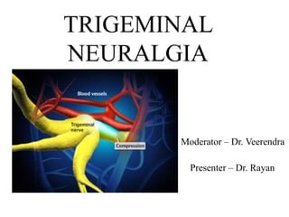
- 1. TRIGEMINAL NEURALGIA Moderator – Dr. Veerendra Presenter – Dr. Rayan
- 2. • Introduction • Historical review • Etiology • Clinical features • Diagnosis • Treatment modalities • Management • Pathogenesis CONTENTS
- 3. INTRODUCTION: • It is the most debilitating form of neuralgia that affects the branches of the fifth cranial nerve. • It is a disorder of the peripheral or central fibres of the trigeminal nerve in which the dominant symptom is pain in the region of distribution.
- 4. Trigeminal Nerve • Largest cranial nerve,nerve of the first branchial arch. • Trigeminal nerve is continuous with the ventral surface of pons by a small mortor root and a large sensoary root. Motor root • Arises separately from sensory root, in motor nucleus within pons. • Fibres travel separately from sensory root. • At semilunar ganglion, fibres pass in a lateral and inferior direction to leave via foramen ovale. • It unites with the sensory nerve trunk of V3 to form a single nerve trunk • Supply • Muscles of mastication • Mylohyoid • Ant belly of digastric • Tensor tympani • Tensor veli palatini
- 6. Sensory root • Comprise the central process of ganglion cells located in trigeminal ganglion. • Located in the Meckels cave,flat crescent shaped. • Sensory root fibres enter the concave portion, and the three divisions exit from the convex portion. • Ophthalmic division (V1) – exits from the superior orbital fissure • Maxillary division (V2) – exits from foramen rotundum • Mandibular division (V3) – exits from foramen ovale
- 8. International Association for the Study of Pain (IASP) : defined trigeminal neuralgia as sudden usually unilateral severe brief stabbing recurrent pain in the distribution of one or more branches of the 5th cranial nerve. International headache society (IHS) : defined trigeminal neuralgia as painful unilateral affliction of the face characterized by brief electric shock like pain limited to the distribution of one or more divisions of the trigeminal nerve. Nurmikko etal; trigeminal neuralgia – pathophysiology, diagnosis and current treatment; British journal of Anesthesia; 87:1; 2001 DEFINITION:
- 9. Painful unilateral affliction of the face, characterized by brief electric shock like pain limited to the distribution of one or more divisions of the trigeminal nerve. Pain is commonly evoked by trivial stimuli including washing, shaving, smoking, talking and brushing the teeth, but may also occur spontaneously. The pain is abrupt in onset and termination and may remit for varying periods – International headache society Nurmikko etal; trigeminal neuralgia – pathophysiology, diagnosis and current treatment; British journal of Anesthesia; 87:1; 2001
- 10. HISTORICAL REVIEW : JOHN LOCKE in 1677 gave the first full description with its treatment. NICHOLAS ANDRE in 1756 coined the term ‘Tic Doloureux’. JOHN FOTHERGILL in 1773 published detailed description of trigeminal neuralgia. ARETAEUS - credited with the first clinical description of trigeminal neuralgia. Siddiqui etal; Pain management trigeminal neuralgia; Hospital physician 2003
- 11. Epidemiology •The annual incidence for women is approximately 5.9 cases per 100,00 women; for men it is approximately 3.4 cases per 100,00 men. •The incidence increases with age. •No known racial or ethnic risk factors exist. Siddiqui etal; Pain management trigeminal neuralgia; Hospital physician 2003
- 12. Etiology • Vascular factors • Mechanical factors • Anomaly of superior celebellar artery • Dental etiology by Westrum and Black (1976) • Infections • Ratners jaw bone cavities (1979) • Multiple sclerosis by Olfson (1966) • Petrous ridge compression by Lee (1937)
- 13. • Intra cranial tumours • Intra cranial vascular abnormalities Compession Distortion • Viral etiology
- 14. Classification •Based on Etiology: Idiopathic Secondary Atypical Post herpetic •Based on clinical features: Classical Mixed Wael Fouad; Management of trigeminal neuralgia by radiofrequency thermocoagulation; Alexandria Journal of Medicine (2011) 47, 79–86
- 15. Theories regarding pathogenesis: •Central – based on similarity of trigeminal neuralgia to focal epilepsy and emphasize the role of deafferentation in the genesis of neural hyperactivity •Peripheral – change in peripheral axons and myelin may lead to altered nerve sensitivity to chemical and mechanical stimuli Siddiqui etal; Pain management trigeminal neuralgia; Hospital physician 2003
- 16. Focal demyelination at the site of compression may also allow electrical spread of excitation between adjacent sensory axons An emphatic short-circuit of this type within the trigeminal nerve might explain the sudden ‘‘electric’’ jolts of pain that characterize the disorder. Rasminsky M. Ephaptic transmission between single nerve fibres in the spinal nerve roots of dystrophic mice. J Physiol 1980;305:151-69.
- 17. Bennetto etal; Trigeminal neuralgia and its management; BMJ 2007; 27:334
- 18. Following trauma, regenerating nerve fibers become relatively depolarized & physiologically more excitable. Spontaneous action potentials originate from multiple sites and single action potentials may evoke sustained after discharges. Rasminsky M, Kearney RE, Aguayo AJ, Bray GM. Conduction of nervous impulses in spinal roots and peripheral nerves of dystrophic mice. Brain Res 1978;143:71-85
- 19. GENERAL CHARACTERISTICS: Incidence: Age: Sex: Affliction for side: Division of trigeminal nerve involvement: 8 : 1,00,000 5th – 6th decade of life Female > male ; 1.6 > 1.0 Right > left V3 > V2 > V1 Bennetto etal; Trigeminal neuralgia and its management; BMJ 2007; 27:334
- 20. CLINICAL FEATURES: Manifests as a sudden, unilateral, intermittent paroxysmal, sharp, shooting, lancinating, shock like pain, elicited by slight touching superficial ‘trigger points’ which radiates from that point, across the distribution of one or more branches of the trigeminal nerve. Pain is usually confined to one part of one division of trigeminal nerve. Pain rarely crosses the midline. Attacks do not occur during sleep.
- 21. TIC DOULOUREUX: Tic douloureux painful jerking It is a truly agonizing condition, in which the patient may clunch the hand over the face & experience severe, lancinating pain associated with spasmodic contractions of the facial muscles during attacks
- 22. Pain is of short duration, but may recur with variable frequency. In extreme cases, the patient will have a motionless face – the ‘frozen or mask like face’. Common trigger zones include: Cutaneous Corner of the lips Cheek Ala of the nose Lateral brow Intraoral Teeth Gingivae Tongue
- 26. DIAGNOSIS: Scrivani, S. J., Mathews, E. s., & Maciewicz, R. J (2005). Trigeminal neuralgia. Oral Surg Oral Med Oral Pathol Oral Radiol Endod. 2005 Nov;100(5):527-38.
- 27. T1-weighted axial image showing an ectatic loop of vertebral artery (arrow) passing anterior to the pons Seth Love et al; Central Demyelination of the Vth Nerve Root in Trigeminal Neuralgia Associated with Vascular Compression; Brain Pathology 8: 1-12(1998)
- 28. Differential diagnosis Diagnosis Important fearures Dental infection or cracked tooth Well localized to tooth,appropriate findings on dental examination Temporomandibular joint pain often bilateral,obvious findings in the joint Persistent idiopathic facial pain Bilateral,extend outside the trigeminal territory,pain is continuous & throbbing Migrane Preceded by aura,nausea, photophobia,phonophobia cervical tenderness Temporal arteritis Common in elderly,temporal arteries maybe firm, tender and non pulsatile on examination
- 30. Dedhia et al; Trigeminal Neuralgia (TGN ) - Pathophysiology and Management; J Anaesth Clin Pharmacol 2009; 25(1): 3-8
- 31. MEDICAL MANAGEMENT: CARBAMAZEPINE: Trade name: Tegretol , Carbitrol Dosage: 100 mg BID Maintenance dose 1200-2400 mg Mechanism of action: Slow recovery rate of voltage gated sodium channels, modulates activated calcium channel activity and activates descending inhibitory modulation system Side effects: Visual blurring Dizziness Skin rashes Rarely hepatic dysfunction, leukemia, Thrombocytopenia, aplastic anemia
- 32. PHENYTOIN: Trade name: Dilantin Dosage: 200 - 600 mg/day Maintainance dose 200-400 mg/day Mechanism of action: blockade of voltage dependent sodium channels. Inhibits the generation of repetitive action potentials. Prefrentially blocks high frequency firing. Canavaro S, Bonicalzi V. Drug therapy of trigeminal neuralgia. Expert Rev Neurother. 2006;6:429-40. It is usually used in conjunction with carbamazepine.
- 33. Side effects: Slurred speech Nystagmus Swelling of lymph nodes Gingival hypertrophy Hypersensitivity Teratogenicity
- 34. GABAPENTIN: It is more expensive than other drugs but has a less side effects. Trade name: Neurontin Dosage:100 - 5000 mg/day Maintainance dose 1800 mg/day Mechanism of action: mechanism unknown but possibly includes blockage of voltage gated calcium channels by binding to α2 subunits Side effects: dizziness, fatigue, weight gain, drowsiness, hepatotoxicity
- 35. Lamotrigine: New anticonvulsant drug Dosage: 25 – 100mg/day BD Maintenance dose 200-400 mg/day Mechanism of action: Decreases repetitive firing of sodium channels by slowing the recovery rate of voltage gated channels Also inhibits the release of the excitatory amino acids like glutamate. Side effects: Sleepiness Dizziness Headache Vertigo Rash
- 36. Topiramate: A sulfamate-substituted monosaccharide, was first identified as an antiepileptic drug. Compared with other antiepileptics, this drug acts at different neural transmission levels such as sodium channels and enhances GABA levels, and has been termed a ‘neurostabilizer’ Dose: 200 – 300mg/day Maintenance dose: 200mg BID Adverse effects: Paraesthesia Dysphasia Fatigue Confusion Insomnia
- 37. BACLOFEN: It is a GABA agonist These drugs reduces the central projection of painful afferent impulses. Trade name: Lioresal Dosage: 10 mg (tds) Maintenamce dose 30- 80 mg/day Side effects: Fatigue Vomiting Ataxia
- 38. TRICYCLINE ANTIDEPRESSANTS: Amitriptyline Doxepin Nortriptyline Imipramine 10 – 300 mg/day 10 – 300 mg/day 10 – 150 mg/day 10 – 300 mg/day
- 39. Botulium toxin It would limit the release of substance P, calcitonin gene- rated peptide and glutamate from presynaptic terminals of the primary sensory neurons, thus counteracting central sensitization Bao-Lin Guo et al; A closer look to botulinum neurotoxin type A-induced Analgesia; Toxicon 71 (2013) 134–139 Injected subcutaneously into the trigger zone (40 to 60 units) The pain recurred five months later and the site was again injected with 100 units
- 40. ACUPUNCTURE Hüseyin Sert, Burhanettin Usta, Bünyamin Muslu, Muhammet Department of Anaesthesiology, Fatih University School of Medicine - Ankara, Turkey CLINICS 2009;64(12):1225-6 • Adverse effects after minimal invasive approaches are paresthesia, facial sensory loss, weakness or paralysis of masseter muscles and, rarely, loss of the corneal reflex • Acupuncture treatment was initiated without making any change to the drug regime. • Acupuncture needles (0.20 x 13 mm needles for the face and 0.25 x 25 mm needles for the other regions) were inserted on the typical areas that are used for trigeminal neuralgia • Every treatment session lasted about forty-five minutes, three times a week • By the sixth week (14 sessions), the patient was completely free of pain • The analgesic effect of acupuncture is due to increased levels of mediators, including endorphin, encephalin and serotonin, in the plasma and brain tissue • Adverse effects are minimal and include bruising or hematoma at the needle site, metal allergy and local infection.
- 41. Treatment resistant trigeminal neuralgia relieved with oral sumatriptan • Sumatriptan is a serotonin agonist, specifically developed to relieve migraine headaches. • Although thecause of migraine is not fully understood, it is thought that a widening of blood vessels in the brain causes the throbbing pain of migraine headaches. • Sumatriptan worksby causing vasoconstriction of these vessels via the • stimulation of serotonin (or 5-HT) receptors. • Sumatriptan mimics this action of serotonin by directly stimulating the serotonin receptors in the brain. • This results in narrowing of the blood vessels and in effective relief of the migraine headache pain JA Moran and A Neligan Treatment resistant trigeminal neuralgia relieved with oral sumatriptan: a case report Department of General Practice, Brookfield Health Sciences Centre, University College Cork, Cork, Ireland and 2UCL Institute of Neurology
- 42. Multiple drug therapies • When a patient only partially responds to a single drug therapy at dosages that evoke side effects, adding a second drug may enhance the therapeutic response
- 43. Peripheral procedures Ganglionic procedure Open procedures SURGICAL MANAGEMENT:
- 44. PERIPHERAL INJECTION: It has been known that injection of destructive substance into peripheral branches of the trigeminal nerve, produces anaesthesia in the trigger zones or in areas of distribution of spontaneous pain. (A) LONG ACTING ANAESTHETIC AGENTS: Bupivacaine with or without corticosteroids may be injected at the most proximal possible nerve site.
- 45. (B) ALCOHOL INJECTION: 0.5 – 2 ml of 95 % absolute alcohol can be used to block the peripheral branches of the trigeminal nerve. Aim is to destroy the nerve fibres. It produces total numbness in the region of distribution of the nerve that was anaesthetized. Complication: Necrosis of the adjacent tissue Fibrosis Alcohol induced neuritis
- 46. (C) Transcutaneous electric nerve stimulation It refers to applying a low voltage electrical impulse to the nerve system via electrodes placed on the skin, one being over painful area and the other elsewhere. The stimulus intensity, frequency and duration are adjusted by the patients to obtain relief from pain Frequency – 10 – 50Hz for 4 to 6 sec Varadarajan; Transcutaneous electric nerve stimulation in trigeminal neuralgia: A review of literature; J Res Dent Sci 2014 ;5:36-41
- 47. The analgesic mechanism of TENS involves: Gate control theory: substantia gelatinosa of spinal cord acts as a gate control system. Activation of large myelinating fibres subserving touch, pressure, vibration is thought to facilitate pre synaptic inhibition of substantia gelatinosa cells in the dorsal horn, thus, reducing pain transmission. Physiological block: As the frequency of stimulation increases, conduction decreases resulting in physiological block. Endogenous pain inhibitory systems: Presynaptic inhibition in the dorsal horn of the spinal cord, endogenous pain control (via endorphins, enkephalins and dynorphins), direct inhibition of an abnormally excited nerve and restoration of afferent input. Varadarajan; Transcutaneous electric nerve stimulation in trigeminal neuralgia: A review of literature; J Res Dent Sci 2014 ;5:36-41
- 48. Advantages: No prostaglandins inhibition, since TENS controls pain by gate control mechanism. Rapid and timely inhibition of pain at peak progression. No adverse effects of drugs Non invasive Short term treatment for 20-40 days as compared with long term medicinal treatment. No need for surgical intervention. Can be used at home with portable machine. Equally effective in post neurectomy and post injection alcohol neuralgia. Singla et al; Role of transcutaneous electric nerve stimulation in the management of trigeminal neuralgia; J Neurosci Rural Pract. 2011 Jul-Dec; 2(2): 150–152.
- 49. PERIPHERAL NEURECTOMY (NERVE AVULSION): Oldest & most effective peripheral nerve destructive method Can be repeated & relatively reliable technique. It acts by interrupting the flow of a significant number of afferent impulses to central trigeminal apparatus. Performed commonly on infraorbital, inferior alveolar, mental and rarely lingual. Disadvantage: May produce Full anaesthesia Deep hypoesthesia Mukram et al; Peripheral neurectomies: A treatment option for trigeminal neuralgia in rural practice; J Neurosci Rural Pract. 2012 May-Aug; 3(2): 152–157.
- 50. INFRAORBITAL NEURECTOMY: (i) Conventional intraoral approach (ii) Braun’s transantral approach Conventional intraoral approach: A ‘U’ - shaped Caldwell – Luc incision is made in the upper buccal vestibule in the canine fossa region. Mucoperiosteal flap is reflected superiorly to locate the infraorbital foramen. Once the nerve is exposed, all the peripheral branches are held with the hemostat & avulsed from the skin surface intra orally.
- 51. The entire trunk is separated from the skin surface is held with the hemostat at the exit point from the foramen & is removed by winding it around hemostat & pulling it out from the foramen. Then it may be plugged with polyethylene plug.
- 52. Braun’s trans antral approach: An intra oral incision is made from the maxillary tuberosity to the midline in the maxillary vestibule.
- 53. The descending palatine branch of the trigeminal nerve is identified & traced to the sphenopalatine ganglion. The maxillary nerve is sectioned from the foramen rotundum to the inferior orbital fissure. The antral mucoperiosteal flap in the vestibule is repositioned & sutured back. A 3 cm window is made in the antero – lateral wall of the maxillary sinus. Posterosuperior portion of antrum is excised to create a posterior window
- 54. INFERIOR ALVEOLAR NEURECTOMY: (i) Extra oral approach (ii)Intra oral approach The extra oral approach: Done through Ridson’s incision After reflection of masseter, a bony window is drilled in outer cortex & nerve is lifted with nerve hook & avulsed from its superior attachment & mental nerve is avulsed anteriorly through the same approach.
- 55. The intra oral approach: Done via Dr Ginwalla’s incision Incision is made along with the anterior border of asescending ramus, extending lingually & buccally ending in a fork like inverted Y. Incision is then deepened on the medial aspect of ramus. The temporalis & medial pterygoid muscles are split at their insertion & inferior alveolar nerve is located.
- 57. The nerve is ligated at two points in the most superior part visible & divided between the ligature. The superior end is cauterized & the lower end is held securely using a hemostat. The mental nerve is also similarly ligated in two points close to the mental foramen & divided between two. The remaining nerve is held at the inferior alveolar end & wound around the hemostat & excised from the canal.
- 58. LINGUAL NEURECTOMY: An incision is made in the anterior border of the ramus slightly towards the lingual side. The lingual aspect is exposed & the lingual nerve identified in the third molar region just below the periosteum. The nerve can be either avulsed or ligated, cut and the ends may be cauterized.
- 59. CRYOTHERAPHY: Barnard first used cryotheraphy in 1981 for the treatment of the trigeminal neuralgia. After identifying the affected nerve , it is then exposed to the cryoprobe intraorally. Direct application of cryotheraphy probe at temperatures colder than -60 C are known to produce Wallerian degeneration without destroying the nerve sheath itself. Nerve is exposed for 2 min freeze followed by 5 min thaw cycle. The freeze – thaw cycle is repeated at least 3 times. Zakrzewska JM, Cryotherapy for trigeminal neuralgia: a 10 year audit, Br J Oral Maxillofac Surg. 1991 Feb;29(1):1-4
- 60. GASSERIAN GANGLION PROCEDURES: PERCUTANEOUS RHIZOTOMY: This is done on the Gasserian ganglion which involves either mechanically or chemically damaging parts of the trigeminal nerve.
- 61. • 1910 – Harris, Tapatas and Hartel introduced percutaneous approaches via foramen ovale • 1931- Kirschner introduced percutaneous electrocoagulation of gasserian ganglion • Three procedures used with variable success rate are • Glycerol injections • Thermocoagulation • Balloon compression
- 62. Technique of needle penetration: The foramen ovale is best visualize with the x – ray tube placed for a submentovertex position. Infiltration of the skin & cheek is done with local anaesthetic agent on the affected side. Three points of Hartel are marked on the side of the face using marking ink.
- 63. First point – a perpendicular line is drawn from the lateral orbital rim to the inferior border of the mandible. Second point – marked at 15 mm lateral to the angle of the mouth on the perpendicular first line Third point – marked at the level of TMJ 2.5 cm from the centre of the external auditory meatus.
- 64. • This line is the line of elevation. • When patient is supine, plane of elevation will be perpendicular to floor • Needle is passed through the second point. • Needle is passed till anterior border of ramus of mandible,then turn the needle medial and upwards to base of skull • Position is confirmed. • Needle is then pushed for half centim eter, final position.
- 65. (A)Controlled radiofrequency thermocoagulation: It was first introduced by Kirschner (1931) & later modified by Sweet (1970). Technique: The patient is sedated with a short acting sedative and vital signs are monitored. The electrode is inserted through the cheek under fluoroscopy into foramen ovale. The patient is awakened briefly to accurately locate the position of the electrode.
- 66. The straight temperature monitoring electrode was used to perform an initial lesion at 60 C for 120 s The patient is awakened for sensory testing The lesion may be repeated at the same temperature and duration if sensory deficit was not evident Facial hyperemia in the distribution of right V2 during RFT. Wael Fouad; Management of trigeminal neuralgia by radiofrequency thermocoagulation; Alexandria Journal of Medicine (2011) 47, 79–86
- 67. Indication: Toxicity of drugs Failure of response to the other modalities Dependence on the drugs for life time Elderly patients Medically compromised patients
- 68. Advantages: Comparative low rate of recurrence Zero mortality Thermocoagulation preserves the motor function of the trigeminal nerve Can avoid major surgical procedure Disadvantage: May cause Anaesthesia dolorosa Loss of corneal reflex Meningitis (rarely)
- 69. (B) Percutaneous glycerol rhizotomy: Glycerol is a neurolytic alcohol which can be used to chemically destroy the nerve root. The injection of 0.1–0.4 ml of glycerol into the trigeminal cistern is carried out under fluoroscopic control
- 70. Advantages: Simple technique Lower incidence of anaesthesia dolorosa Complication: Post operative headache, nausea, vomiting Meningitis Post operative herpes simplex perioralis
- 71. (C) Percutaneous balloon compression: This is a mechanical means of destruction of the trigeminal nerve introduced by Mullan & Lichtor in 1980. Technique: A no. 4 Fogarthy’s catheter is introduced with fluoroscopic guidance. A 0.5 - 1 mm balloon is inflated for 1 – 6 minutes.
- 73. OPEN PROCEDURES ( INTRACRANIAL PROCEDURES): (A) Microvascular decompression of the trigeminal nerve sensory root: Procedure popularized in 1967 – 1976 by Jannetta. Most commonly performed intra cranial open procedure. The root is examined under the microscope
- 74. A compressing branch of the superior cerebellar artery will be seen medial to the nerve at the root entry zone. Incision is made over the mastoid area
- 75. Then the trigeminal nerve is freed from the compressing / pulsating artery. After freeing the nerve, the nerve is separated from the artery by placing a piece of Teflon between them.
- 76. Non absorbable insulating sponge may also be placed.
- 77. (B) Trigeminal root section: (a) Extradural sensory root section: It is also known as the subtemporal extradural retrogasserian rhizotomy. It is no longer used now. In this, sensory root is divided, sparing the motor root, as close to the brainstem as possible. Disadvantage: Profound sensory loss High incidence of anesthesia dolorosa
- 78. (b) Intradural rhizotomy: This is an intradural procedure that is done when the pain recurs after MVD. This is usually done in the posterior cranial fossa. It can be selective or complete. (c) Trigeminal tractotomy: It is also known as the medullary tractotomy. This is not usually done. The descending tract of the trigeminal nerve is sectioned at the junction of the cervicomedullary region.
- 79. STEREOTACTIC RADIOSURGERY (GAMMA KNIFE): The radiation is aimed at the proximal nerve and root entry zone in the pons The gamma knife projects 201 very fine beams of gamma rays at dose of 70 – 90Gy( generated by radioactive cobalt) through skull and brain It has been shown to affect abnormal ephatic transmission but not normal axonal conduction Siddiqui etal; Pain management trigeminal neuralgia; Hospital physician 2003
- 80. Conclusion •Trigeminal neuralgia has long been recognized by the medical professionals •However it is still an enigmatic disorder, and its management remains controversial.
- 81. References • Holguin J et al; Suprameatal extension of retrosigmoid approach for microvascular decompression of TN; IJOS 2015: 13-16 • Yasushi L etal; Neuronavigation assisted decompression of TN: Interdisciplinary neurosurgery 2014: 38-40 • Koopman J et al; Nationalwide study of 3 invasive treatment for TN; IASP 2011: 507-513 • Bennetto L et al; TN and its management; BMJ 2008:334 • Chole R et al; Drug treatment of TN: JOMS 2007:40-45 • Nurmikko et al; TN- pathophysiology, diagnosis and current treatment; BJA 2001: 117-32 •Textbook of Maxillofacial Surgery- by Peter Ward Booth, 2nd Edition, Vol 2 Principles of Neurological surgery- by Richard G. Ellenbogen, 3rd Edition. Principles of Maxillofacial Surgery- by Larry J. Peterson Textbook of pharmacology- by Goodman & Gillman
- 82. • Rasminsky M, Kearney RE, Aguayo AJ, Bray GM. Conduction of nervous impulses in spinal roots and peripheral nerves of dystrophic mice. Brain Res 1978;143:71-85 • Rasminsky M. Ephaptic transmission between single nerve fibres in the spinal nerve roots of dystrophic mice. J Physiol 1980;305:151-69. • Wael Fouad; Management of trigeminal neuralgia by radiofrequency thermocoagulation; Alexandria Journal of Medicine (2011) 47, 79–86 • Siddiqui etal; Pain management trigeminal neuralgia; Hospital physician 2003 • Joffroy A et al; Trigeminal neuralgia pathophysiology and treatment; Acta neurol beig 101: 20-25: 2001 • Zhou X et al; Comparision of nerve combing and percutaneous radiofrequency thermocoagulation in treatment of TN; Braz j otorhinolaryngology; 2016: 2:1 • Seo M et al; Gabapentin therapy in patients with orofacial neuropathies; Oral science international; 2011: 8:17-19 • Elsotouhy A et al; Tic douloureux in patients with incidental intracranial meningioma; Egyptian j of radio neu med 2015: 46; 455-460
- 83. Dedhia et al; Trigeminal Neuralgia (TGN ) - Pathophysiology and Management; J Anaesth Clin Pharmacol 2009; 25(1): 3-8 Qiang-ping et al; Topiramate versus Carbamazepine for the Treatment of Classical Trigeminal Neuralgia; CNS Drugs 2011; 25 (10): 847-857 Jørgen Degn : Surgical treatment of trigeminal neuralgia. Results from the use of glycerol injection, microvasculardecompression, and rhizotomia Acta Neurochir (2010) 152:2125–2132 Giorgio Cruccu; Refractory Trigeminal Neuralgia; CNS Drugs (2013) 27:91–96 Xinjie et Al; Effect of radiation dose on the outcomes of gamma knife treatment for trigeminal neuralgia: A multi-factor analysis; Neurology India | Jul-Aug 2014 | Vol 62 | Issue 4 E. Besi et al; Comparison of tolerability and adversesymptoms in oxcarbazepine and carbamazepine in the treatment oftrigeminal neuralgia and neuralgiform headaches using the Liverpool Adverse Events Profile (AEP); The Journal of Headache and Pain (2015) 16:81
- 84. Wenyao Hong et al; Clinical features and surgical treatment of trigeminal neuralgia caused solely by venous compression; Acta Neurochir (2011) 153:1037–1042 Carlos Zúñiga et al; Acute Treatment of Trigeminal Neuralgia With Onabotulinum Toxin A; (Clin Neuropharm 2013;36: 146–150) Agrawal et al; Peripheral Neurectomy: A Minimally Invasive Treatment for Trigeminal Neuralgia. A Retrospective Study; J Maxillofac Oral Surg. 2011 Sep; 10(3): 195–198. Varadarajan; Transcutaneous electric nerve stimulation in trigeminal neuralgia: A review of literature; J Res Dent Sci 2014 5:36-41.
Editor's Notes
- International classification of headache disorders 2
- Sensory root resection
- Iminiostilbine
- hydantoin
- Foetal hydantoin syndrome – cleft lip n palate microcephaly and hypo plastic phalanges
- Neurotoxic protein. Prevents release of neurotransmitter acetylcholine at NMJ causes flaccid paralysis
- 1965 Ronald Melzack and Patrick Wall
- Nerve stump neuromas
- Thaw allows for longer vascular stasis and longer exposure to toxic solute levels within the cells Double freeze thaw cycle technique
- anesthesia dolorosa- deafferentation pain,constant burning aching