Vip Call Girls Anna Salai Chennai 👉 8250192130 ❣️💯 Top Class Girls Available
340 469
1. MULTTTOCAL MoTOR NtUuOpArHy (MMN)
Description . A progressing disorder affecting motor nerves, rcstrltirrl.l irr wr,,rhrr,..,..
. The disorder is autoimmune, and as such, often resp<tnds lo tnurrrrrrr
modulating therapy.
Pathophysiology . MMN is an autoimmune disorder, although the cause of the irrrnrrrrr,.
attack is not known. Canglioside antibodies have been implicatctl rrr
some cases.
. Demyelinating changes predominate in the motor nerves, althouglr
there is some axonal drop-out. Conduction block is an important
differentiating feature of MMN.
Clinical findings . MMN presents with progressive weakness, often with cramping thal
affects peripheral nerve distributions in a patchy and asymmetric
fashion, e.g., radial distribution producing wrist drop and peroneal
distribution producing foot drop.
. Sensory symptoms are absent.
. Fasciculations can be seen along with the cramps.
. Re{lexes are normal or slightly reduced initially, but become absent
with progressive disease.
Laboratory studies . Routine labs are normal.
. CMI ganglioside antibodies are seen in about half of patients, and arr
supportive of the diagnosis but not required.
. lmaging is normal and often not needed.
. NCS and EMC show conduction block-where there is attenuation of
conduction of the motor nerves through selected nerve segments.
Sensory conduction through the same regions is normal.
Fasciculations may be seen, but denervation is not prominent.
Diagnosis MMN is suspected when a patient presents with progressive
asymmetric weakness without sensory abnormalities. ALS is usually
the first thought in the clinician's mind, but exam does not show
corticospinal tract signs (hyperactive tendon reflexes and up-going
plantar responses), and NCS and EMG show conduction block rather
than extensive denervation.
CMI antibodies are supportive of the diagnosis, but not diagnostic
and not required for diagnosis.
Differential
diagnosis
Amyotrophic loterol sclerosis. The main differential diagnosis
presents with progressive asymmetric weakness, but there are signs of
corticospinal tract dysfunction, and NCS and EMG show widespread
denervation without significant conduction abnormalities.
Mononeuropothy multiplex. From many causes, can present with
asymmetric weakness.
Management . lVlC is first-line therapy for most patients.
. Cyclophosphamide is used when patients cannot take lVlC or do not
respond.
. Corticosteroids are not effective and are not used.
Clinical course Patients can have substantial improvement with lVlC treatment, so it is
important to differentiate this from ALS, which essentially is untreatable.
Mttlltlr)( (rl N4olot Ncut()l)(ll lty
tlrr
( ('rvr((rl l((l(lir rrloprrllty
lll)lloli
,l,llysiolt>8y
('r.l{vlcAt. I{AD!-CU!
Cervical nerve root damage can result in any combination or sensory
loss, pain, or motor loss confined to a single nerve root distribution.
Causes of cervical radiculopathy are multiple, and include:
Disc disease
Osteophyte formation
Tumor
lnfection
Diabetes
The common pathophysiology is nerve root impingement or
infiltration with resultant damage to the myelin and axons.
,rl findings Clinical findings depend on the level of the lesion. Most patients
have neck pain that radiates down the arm in the dermatomal
distribution. Weakness may be present, but is not invariable.
Rash over a portion of the involved dermatome suggests herpes
zoster. This usually has a characteristic vesicular appearance.
lndividual nerve root symptoms and signs are as follows:
C5 rodiculopothy produces sensory deficit in the radial forearm.
Motor loss is in the deltoid and biceps. Biceps reflex is
decreased.
C6 rodiculopothy produces sensory deficit in digits t and 2 of
the hand. Motor deficit is in the biceps and brachioradialis.
Biceps reflex often is decreased, and brachioradialis reflexes may
be as well.
C7 rodiculopothy produces sensory deficit on digits 3 and 4 of
the hand. Motor deficit is in the wrist extensors and triceps.
Triceps reflex may be depressed.
C8 rodiculopothy produces sensory deficit on digit 5 of the hand.
Motor deficit is in the intrinsic hand muscles-both median and
ulnar innervated. Reflexes are normal.
rratory studies Routine labs are normal.
lmaging with MRI can show nerve root impingement or
inflammation.
NCS is normal. EMC may show denervation in the distribution of
the nerve root, but is not always present.
LP can be performed for neoplastic meningitis, but is of extremely
low yield in patients without known cancer.
lnosrs Cervical radiculopathy is suspected when a patient presents with
pain and/or sensory loss in the arm with neck pain.
lmaging can show a structural cause in many patients. ln others,
diabetes and herpes zoster have to be considered.
Zoster is supported by the development of the rash, which may be
minimal and may occur days after the onset of symptoms.
Diabetic radiculoneuropathy is suggested by the diagnosis of
diabetes and the absence of a structural cause seen on studies.
540 DISORDERS-NEUROMUSCULAR DISORDERS NEUROI4USCULAR 54I
2. Differential diagnosis
Management
( t't'vit rtl l(trrlit rtlolrrrtlry tt,ru*tt
CERVIC.AL RADICUTOT,ATHY.-<onI,d
Brachial plexopothy. Can produce motor and sensory [irrtlirrgs rrr
one or both arms. However, the symptoms span dermatonr,rl
distribution.
Peripherol mononeuropothy Especially the radial nerve, periplrt'r,rl
mononeuropathy can mimic cervical radiculopathy, e.9., C7 lesion
However, examination of proximal and distal musculature helps to
differentiate a peripheral from a nerve root lesion. Also, most
peripheral nerve lesions span root distributions.
Nerve root compression from disc or bony element usually can
usually be treated conservatively. Treatment options include:
Physicol therapy is extremely helpful for reducing pain and
restoring function. This should be offered to almost all patienls
Muscle reloxonfs are used if there is paraspinal muscle spasnr
that contributes to the pain.
Anti-inflommotory agents are given to most patients and can
reduce pain.
Epidurol corticosteroid injections are considered if conservativt.
measures are not helpful.
Surgicol decompression is considered especially if there is
weakness or refractory pain.
Herpes zoster is treated with analgesics and antiviral medications.
Anticonvulsants often are often used as an adjunct for control of tht,
neuropathic pain.
Tumor infiltrotion is treated with local or neuraxis radiation.
lntrathecal and systemic chemotherapy also often are given.
Corticosteroids also are helpful for the pain of tumor infiltration.
Diobetic rodiculoneuropothy produces neuropathic pain, which can
be treated not only with analgesics but also by anticonvulsants suclr
as gabapentin. These agents are less helpful for the mechanical pairr
than the neuropathic pain.
Most patients improve with treatment, although not all. lf the cause n
compressive, there is a high remission rate with medications and
therapy, although there is the possibility of recurrence of the pain at ,r
later time.
542 DISORDERS-NEUROMUSCULAR DISORDERS-NEUROMUSCULAR 54I
( r,tvit rrl l{rrrli< trloprrllry
l)t.rrtt.rlorrrr.s art<l Myololncs of Uppcr Limb
,,t,.',, lr.It,rtt, rlcrrr,rrr,rlrorr (,1 tl(,illl,tlr)[t(.
,,,,,rrlrrrri lo [r,r,r].rrr.ur(l (;,rt('ll) sltorln.ts distinct
rlr ri,rrt,, I lrlt rs ,rr lrr,rill,r onsirk'r.rltle ovcrlap
tr1r,r,rr ,rlj,rr cnl (l('ll.ll()ntcs. An alternative
rr,rt()rr( nr,r1r is Ih,rl providcd by Foerster.
( .l lo I I Scnsory Representation Anterior view
Posterior view
au
{, *t l-dffi
cz,-nl, -:]
t *+*
t' . -. ,t
c4 '.l.t**-,r
C5
C5
I
C8
.,'
i
C6
r;,-n qr-:-r'.'-
' ".-ca lr,-';: '
I Motor lmpairment Related to Level of Cervical Root Lesion
Hcrniated disc
comprcssing
nerve root
3. I ltotrtt it l(rt<lilttloprtllty
THORACIC RADICUT.OPATHY
Description Thoracic nerve root damage can result in pain, predornirr.rrrlly rrr llrr.
chest.
Pathophysiology . All of the pathologies that can affect the cervical spine can alfcr I
the thoracic spine. ln general, disc disease and osteophyte
formation are less common than they are in the cervical and
lumbar spine.
. Herpes zoster and diabetic radiculoneuropathy are more imporl,rr
causes.
Clinical findings . Thoracic radiculopathy commonly produces unilateral chest wall
pain, which begins near the spine and radiates toward the front,
following the dermatome.
. Rash in the distribution suggests herpes zoster, especially when
vesicular.
. Motor deficit is not expected with thoracic radiculopathy.
. Reflex abnormalities are not expected, unless there are myelopatl
findings due to the cause of thoracic radiculopathy, e.g., disc or
tumor with nerve and spinal cord compression.
Laboratory studies . Routine Iabs are normal.
. lmaging with MRI shows a structural cause in most patients. Wher
imaging is normal, diabetes and herpes zoster have to be
considered.
. NCS and EMC are of little value in diagnosis of thoracic
radiculopathy and are not routinely performed.
Diagnosis Thoracic radiculopathy is suspected when a patient presents with
sensory loss and/or pain in the chest. Careful exam confirms the
distribution of the symptoms within one nerve or dermatome
distribution.
MRI can show a structural lesion. lf this is normal, diabetes and
herpes zoster have to be considered.
LP can be performed to look for neoplastic meningitis, but is of
extremely low yield in patients without known cancer.
Differential diagnosis . Lung disorders. Can produce pain that may be mistaken for thorar
radiculopathy, especially pleural-based lesions, e.g., mesothelioma
and other tumors, infections, or inflammation.
. Cordiac poin. Rarely confused with thoracic radiculopathy.
Management Treatment of thoracic radiculopathy does not greatly differ from
treatment of cervical radiculopathy.
Thoracic radiculopathy is not associated with perceptible motor
deficit, and, therefore, pain is the main symptom to be addressed.
Clinical course Most patients improve with time, regardless of cause. Long-term use
of agents for neuropathic pain occasionally is needed.
544 DISORDERS-NEURON/USCULAR DISORDERS-NEUROMUSCULAR 545
Iltotrtr it l(rrrlit trloprrllry
Spirr.rl Norvr' ()rigin: Cross Scction
'r'r liorr lhrorrth lhor.rcic vertebra
lt o| Ilrlr'lrr,r
I Lt,pt, ,me n inges
Fat in epirJur.r l sp.rcc:
Sympathotic 5;anglion
Ventral ro()1
Whito .rnd grav ranri
cotnrl u n ic;t ntt:s
Spinal nervt:
Ventral r.rmus
(intorcost.rl nervei
Dorsal ranrus
Spinal sensory
(dorsal root) ganglion
Dorsa I rool
I ;:lerr l horn of
gray m.rttcr oI spinal corcl
lnternal vertebral
(epidural) venous plcxus
4. LUMBOSACRAL RADICULOPATHY
Description Damageto the lumbar or sacral nerves can producc s('nsory ()r nrrl
deficit affecting the lower back, pelvis, and legs.
Pathophysiology The same causes of cervical radiculopathy can affect the
lumbosacral nerve roots.
Disc disease and osteophyte formation are the most common
Tumor and infection are less likely.
Clinical findings Lumbosacral radiculopathy causes pain in the back that can radi,rlr.
down the hip and legs in a dermatomal distribution. Weakness c,rrr
be present, and of great localizing value.
Findings with individual nerve roots are as follows:
L2 rodiculopothy-sensory deficit on the lateral and anterior
upper thi8h. Motor deficit in the psoas and quadriceps. No rt'll
abnormality.
L3 rodiculopothy-sensory deficit on the lower medial thigh.
Motor deficit in the psoas and quadriceps. Knee reflex is
-
reduced.
L1 lodigulgpothy-sensory deficit on the medial lower leg. Morr
deficit in the tibialis anterior and quadriceps. Knee refleiis
decreased.
L5 rodiculopothy-sensory deficit on the lateral lower leg. Mot<r
loss of the peroneus longus, tibialis anterior. No reflex
-
abnormality.
S.l rodiculopothy-sensory deficit on the lateral foot involving
digits + and 5. Motor deficit involving the gastrocnemius. Anlle
reflex is decreased.
The unlisted roots-LI and S2-S4-can be involved, but much less
commonly than the ones listed.
Laboratory studies . Routine labs are normal.
. MRI shows a structural cause in most patients. lf this is negative,
diabetes, herpes zoster, and tumor are considered.
. CT of the lumbar spine is less sensitive than MRl, but has to be
performed when MRI cannot be done (e.g., pacemaker). lntrathec
contrast dye improves the diagnostic sensitivity of the CT.
. NCS is normal. EMG may show denervation in the appropriate
nerve root distribution if there has been significant involvement of
the motor nerves.
. Lumbar puncture (LP) can be done to look for neoplastic
meningitis, especially with polyradiculopathy, but is of low yield in
the absence of known cancer.
l.tttttl rr ).(t( I ( tl l(r rrlir rrlr )l )( rl lly
lr',
I rrrrrlrr)s(l( r(rl l{rtrlir ttlolrrrtlry
l.U M ROSACRAI. RADICULOPATHY--cont'd
Lumbosacral radiculopathy is suspected when a patient presents
with sensory and/or motor symptoms in the leg. The presence of
back pain is supportive, especially of structural causes.
MRI shows a structural lesion in most cases. lf this is normal,
diabetes, herpes zoster, and tumor are considered.
A vesicular rash in a dermatomal distribution suggests herpes
zoster.
Laboratory signs of diabetes support the diagnosis of diabetic
radiculoneuropathy, although this does not mean that some other
cause is not present.
NCS and EMG can support the diagnosis of radiculopathy if there is
Iocalized denervation.
t l,'rential diagnosis Lumbosocrol plexopothy. Can produce pain in the leg, which can
also involve the hip and back. Weakness is common. This disorder
affects muscles of more than one dermatome, however.
Examination and EMG can make this distinction.
Peripherol mononeuropothy. Can mimic radiculopathy, e.g.,
peroneal neuropathy and L5 radiculopathy can both produce foot
drop without reflex changes. Differentiation is by examination and
EMG.
Management of lumbosacral radiculopathy does not differ from that
of cervical radiculopathy.
Most patients improve. At least
-l5o/o
oI patients with lumbosacral
radiculopathy improve with conservative care, although some will
eventually still come to surgery.
546 DISORDERS-NEUROMUSCULAR DIOIiI )I IIS NI IJI{OM(JCUIAR
'47
5. l.tt tttI rosrrr't rrI l{rtrlit trloIrtrI Iry,,tt,,t,,t
l)ain l).tllcrns in Lurnlr.rr l)ist.ase
Radicular pain due to nerve root compression
Nervt,root
comprcssed by
enlarged iac:ct
Rad i cu lar
{ 1,.rin.p.rrterns
(rnBlL'seqmr.nt
dislributirin)
L-omprc,ssion oi specific nervc roo[
results in p.lin scns.ttion in radicular
pJlleilr .l)c( iii, trr di.lril,utiorr ilr
that particular ncrve ro()t
"f
r )r'.,( lll)lton
t,rtlrophysiology
rlrrrical findings
lltrtr ltirtl l'lcxr)lxrtlly
IIIIACHIAL PLEXOI'ATNY
Damage to the brachial plexus can produce pain, sensory loss, and/ot
weakness in one arm. Some plexopathies can be bilateral
, ,,, ,
t*l'.iu#
. ir',
,,t
. The brachial plexus is formed from the individual cervical and
upper thoracic nerve roots. The nerves diverge and reconnect to
form the nerves of the arm. especially median, ulnar, radial, and
musculocutaneous. There also are other minor nerves supplying
shoulder muscles, and these are shown in the diagram.
. Causes of brachial plexopathy include: trauma, plexitis, tumol
radiation, and bleeding.
Clinicat findings depend on the cause of the lesion and the precise
location. Cenerally, there is pain and/or sensory loss in the arm.
Weakness can develop in muscles innervated by the involved portion
of the plexus.
Upper plexus /esions produce sensory andlor motor deficits
affecting the distributions of the C5 and C6 nerve roots. Deltoid
and biceps are especially affected, with sensory change that
extends below the elbow to the hand.
Lower plexus /esions produce sensory and/or motor deficits
affecting especially the C8 and Tl nerve roots. Median and
ulnar-innervated muscles are affected, with hand weakness, and
sensory symptoms involve much of the palmar hand, ulnar
aspect of the dorsal hand.
Specific findings with individual causes include:
Troumo produces variable damage to the plexus. Stab wounds
can affect almost any portion, but the upper plexus is more
exposed. Weakness is prominent early, soon followed by
neuropathic pain. Upward traction of the shoulder can stretch
the lower plexus. Downward traction of the shoulder can stretch
the upper plexus.
Brochiol plexitis presents with pain in the shoulder and arm that
eventually improves. During this phase, weakness develops,
which has slower improvement. Upper plexus is mostly affected.
. Tumor can be compressive from the lung or infiltrating from
cervical lymph nodes. Tumor presents with severe pain, often
weakness, and Horner's syndrome. Lower plexus is mainly
affected.
Rodiotion theropy to the neck and chest produces dysesthesias
that are uncomfortable, but not really painful. Weakness can
develop. The upper plexus is predominantly involved due to
thinness of the tissues in the region of the upper plexus.
Bleeding into the neck and plexus from trauma and from
bleeding disorder presents with weakness and motor loss in the
arm, often with a palpable hematoma in the supraclavicular
area. Pain may be present, but is less prominent than with
tumor or plexitis.
548 DISORDERS-NEUROMUSCI]I AR DISORDERS NEUROMUSCULAR'49
6. Itl(t( lll(tl I'lr,xollrtllty , t,nunuttt
BRACHIAL PLTXOPATHY*cont'd
Laboratory studies . Routine labs are normal. There are no reliable markcrs lor lrr.rr lrr,rl
plexitis, although antinuclear antibodies (ANA) and ESR ottt'rr ,rrc
checked.
. MRI is able to show structural cause in a minority of patients,
chiefly tumor or signs of trauma. Brachial plexitis and radiation
plexopathy are not associated with reproducible findings on studit',,
. NCS usually is normal, although the sensory and motor action
potential can become reduced in amplitude after t-2 weeks. EMG
can show signs of denervation after 3-4 weeks.
Diagnosis . Troumo as a cause for brachial plexopathy is evident from the
inciting event. EMG can help to localize the lesion. lmaging may bc
normal with stretch/traction injuries but may show denervation
hematoma and tissue disruption with penetrating trauma.
. Brochiol plexitis is suspected when a patient develops pain in an
arm and no structural cause is identified. Subsequent development
of weakness as the pain improves supports the diagnosis.
. Tumor infiltrotion is suspected with severe shoulder and arm pain
with or without weakness. lmaging shows tumor compression or
infiltration in the region.
. Rodiotion plexopothy is suspected when a patient develops
dysesthesias in the arm months after known radiation therapy.
lmaging does not show a structural cause.
. Bleeding into the plexus can be seen on imaging and suspected
from exam.
Differential diagnosis . Cervicol rodiculopothy produces pain in the arm, but the lesion
affects only a single nerve root, unless there is polyradiculopathy.
, Mononeuropothy of the upper extremity can produce pain and
weakness, but the deficit is distal to the plexus, which may be
evident on exam or EMC.
Management . Tumor can be treated with surgery chemotherapy, andlor radiation
therapy. Surgery can make the plexus damage worser so this is less
commonly used than radiation therapy and chemotherapy.
. Brachial plexitis often is treated with corticosteroids, although this
has not definitively been proven to be helpful.
. Management for the other causes is supportive.
Clinical course lmprovement depends on the cause of the plexopathy. ldiopathic
plexitis commonly improves, with most having resolution of the pain
and eventual improvement in strength.
55O DISORDERS-NEUROMUSCI.]LAR DISORDERS-NEUROMUSCULAR 557
l!trrr ltirtl l'lcxollrtlltY
lh.rr hi,rl l'k'lrts: St ltt'trl.r
i l,,l, i 'rt.rl r,,rrtlr,rrtltott rltrrlr'tl
I r, lrr, rl lrl.rrr', lr.r. l.rrtir'( I
, ,,nlrlrrllr)rr lrrrt l.rr k' I l.
lir llrrlrl 1,1,'rrt. l,r< I(s ( 5 l)ul
lr l L', ottltilrttliott 31ruo 3".,,';.'i;1n,7,/ r/r,":,:'
Suqrr,rrtaltul.tr lirPhnni' /' / -l
nerrc r( 1, 6 ncrv(' / C5.
:i&.'
rpr('l( ,, (), Y('r(' / C5.
ll;:'l,tl::, *2,
,.0*l .y'
.
*i'dg"
.--'*,
Yl,,*,,,
-lsl
ril) lt,'l.nHust,'lli
sa
6a,' il'l:i,::''"
1c5.6 7,rlr
'rll,rr1 ,/
r,,'rr r' (C5, {,) /
ll.rrlr,rl trcrvt /
r , r.,7,1,I1lt
lt'cli.rn netvt' ./
c5, u,7,t,,I1t/
I'lrtrr nt,rvt, __--
C7. 8,f 1t
-
lnconstanl contribution
r.t iritJiin-{roi i
",u"
ncrvt'(C5, 6, 7)
pectoral ncrve (CU, l 1 )
Medial cutansrus nt'rve of .rrm (T1)
Meclial cutancous ncrve oi iorcarnr (C.8, T1 )
LJppcr sultscapular ncrvc (Ci5, 6)
Lower subscallular nerve (C5. 6)
Thora< odorsal (micldlc sullscapul'rri ncrvc (C6' 7' tl)
.a ; .,'
I atcral pet krral
^r$$""'o''i ,
7. I rrrrrlrr )(t( lltl l'lt'xoprrl lty
't:
lt.l
ul
dermatomal
ht,
rith
the
Description
LUMBOSACRAL PTEXOPATIIY.
-
Damage tothe lumbosacral plexus in the abdomen ((ur l)ro(hr((, l(.
sensory and motor symptoms. The differential diagnosis is sirrul,rr tr
that of brachial plexitis.
. Lumbosacral plexitis has similar causes to brachial plexitis; howr,v
r.adiation plexopathy is uncommon, and idiopathic plexitis is lt:ss
likely.
. Diabe.tic amyotrophy. is sometimes discussed under plexopathy, lrr
is really a polyradiculopathy
Pathophysiology
Clinical findings Lumbosacral plexopathy_is most commonly due to tumor. Bleeding,
trauma, and idiopathic also are considered.
Clinical features of individual entities are as follows:
Tumor com.p.ression or infiltration of the lumbosacral plexus
presents with severe local and radiating pain into the leg.
Patients typically have a known history oi renal or
gastrointestinal cancer.
Troumo can produ.ce direct damage to the plexus, although in
this case, there is likely to be significant direct abdominaiorgu,
damage. Pain in the abdomen and legs can be seen.
Lumbor plexitis is uncommon, and presents with hip and leg
pain, followed by weakness.
Bleeding into the plexus from femoral stick for angiography or
trauma produces a block of axonal transmission, r6su-lting in
decreased sensation and often weakness that spans derriatom
distributions.
Laboratory studies . Routine labs are normal.
. MRI can show a structural lesion or layered blood, if present, in tl"
paraspinal region.
. EMC can show denervation in a distribution appropriate to the
deficit; however, 3-4 weeks may elapse beforb'the EMC becomes
abnormal, and a normal EMG does not rule out the diaenosis.
Diagnosis . Lumbosacral plexopathy is suspected when a patient presents wit
parn and/or weakness of one leg, often associated wiih pain in tt
flank region or abdomen.
. lmaging can confirm a structural lesion. lf structural imaging is
normal, then tumor infiltration is Iess likely, but not rulei o"ut.
. EMG is initially normal, but subsequently becomes abnormal 3-4
weeks later, and the distribution oi the findings can confirm the
localization to the lumbosacral plexus.
Differential diagnosis . Lumbosocral rodiculopothy. Can present with weakness and/or
sensory deficit.in one leg. The pain can radiate, suggesting
radrculopathy, but the symptoms and signs are confined to one
nerve root distribution.
.
liob.elic omyotrophy. Presents with pain and subsequent weakne
involving, mainly, the quadriceps. The clinical preseniation can be
indistinguishable from lumbosacral plexitis.
552 DISORDERS-NEUROMUSCULAR
weakness
DISORDERS-NEUROMUSCULAR 555
Ltttttl rt)s(r( t(rl l'lcxoprtl lty
. Marr,rgr:rrrent of lumbosacral plexopathy is largely supportive unless
treatable tumor is identified.
. Lumbosacral plexitis often is treated with corticosteroids, although
the benefits have not been proven.
. Hematoma eventually absorbs, resulting in return of the function of
the plexus. Evacuation seldom is necessary.
Most patients improve, but the prognosis depends on the cause.
ldiopathic plexitis results in significant improvement in most Patients.
8. l.tttttllrtstt< trtl l'lcxolrrrllty,,'tttt,t1t.1t
Lumbar l'lt'xtrs
Schema SuhcLrslal ncrvL' (T I 2 )
Whitc and gray rami communicantcs
lliohypogastric n"*" =- ...
-'"-
..,
,.:,
I I ioi n gu i na l nerv€r--------f; .,,
{
Clcnitofenroral ncrvc
Latcral cutanc-,ous
rt)
t1
L2
t3
nene,,f thish--___-_
Crav rami a()mmlrniaantes
Muscu la r bra nchcs
to psoas and iliacus muscles
Aa c essory
Sacral and Coccygeal Plexuses
Schema
Vcnl ra I
rami oi
sp in,r I
nervos
Anterior division
Posterior division
umbosacral trunk
Anterior division
Posterior division Cray rami communir ,rr,t,
inferior hypogastric
Superior gluteal nerve
J::::H#:
Nerve to quadratus femoris (and inferior gcn
Nerve to obturator internus (and superior
I54 DISORDERS-NEUROMUSCULAR DISORDERS-NEUROMUSCULAR 555
r lnosts
( r tltr rrl lllrrcss l'olyttctu()l)(rtlty
ill)lt{)n
r, rplrysiology
r, .rl f indings
lRtTrlAr. il.r.N[ss T oLYNEUROPATHY (CrP)
A common cause of weakness and failure to wean ICU patients from
their ventilator
The etiology of CIP likely is multifactorial. ICU care, critical illness,
corticosteroid administration, and paralytic administration are all risk
factors, although they are not all necessary for CIP development.
Patients develop weakness with decreased tone after at least I
week, and usually 2 weeks, of ICU care. There may be sensory
symptoms reported when the patients are questioned; however,
pain is not a common feature.
The weakness often manifests as a failure to wean from the
ventilator.
rratory studies Routine labs are normal, or only show abnormalities associated
with the underlying disease. Creatine Kinase (CK) often is measured
to look for myopathy, and is normal or only mildly increased.
lmaging is normal, and often not necessary when electrophysiologic
studies have demonstrated the neuropathy.
NCS and EMC show polyneuropathy with mainly axonal features;
widespread denervation is seen.
CSF is normal or shows mildly elevated protein.
. CIP is suspected when a patient recovering in the ICU has slow
weaning and is noted to have flaccid weakness, even in the
absence of sedatives and paralytics.
. Diagnosis is supported by lack of markedly elevated CK, axonal
neuropathy identified on NCS and EMG, and absence of other
identified abnormality from study.
rrential diagnosis Criticol illness myopothy. A closely related condition with some of
the same features. This is difficult to distinguish from CIP without
biopsy. EMC can give some guidance, but may not be definitive,
especially early in the disease process. Sensory examination in this
clinical setting is imperfect, and many patients have other medical
reasons to have polyneuropathy confounding the use of this as a
distinguishing feature.
Acute inflommotory demyelinoting polyneuropothy (Guillain-Borre
syndrome). Presents with weakness, which can develop in an ICU
setting. This occasionally is missed when a patient with known
congestive heart failure (CHF) or chronic obstructive pulmonary
disease (COPD) presents with weakness that is presumed to be
due to their medical illness, and the neuropathy is not noticed. NCS
and EMG show demyelinating changes, and CSF protein is elevated.
ragement Treatment is supportive. lf AIDP has been eliminated as a possibiliry
immune-modulating therapy is not of proven value.
Physical therapy and occupational therapy are of tremendous help.
Medical management includes minimizing corticosteroids and
paralytics. This general approach may not only be helpful for the
patient but also for lowering the risk in other patients.
ical course . Patients make a dramatic recovery from ClP. Patients who are
quadriplegic regain strength, and ultimately ambulation, with time
and care.
. About 50o/o have total recovery.
9. ( Iiltr rtl lllrtr,ss l,olyttr,tttoprrllry ,,,ttt^t,,t
History
Vlolot Nr,urorr I )isr,rrst's
ovlRvlt:w of Mol'oR NEURoN DISEASES (MND)
. Motor neuron diseases produce weakness through degeneration of
the upper and/or the lower motor neuron.
. They are pure motor disorders, without sensory symptoms or signs.
. Progressive weakness in the ICU
. Ask about sensory symptoms
. Ask about a history of DM, cancer, or other causes of neuropathy
. Ask about medications and other exposures that can cause neuropathy or myopathy
(certain antibiotics, statins, alcohol)
,rnbined upper and
ruer motor neuron
5ease
The upper motor neurons are those whose axons make up the
corticospinal and corticobulbar tracts. Their cell bodies lie
predominately in primary and secondary motor cortices. They
directly activate lower motor neurons, which primarily lie in the
anterior horn of the spinal cord gray matter and in brainstem
motor nuclei.
These disorders typically are painless and slowly progressive.
Symptoms of weakness, spasticiry and increased deep tendon
reflexes predominate.
. The lower motor neurons have their cell bodies in the anterior
horns of the spinal cord and in the brainstem. They form the long
motor axons, which supply the muscles of the entire body.
. Lower motor neuron disorders cause weakness without spasticity,
unless combined with upper motor neuron dysfunction. Decreased
tone and decreased reflexes are seen.
. Degeneration of both the upper and lower motor neurons is
usually idiopathic. ALS is the principle.
. The upper motor neuron degeneration causes weakness especially
distally with atrophy of the intrinsic muscles of the hands.
. The lower motor neuron degeneration causes spasticity of the legs
with impaired coordination. There are no sensory deficits.
. The differential diagnosis consists of simultaneous unrelated upper
motor neuron damage (e.g. spondylosis) plus lower motor neuron
damage (e.g. neuropathy).
. Motor exam - weakness may be proxlmal (myopatiry), Uistal lnerropa-hy1, o, botf,. Sensory exam - any sensory loss and distribution *-distal, proximal, or spinal level.
. Rellex exam - absent DTRs suggests AlDp, ClDp, Clp; increased suggests spinal or
cerebral cause
. Lab: CPK, aldotase, myoglobin, TFIs, Brz,
' Ncs & EMG: can differentiate neuropathy, myopat-hy, neuromuscular transmission defect
' Muscle biopsy: confirmation of certain myopathies and supportive of some n"rroprt ,"". Nerve biopsy: confirmation of certain neuropalhies
. Weakness, latigue
. No sensory delicit
. Dx by NCS and lab
. Weakness and latigue
. No sensory deficit
. Dx by NCS and lab
Lambert-Eaton Myasthenic
syndrome
. Weakness, fatigue
dry mouth. No sensory loss
. Dx by NCS and tab
Myopathies
Critical illness myopathy
. Weakness and decreased
tone
. Dx by EMG and tab
. Confirmed by biopsy
Rhabdomyolysis
. Weakness often with
muscle pain
. Dx by lab (including lCpK)
lnflammatory myopathy
. Weakness 1 muscle pain
. Dx by EMG and lab
. Confirmed by muscle biopsy
Neuropathies
Critical illness
polyneuropathy
. Weakness, decreased tone
. Loss of DTFIs
. Dx by NCS, EMG, lab. Bx?
AIDP
. Weakness, decreased tone
. Often with pain
. Loss of DTRs
. Dx by NCS, lab, LP
CIDP
. Weakness, decreased tone
. Loss of DTRs
. Dx by NCS, lab
. May need nerve bx +/o Lp
556 DISORDERS-NEURON/USCULAR
DISORDERS NEUROA/USCULAR T57
10. Motol Nt'tttott I )ist'rtst's,,,,t,,r',t
?
l't itttrrty Lrrlt,rrrl St lt'r'osis
| ,, .r
t rlrr
.tr,'fi:
.-r'
(
Midbrairr
s 11)
{)
,'A
d
"d
.#
Basis
pedunculi
pontrs
t1
t_*
'{
i:
t:j
Motor system
Fibers oriSiinate in motor cortex .rttr I
descend via posterior limb of inlorrr,,l
r:apsule to basis pedunculi of mirllrr,rr',
Longitudinal bundlcs branch upon
entering basis pontis ancl rejOin lO
cnter pyramids of medulla
At lowcr medulla, bulk of fibers t r,,'
median planc to form lateral
corticospin.rl tract; somo [ibcrs
continue downward in the ipsilatcr,rL
l,rlcr,rl ,,rtlir oPin,rl Ir.r( lj r)lher.
rles, ending ip.ilaler,rl in lh(',rnlen,,r
()rticospinal tract
Synapsc occurs at spinal Ievel: Lalcr,rl
corticospinal fibers synapsc on
ipsilateral anterior httrn cells; antt'ri,,r
corticospinal iibers synapse on
contralateral antcrior horn cells
- -, l,-'
**d*...
Pyramids
t"
q
tq
Ahove mid-
thorar:ir:
level
,;iJ:r::"'-
Belolv micl
thoracic ..
lr:vel ..:
i*i*;*r"
)gement
Spi na I
corcl
i.,,
{,'
Anterior c0rticospi nal tract
Lateral cortircspinal tract
tl
"tlt"#}
|ll)lron
r1rltysiology
,rl findings
pRIMAnY
$rERAr Scrrn0$rl
x1fg$,"r.ir" rp.rti. p
Cause is unknown, but is a degenerative condition with no known
triggers
This rare disorder usually presents as slowly progressive spastic
paraparesis, which eventually progresses to include the upper
extremities.
It sometimes is considered a variant of ALS. ln many cases that are
followed for long periods of time, some lower motor neuron
involvement eventually develops.
Age of onset usually is in the fifth or sixth decade and equal
incidence between the sexes.
atory studies . Routine labs are normal, multiple labs are performed to look for
reversible causes.
. lmaging of the spine is normal. MRI usually is performed.
Myelography can occasionally show structural abnormalities not
seen on MRI.
losts PLS is considered with a patient has progressive paraparesis and
imaging studies do not show a structural cause.
PLS is a diagnosis of exclusion, only after ruling out multiple
sclerosis, hydrocephalus, cervical spondylotic myelopathy, B,,
deficiency, adrenomyeloneuropathy, HTLV-l infection, Lyme disease,
and other identifiable causes of gradually progressive myelopathy.
ential diagnosis Amyotrophic loterol sclerosis. Considered in the differential
diagnosis. Some patients, when followed over long period of time,
will eventually develop lower motor neuron findings. Before this,
PLS patients do not have denervation on EMC.
Spinol cord /esron. Always in the differential diagnosis. Spondylotic
myelopathy features prominently in the differential diagnosis. Other
causes include tumors, vascular malformations, and disc disease.
The absence of back pain and imaging abnormalities favors pLS.
Tronsverse myelitis. A demyelinating disorder related to multiple
sclerosis. Myelopathy can occur with TM or MS, and typically
Progresses over a few days without a later progression.
There is no known treatment for the disorder itself, but spasticity may
be partially ameliorated with the use of baclofen or tizanidine.
al course Patients progress slowly. ln most, the symptoms remain confined to
those of the spinal cord. However, in some individuals, there is later
development of lower motor neuron degeneration.
558 DISORDERS-NEUROMUSCULAR
*:w*'
DISORDERS-NEUROA/USCULAR I59
11. Prlmnry Loterol Sclerosls rurtttnut!
Second thoracic
lascit ulus grar ilis._
[-asciculus ( unedlus------------1
Dorsolateral [ascir ulus-
rl issauer's Zoner
. ;-Marginal zpnt'
Lateral corticospinal tract j-----u o
*
' t
Subiontio gcl.rlirros,r
Nur leus pr4rriu.
Rubrospinaltrdcl I i "*, -.------------- Nucleus dorsalis o[ ( l,rr l'
Lateral horn
Ventral :pinocert.bellar lract
-
:iJ,IJ,l..ri',,.,,,,,.,,
Anterolateral system---l:-- .r ru----7-::t;':':il;;
,' * ,1,, Sub5iantia gelatinosa
l,rt'r.rl r.rtirrrspinrl lr,:tt--- i,1) Nutleuspropriu:
1( ntrnl suinocerebellar tract --*-. lX N. @ -- ,f-:Jruil;;
Anlerulalcral svslem- Vttt
#*--=---,Spin,,thalamit trartand )
-lowermulornouronsspinoreticular trac0 ., , . l,,t I I .:,:, in anterior horn
l,rlt,ral (medullary) reticulospinal rr^rt--J7/ | | |
Lateral vestibuloi pinal tractJ/ | | nlnt"rio, *hite commissure
Medial (pontine) reticulospinal tract /
| ,Vedial longitudinal fasciculus
Anterior corticospinal tract
First lumbar
Prlmory Ltrterol Sclerosls onnntkt
Anterior corticospinal trJct
-------
Marginal zone
.f Substantia gelatinosa
Nut leus proprius
ffi1i:,f:i:*,ilI:I;, -l xK '1i cerrcor mn
spinorericurar rracr , ,l ) f:;,'"['Ji:,,';'""''
Lateral rmedullaryr retir ulospinal nact----,/// / | ' I I'
Laterai vestibu lospinal traa--J ,/ | | ,qhterio, *nite commissure
Medirl rpontiner retitulospinal lratlJ I l4bdial longirudin,rl last iculus
I ighlh llxrr.rcir'
l.rr, rr ulrrrlir,rr rlr.--1
| )rr1s,rl.rlr.r,rl l.rsr ir ulrr'-------1
lisr,rttlr's,/rrttt.r
I )ors,rl spirtrx t'ru'lx'llar tract + I
| -- - nm
Anterior(orti(ospinJl lra( I , *o
Descending monoamine axons
(noradrenergic, serotonergic)
Descending fibers from hypothalamus
and brain stem to spinal cord
Fasciculus gracilis
Dorsolateral fasciculus
(Lissauer s Zone)
Dorsal spinocerebellar tract-.,
Lateral corticospinal tract+ r i
i" Perkins
}{5. MTA
Third lumbar
[rs, ir ulus grat ilis-------1
Dorsolaleral lascir ulus
tlirsauer's lone, a
,
llnr
,IV
Rubrospinaltract------:-"*'V-l]..,x,ll..+-Nrc]eusdorsaIis
Anterior corticospinal tracl
35O DISORDERS-NEUROMUSCULAR DISORDERS-NEUROMUSCULAR 16I
12. HEREDITARY SPASIIC PARPARTSIS (HSP)
Description Also known as familial spastic paraparesis and Strurrlpt'll lott,tttt.
syndrome, this heterogeneous grouP of disorders prodtttcs
gradually progressive spastic weakness, usually confined to tlrc
lower extremities.
The degree of weakness is variable. Numerous families have lrt','t
described.
Pathophysiology Causative genes have been identified on chromosomes x, 2, 3, t
11,12,14,,l5, and 19.
The most common mode of inheritance is autosomal dominant,
although X-linked and autosomal recessive inheritance has beerr
described.
Clinical findings Patients present with progressive weakness, usually confined to
legs.
Exam shows spasticity, with increased tone in the legs and upgr
plantar responses. Cait is stiff and awkward. Balance clearly is
impaired.
Laboratory studies . Routine Iabs are normal.
. lmaging of the spine shows no abnormalities.
. EMC shows no abnormalities on routine testing.
. Evoked potentials have been reported to show impaired conduct
of the ascending sensory axons through the cord.
Diagnosis Diagnosis largely is based upon positive family history in the
appiopriate clinical setting. The clinical finding of extremely brisk
reflexes, brisk abdominal reflexes, and downgoing toes to Plantar
stimulation are strongly suggestive of this disorder.
MRI of the spine is performed to look for structural cause and is
negative.
Labs for other causes of myelopathy, including B,, deficiency and
HTLV-1, are commonly performed and would be negative in HSP.
Differential diagnosis Cord compressron. From any cause, can have identical clinical
presentation; although this would not be expected to be familial.
8,, deficiency. Can produce myelopathy. Not all patients will havr
other neurologic and hematologic stigmata of 8,, deficiency.
HTLV-t-associoted myelopothy. A progressive myelopathy that ca
have a similar presentation.
Management Treatment is symptomatic and family counseling is advisable.
Antispasticity agents such as baclofen, tizanidine, and the
benzodiazepines, are helpful for management of the spasticity.
Therapy and continued activity are important for maintenance of
continued mobility.
Clinical course The neurologic deficit is progressive. There are no treatments that
alter the course of the weakness.
I ltrlrltltrr y S;rrtsltt l'tttrtlrrltt'sis
.i I tl
lrill
rll,l
llllV I Assor rrrlt'rl Mycloprrllty
tlr,'
oilrll lnosts
l,,t:
,rgemen
ave
can
rllr( )n
,1rlrysiokrgy
,rl tindings
IrTLV,r A:isOCrAr[D MYTLOPATHY (HAM)
rNoPtcAt. S/A{IIC PAITESIS oSP)
A pirjryrirg illr"tt
""d"
. HTLV-1 is the causative agent for adult T-cell leukemia and for TSP.
lnfection results in inflammatory infiltration and degeneration
affecting the spinal cord and brain white matter.
. HAM presents as a gradually progressive, often dysesthetic spastic
paraparesis, with age of onset typically after 30. The arms can be
affected, as well, although less so than the legs.
. ln contrast to most other pure upper motor neuron conditions,
bladder dysfunction commonly is seen.
,rtory studies Routine labs usually are normal.
HTLV-l testing is confirmatory of infection
HAM is suspected in a patient with myelopathy without a structural
cause having been identified. ln these patients, MS, B, deficiency,
and HAM should be considered and tested for.
Diagnosis rests upon HTLV-l-specific antibody or PCR testing of
blood and CSF.
lmaging is normal.
CSF often is obtained and shows no specific abnormalities.
ential diagnosis . Multiple sc/erosrs. Can present with myelopathy with a subacute
onset.
. 812 deficiency- Can produce myelopathy, and the patient may have
none of the other hematologic or neurologic stigmata of 8,,
deficiency.
t Atthough there are a plethora of agents now available for treatment
of HTLV-Ill, there is no known treatment for HTLV-|.
lmmune-modulating therapy occasionally is helpful-plasma
exchange and corticosteroids have been tried with only temporary
benefit.
Management of spasticity is supportive. Baclofen, tizanidine, and
diazepam commonly are used.
al course Progressive symptoms are expected, and there is no treatment that
alters the course of the disease.
562 l)t!( )tit)t lis NtLlRol/LlscUtAR DISORDERS NEUROMUSCULAR 565
13. SPINAI. MUSCULAR AINOPHY (SMA)
Description Progressive lower motor neuron degeneration, predorrrin.rrrlly rrr
childhood
Pathophysiology SMA is a genetically transmitted disease with predominantly
autosomal recessive transmission, although rare X-linked and
autosomal-dominant cases have been described.
The primary causative gene is the SfflN-/ gene on chromosome
a gene unique to humans. Absence of SMN-l protein function
leads to premature death of spinal motor neurons.
Clinical findings Spinal muscular atrophy is one of the leading causes of childhood
neurologic disability-with an incidence of 1/6000-1 /9000 births.
Age of onset is correlated with amount of protein function and is
divided into four broad categories:
Eorly-onset type I , usually evident in infancy, also is known a
Werdnig-Hoffmonn disease. Severe generalized flaccid weakn
is noted, leading to death by the age of tuvo-usually as a
complication of respiratory failure.
When symptoms arise in childhood after the age of l8 montf
type lll, or Kugelberg-Welander diseose, is diagnosed. Symptr
are identical, but severity is milder and patients usually surviv
well into adulthood.
Type ll is intermediate in onset and severity between types I i
lll, but there is no corresponding eponym. ln Type ll SMA, on:
is behnreen 6 and l8 months. Patients never walk, but surviva
into adulthood is common.
SMA type lV, or adult-onset spinol musculor otrophy, is
exceedingly rare and not associated with the SMIVJ gene.
Gradually progressive limb-girdle weakness with onset after a
20 is seen, along with prominent fasciculations and lack of
respiratory muscle involvement.
None of the patients have upper motor neuron findings.
Laboratory studies . Routine labs are normal.
. lmaging is normal. MRI often is done to look for a spinal cause
Iower motor neuron degeneration.
. EMC shows denervation, commonly with acute and chronic
features.
. Muscle biopsy shows denervation, and commonly is done to loc
for myopathies, which occasionally can be difficult to distinguish
from SMA, especially in the very young.
Diagnosis Electrodiagnostic testing, muscle biopsy, and genetic assays are
helpful in diagnosing these disorders.
Spirrrrl Mrrs< ul(u n ltl)l)lty
t rrllcrr.rrli.rl diagnosis
Spittrrl Mrrscrrltu' Atnrplry
SPlNAl. MUSCULAR ATROPHY (SMA)*cont'd
- Antyotrophic loterol sclerosis. A degeneration of the upper and
lower motor neurons. ALS is not seen in youth. Also, corticospinal
tract signs are not seen in SMA.
. Metobolic myopathies. Can present in childhood with weakness.
They are mainly differentiated by muscle biopsy, although some
have manifestations on blood testing.
rown as
weaknes,,
SA
months,
Symptorrr',
survtve
/Pes I ar)(l
/lA, ons('l
survival
Iook
very
a8('
. Treatment is supportive. Patients will have the need for therapy,
training in adaptive skills, and medical devices later in the course.
. There have been no treatments found effective for limiting the
neuronal degeneration.
Weakness is progressive, although very slowly for some.
364 DISORDERS-NEUROfulUSCULAR
DISORDERS-NEUROMUSCULAR 365
14. Spittrrl Mttsr ttlrtt Al trrlrlty,,,ttr,t,,l
f Arrryolroplrtr l rrlt'trrl Sr lt'rosis
Wcrrlrrig-l lo[[lrr.rnrr I )isc,rsc
Muscle biopsv specimen shrxving groups
of smarll atrurphic rrusclc fibcrs and
arcas of normal or enlarged iibers
igroup atrophy). (Trichrome stain)
:.,...
lni.rnt u ith l) pir ,rl lr.ll .lt,rP.rl th,,r.rr ir.g-h,g ':l
p,,ru ,. , nd :1ug hrr,rll,, p,,.,,,", .irrrplil,i,,r'. ::ll:,:;:''1.::llll, J
.alrocar(togrJnt
,,,,
Erec*omyosraphy (molor ;r' ;:;
"",,* "r,,,"",,;l:rtrucardiogrrnr,
il,{t '#,
llov rvith much t {
fr,1,;1- ,,t{-[,iitHr;::*
werdnig-Ho{tmann disease {
,i r ll )l t( |t I
I roIrlry',i1;l6gy
rr, ,rl tirrdings
AMyo r H()r,r il(: r:ll"llltll9tERosrs (ALs)
Also l<rrowrr as "Lou Cehrig's disease," this is the prototypic motor
ncuron disease.
Degeneration of the upper and lower motor neurons is of unknown
cause.
The usual clinical pattern is one of progressive painless generalized
weakness, often asymmetric at least at onset. Fasciculations and
muscle cramping are often seen both clinically and
electrophysiologically.
Because motor neurons both in the brain and spinal cord are
affected, there is a combination of upper-motor neuron and lower-
motor neuron signs on neurologic examination.
Bowel and bladder functions are spared, but brainstem motor
function and respiration are always eventually affected.
The disease typically strikes adults in the fifth decade and beyond,
although age of onset varies. There is no difference in incidence
between sexes.
As the disease progresses, significant weight loss occurs as a result
of loss of muscle mass. Disease duration is a dismal 2-5 years,
and if there is significant brainstem involvement at the time of
diagnosis, duration is much shorter.
oratory studies Routine labs are normal.
lmaging shows no significant cord compression or lesion.
EMC shows widespread denervation changes, as does muscle
biopsy.
Muscle biopsy shows denervation, but is not needed for diagnosis
in most patients.
rqnOSlS ALS is a clinical diagnosis. The combination of progressive weakness
with fasciculations, atrophy, and upper motor neuron findings
supports the diagnosis.
lmaging of the cord usually is needed to rule out cervical
myelopathy.
EMG is needed to document the lower motor neuron degeneration
in at least three extremities.
[erential diagnosis Cervicol spondylosis. Can produce progressive weakness of the
arms, with lower motor unit dysfunction in the hands and upper
motor unit dysfunction in the legs from cord compression. lmaging
is needed for diagnosis. Some patients with ALS undergo cervical
decompressions when there is doubt about the diagnosis, but at
least some of this is unavoidable.
Myosthenia grovis. Considered in patients with progressive
weakness without sensory symptoms. Ptosis and diplopia are not
expected in ALS, whereas they are common in MC.
Multifocol motot neuropothy. An autoimmune disorder characterized
by weakness without sensory changes.
3s;,r-{
iff:
566 DISORDERS-NEUROMUSCULAR DISORDTRS_NTURON/USCUI AR 567
15. Management
Clinical course
Atttyrrlt'ollltit l.ttlt'tttl St lt'trrsis tttti,,tt
AMyoTROPHIC LATERAL SCIERoSIS (ALS)--<ont'd
Riluzole is the only medication approved for treatment ol ALS irr
the United States. Not all patients take riluzole, due to uncertarrrly
that the balance of benefit, cost, and adverse effects favors
administration.
The remainder of treatment is supportive and palliative.
Progression is expected, with patients losing independence.
Patients should decide about whether they want tube feedings arrrl
intubation with mechanical ventilation Prior to these issues
approaching crisis. Some patients may decide that they want to
have complete support, whereas others will decide to withhold
some or all of these supports. Most physicians feel these decisiotr',
are within the rights of the patient, and it is wrong to impose
decisions on them.
Fine movements of hand impaired; prominent metacarpal
bones indicate atrophy of intcrossei muscles
Weak, dragging gait;
foot drop or early
fatigue on walking
Clinical signs
AtttyoIlo1rlrit l.rrIt'r'rrI S< lt,r'o.sis
Y
368 DISORDERS-NEURON/USCULAR DISORDERS*NEUROMUSCULAR 569
16. l)olto rut(l l'osl 1rolio Syrrrltonrr,
PARALYTIC POLIO AND POST.POTIO SYNDROMT
Paralytic polio has virtually vanished. Patients present
asymmetric paralysis.
l'olto rrnrl l'}osl polio Sytrrllotrrc
I'Al(Al.Y I l( l'()l.l() ANI) POST-POlfO SYruOnOfvfi-contid
lrrolyti< lxtlio is suspected when a patient presents with muscle pain
followed by asymmetric weakness in association with a febrile illness,
which begins days to weeks prior to the neurologic symptoms. This
prodromal phase is typical of a viral illness with fever, sore throat,
headache, nausea, and muscle aches. A virus can be detected in stool
samples and also can be obtained from throat culture done early in
the course. lsolation of virus from the CSF is rare.
Post-polio syndrome is suspected when a patient with a history of
polio has progressive weakness. EMG shows denervation- No other
cause of neuronal degeneration is identified.
Parolytic polio. Essentially eradicated from the world as drr irrft.< trorr.,
disease, it is caused by a picornavirus transmitted by the fecal or,rl
route. Remaining polio outbreaks usually are due to politically
motivated boycotts of worldwide vaccination programs. Extrenrt.ly
rare cases of polio occur as a result of the oral polio vaccine at ,i r.,r,
of about I case per 2.5 million exposures.
Pgst-polio syndrome. The exact cause of this syndrome is unknowrr
There is no reported evidence of viremia in these cases. Acceptarrr .
that the syndrome exists is not universal.
. Creater than 95o/o of persons infected with polio will experienct
asymptomatic viremia and spontaneous clearing. Fewer than lrXr ol
exposed persons develop neurologic symptoms, although 2o/o 3t't,,
will develop a viral meningitis, and another I0o/o will have a brit,l
flulike illness.
.
WlSn neurologic symptoms develop, they do so following a brit,l
flulike prodrome; followed by severe generalized myalgiai with
focal, often.asym_metric, fasciculations; which is then followed by
weakness that often is severe. The legs often are most affected,
although any muscle or region can be involved, including
diaphragm and bulbar muscles.
. Recovery typically is incomplete, with atrophy and asymmetric
weakness that often is permanent. The remaining motor neurons
will undergo axonal sprouting so that partial reinnervation occurs,
Ieading to some degree of recovery. This results in very large molor
units noted in electromyographic testing.
Post-polio syndrome:
. Occasionally a syndrome develops in former paralytic polio victinrs
several years following the initial attack.
. Patients typically complain of diffuse myalgias and recurrence of
weakness in muscles that were affected in the initial attack.
. The lag between the initial attack and development of so-called
post-polio syndrome often is measured in detades.
lololytic polro. Routine labs are normal. lmaging is normal or shows
inllammatory changes in the spinal cord with- high-resolution images
CSF shows pleocytosis, which is initially polymorphonuclear, then"
evolves to lymphocytic.
Post-polio syndrome. The diagnosis is more secure when new
denervation can be discovered by electromyography in the absence
of any other cause. This finding has suggesied the hypothesis that tlr,
remaining pool of motor neurons has begun to age, fatigue, or
otherwise wear ou_t prematurely as a result of dramatically increased
workload. ln the absence of definitive EMC abnormalities, there is
hesitation to suggest this diagnosis.
t , tlorential diagnosis
I L rlraSement
r rnical course
Pathophysiology
Clinical findings
Laboratory studies
Peripherol neurcpothy. ln a patient with previous polio, it can
produce what appears to be the post-polio syndrome. AIDB CIDB
other immune-mediated neuropathies, and idiopathic peripheral
neuropathies superimposed on the chronic axonal damage all have
to be considered.
Motor neuron drseoses. lncluding ALS, are considered in the
differential diagnosis of post-polio syndrome. Corticospinal tract
signs suggest the more ominous diagnosis.
Porolytic polio.There is no treatment for the underlying infection.
Therapy and support are the mainstays of management.
Post-polio syndrome. Support and therapy are needed for these
patients, with an aim to maintain mobility and functionality. Some
have expressed concern that exercise may worsen the function in
these patients, but there is no evidence of this.
. Most patients with paralytic polio eventually improve, although the
recovery is protracted and incomplete.
. Post-polio syndrome is a chronic condition and requires continued
activity.
570 DISORDERS-NEUROMUSCULAR
DISORDERS-NEUROMUSCULAR 577
17. l'olio rrrrrl I'osl-polio Sytttltf rtttc t trtttnut,tl
Paralytic residua of
spinal poliomyelitis :$a
c
i
I
1
I
j
#*
']i /,
il NctttotIttts( ul(Ir' Ittrtt lirlrr [)isortlcrs
Multiplc crippling
deformities:
contracturos,
atrophy, severe
scol iosis and
equ r novarus i
i-
,f
d
,1,, ,
I
1' 'Y'+'"
,, ,il
.hl. l*.Jr
ur{{$d -
I r)mmOn
l, dtur€S
( oll.]mon
llatures
Myasthenic
(l ambert-E
',vndrome
lIotulism
Gcnu
recu rvatum,
atrophy
of limb
,#.
, .l
t)rt f
,bi*' 1 .-.
, il..ttt;
'ii.i '
t],
)
I
fl
1
,.,f]
il!'
N'ly
ovrl
st riptiorr
vlly 9r NIUI
These disorders cause weakness by interfering with the transmission
from the motor axon to the muscle fiber.
hophysiology Three disorders comprise the most important neuromuscular
transmission problems:
Myasthenia
Myasthenic (Lambert-Eaton) syndrome
Botulism
res
clinical . Patients present with weakness and often autonomic symptoms,
indicating a deficit in cholinergic transmission.
. Fatigability is common, with a significant drop-off in strength with
repetitive activity.
res
laboratory . There are lab tests for these disorders, discussed on the following
Pages.
. NCS shows abnormal responses to repetitive stimulation, with
changes that differ depending on the disorder.
Disorder Essentiol Feotures
asthenia Antibodies to the acetylcholine receptor produce impaired
neuromuscular transmission.
Ocular myasthenia is weakness confined to the extraocular muscles
and eyelids.
Myasthenia gravis is generalized weakness in addition to ocular
weakness.
tc
Eaton)
Lambert-Eaton myasthenic syndrome (LEMS) often is related to
cancer, particularly of the lung. Patients present with weakness and
fatigability, and typically have autonomic symptoms.
. Exposure to the toxin Clostridium botulinum produces failure of
neuromuscular transmission.
. Patients have weakness that progresses rapidly and has a slow
recovery.
q: 'r,t61,,
riffi'
572 DISORDERS-NEUROMUSCULAR DISORDERS-NEUROMUSCULAR 575
18. Nctttoiltttsr'ttlrtI f ttttt tiott I)isotilcls tt,rtrrlrlt
N cu rom uscu l.t r Tra nsrl ission
t My<rstlrt'rritr
Myclin shcath
Neurilemnra
;- Axoplasm
I
Mv.iib,il, //
[
Synrptit r left
Bascment mt:nrl rr,rr,,
Sarcolemnr.r
ttr
Nucleus ol
musclc ct'll
Postsynaptic
menr b ra ne
!1..
&i ir:i
ia.'ii 'Ili
il"tli' r.
i$ {l
:(1i:"..ir
lunclional fold
Sart,rplr:m
Acetylcholine receptor sites
rboratory
lr agnosis
-z
5thuenn ccll
-.,/-
Mitorhondria
Basement membrane
Ntrcleus of Schlvann ce]l
Pres) nrptic nreml;reno
Active zone
Sl,naptic vcsiclcs
Il'
,( ill)lton
lrophysiology
MYl|I$n$
-
ALrto.rrtibodies against the acetylcholine receptor produce weakness
thdt can affect the entire body or only eye movement.
Autoantibodies bind to the acetylcholine receptor and cause
increased receptor degradation. The combination of the binding and
the turnover effects results loss of receptor so that an action
potential in the motor neuron does not always result in an action
potential in the muscle fiber. The normal l-to-l transmission from
motor axon to muscle fiber breaks down.
The cause of the autoantibodies is not known. The thymus is
implicated in the inception and generation of the autoantibodies.
Thymoma is present in some patients with myasthenia.
rical findings Myasthenia presents in hruo ways-ocular and generalized
(myasthenia gravis).
Oculor myosthenro is characterized by ptosis and weakness of eye
movement that cannot be explained by a single ocular motor nerve
or muscle lesion. Both eyes are affected, although not equally so.
Patients with pure ocular involvement at onset usually remain pure
ocular; however, some will progress to generalized myasthenia.
Myasthenia grovis (MG) is characterized by weakness not only of
the ocular muscles, but also of bulbar and extremity muscles.
Dysarthria, dysphagia, and weakness with arising from a chair are
some of the common symptoms.
There are no sensory findings.
Weakness tends to be better in the morning and worse later in the
day.
studies Routine labs are normal. Myasthenia antibody testing shows
abnormalities in most patients.
EMG often shows no abnormalities on routine testing, but repetitive
stimulation usually shows a decremental response from the muscle
with stimulation rates of 5/sec. Single fiber EMC shows increased
jitter.
CT of the chest may show thymoma.
Myasthenia is suspected when a patient presents with diplopia and
ptosis. Bilateral symptoms are more commonly myasthenia than
unilateral.
Ceneralized weakness, especially with dysarthria and dysphagia,
along with the bulbar weakness, supports the diagnosis of
myasthenia gravis.
Myasthenia antibodies can confirm the diagnosis.
Chest CT is done on patients with myasthenia to look for thymoma.
574 DISORDERS-NEt]ROMtJSCtJLAR DISORDERS-NEUROMUSCULAR 575
19. Differential diagnosis
Management
Myrrst ltr,tt irt, t,tturrt tl
. Myosthenic (Eoton-Lambert) syndrome. A paraneoplastit syrrtlroIrrr.
characterized by diffuse weakness and autonomic deficits.'
Paraneoplastic antibodies can help to make this differentialiorr. I M(,
special testing also can suggest myasthenic syndrome rather llr,rn
myasthenia, although not all EMC machines have the capabilily ot
this testing.
. lnflommotory ryyopothy. Patients with polymyositis can presenl wrrl,
weakness and fatigue without sensory deficit. EMC shows
myopathic features. CPK is elevated.
'
. Weakness is.managed by aceytlcholinesterase inhibitors. Long-tt,r rrr
therapy is with immune suppiession. Crisis is managed with llasru,,
exchange or lVlC.
. lmmune suppression is a cornerstone of treatment with
myasthenia, and mainly is used for people with generalized
myasthenia-m.yasthenia gravis. Corficosteroids, such as prednisorrr,,
a.re begun as daily dosing, and ultimately adjusted to an alternatirrll
day dosing schedule. The dose is taperei, so that most patients .rr,,
able to get.to a low.alternating-day'dosing schedule. Stronger
agents, such as azathioprine and other chemotherapy agenls, also
are used, but are less common.
. Acetylcholinesterase inhibitors are used for most patients with
ocular myasthenia and myasthenia gravis. These are purely
symptomatic and not disease-altering. They inhibit the breakdowrr
of acetylcholine at the neuromusculai junction, thereby improving
transmission.
. Thymectomy is performed for patients with thymoma, and in
patients who have not had adequate response to immune
modulation treatment.
. lntravenous immuoglobulin (lVlG) is used for some patients in
crisis, and can help to improve strength. The risk is relatively low
with close monitoring of the patients.
. Plasma exchange (PE) is used for patients with crisis, and can be ol
tremendous benefit. PE often is often used if IVIG is not tolerated,
contraindicated, or ineffective.
My,rsllx.rri.r (,r,rvis: ( lirrical M.rnifcslationsMYASTHENIA*cont'd
Ptosis and weakness of smile
are common early signs
lmprovement after
edrophonium chloride
Mytrstltt,rrirr
&-
i&
it
lmprovement is expected, although most patients are maintained
on a low-dose corticosteroid aftei their initial tapering.
Crisis may develop requiring hospitalization, administration or lvlC
or PE, and/or transient increases in corticosteroids.
I
I
']/it Itrl
l{/)tl
il il /
/rt /.dtJ,!
3O.k 1O.k
Regional distribution
of muscle weakness
Patient with chin
on chest cannot
resist when
physician pushes
head back
r- t't r{,V
I L;Y,.r
.8 t',t' ' '
576 DISORDERS-NEUROMUSCULAR DISORDERS-NEUROMUSCULAR 377
20. LAMBERT-EATON MYASTnENIC SYNDROME (t.tMS)
Description Autoimmune disease affecting the neuromuscular juncliorr
Associated with cancer, especially small cell lung cancer
Pathophysiology . LEMS is a prototypic paraneoplastic condition. The most cornrn()
associated cancer is small cell lung cancer.
. Most patients have antibodies to voltage-gated calcium chanrrt'ls
(vccc).
. The deficit is in transmitter release.
Clinical findings . LEMS presents with a weakness and fatigue, mainly affecting
proximal muscles.
. Autonomic symptoms, including dry mouth, constipation,
impotence, and bladder dysfunction, are common.
. Tendon reflexes are decreased.
Laboratory studies . Routine labs are normal.
. Paraneoplastic panel shows antibodies to the voltage-gated calcir
channel.
. lmaging of the brain and spine is normal. lmaging of the chest ir
done to look for neoplasm, and often is positive.
. NCS and EMC show an incremental response to repetitive
stimulation at high rates.
. Positron emission tomography (PET) can be performed to look f<
cancer.
Diagnosis LEMS is considered when a patient presents with proximal
weakness. Myopathy usually is considered first, but EMG does r
show myopathic features and CK typically is not elevated.
Paraneoplastic panel is ordered, whether or not there is a knov
cancer.
Diagnosis is confirmed by NCS, EMC, and the VGCC antibodies.
Vigilance for cancer must continue after diagnosis-many montl
may elapse between development of any paraneoplastic condit
and diagnosis of the cancer.
Differential diagnosis . Myosthenio grovis. The main differential diagnosis. Patients presr
with weakness and abnormal response to repetitive stimulation,
the pattern of abnormality is different. Also, there are different
results of antibody testing with MC, as opposed to LEMS.
. ALS. Considered in patients with progressive weakness, although
the weakness is more prominent distally. Also, corticospinal tract
signs are seen in ALS, but not LEMS.
Management . Guanidine, diaminopyridine, and pyridostigmine are used to
improve strength of patients with LEMS, especially for disease
is not associated with malignancy.
. Immune-modulating therapy often is used, including corticost(
lVlG, or plasma exchange.
Clinical course LEMS is a chronic condition, requiring continuing treatment.
Vigilance for the late development of cancer is needed.
l.rttttlrct t l.ttlott [VlytrslItr,tIir Sytttltolrrt,
rly
I rrrrtlrr,rl I rrlott Myrrslltt'ttir Syttrllotttc
X r,l,lilrn shorving large
tunror in hilunr of lung
Acct,vlcholinc lACh) rclcast:
at neuromuscular. junction
rlt:creased; sparse, disor
g.rnizcd activcr zoncs
ior ACh release
f
a.
5
a
-
Nerve axon
Synapti c
vcsic les
i'
j;
flrl
Ir
I]
oes nol
known
,dies.
nonths
onditiorr
Presenl
tion- lrrrl
ease thal
icosteroitl',
,aij,-::i,
!"--;
*l= -r
) !--t1
it riLr Lrltv in c limbing stairs or
rrg from chair otien earl,v
lrtom due to weakness
r'lvic girdlc muscles
l ow-amplitude
r (,sponse
Dryncss of mouth due to
decreased saliva secrction
Rostccl I Excrcise
muscle | 10 seconds
l<---------->
Electromyography with voluntary exercise
Each tracing represents -i superimposed action potentials evokod by stimulation
3 minrJtes
after exercise
7 Drop-uff
/ greater th.rn
, rt rest
-,-
Depressed
response
3 seconds
aftcr excrcisc
Creatly increased
rcsponse (over
200'1,); no drop-off
at 3/second
I0 minutes
a{ter exerclse
Return to
rested response
7 Slight
l/ drop-cft
578 DISORDERS-NEUROMUSCULAR DISORDERS-NEUROMUSCULAR 579
21. BOTULISM
Description Paralyzing illness due to toxin ol C. botulinum strains
Pathophysiology . Botulinum toxin is produced by clostridium strains in anacrobic
conditions.
. The toxin binds to the presynaptic terminal and prevents releasc
acetylcholine from the terminal.
. Exposure to botulinum toxin is from food or wound infection.
lnjections of botulinum toxin can produce localized weakness, b
systemic botulism does not occur at therapeutic doses.
Clinical findings Botulism presents with autonomic symptoms, including nausea,
abdominal cramps, diarrhea or constipation, followed by
generalized weakness. Weakness of ocular motor and bulbar
muscles also is present. Pupillary constriction is impaired as well
Laboratory studies Routine labs are normal.
lmaging of the brain and spine is normal. lmaging often is
performed of the brain when there are bulbar signs; of the spine
when extremity weakness predominates.
NCS and EMG show reduced amplitude of the compound motor
action potential. Special testing may show an incremental respon
to high rates of repetitive stimulation and an augmentation of
response with exercise. The response often is patchy, with some
muscles being normal.
Diagnosis . Botulism is suspected when a patient presents with rapidly
progressive weakness in the setting of autonomic symptoms sh
predating the weakness.
. Diagnosis is supported by the typical electrophysiologic studies.
. Toxin can be assayed in body fluids and foods. lsolation of the
bacterium without the toxin does not make the diagnosis.
Differential diagnosis Myasthenio grovrs. Presents with ocular motor weakness and
generalized muscle weakness. Autonomic symptoms are not
prominent. Also, the NCS and EMC findings differ with slow and
fast repetitive stimulation.
Lombert-Eoton myosthenic syndrome. Presents with weakness a
autonomic symptoms, although this is not a fulminant presentat
as with botulism.
Management . Supportive treatment is essential and typically can be difficult.
. Antitoxin is administered whenever possible, and is available
the CDC.
. Antibiotics are used for patients with wound botulism.
Clinical course Most patients improve with treatment, although the improvement
protracted and incomplete.
llolrr lisrrr
( h)'Iti(IiutD botulinum
rvirlt,lv rlistributed in nature;
r ,rr r it's hc.rt-rcsistant spores
{.'{k"-".***
',
I rtrxluced into
, rrlrrurperly
1
't
I'scrved
1, x)(lS
Tox i ns
absorbed
from
intestine
Specific organism
not isolated. Algae
that redden water
carry it into
shellfish (clams)
Causative organisms of each o{ these
discases, or exotoxins produced by
them, arc intrcduced into Cl tract
Cl upset of variablc
severity occurs in I
all ] diseases i
llot rt Iistn
Cigualcra
"r,j
Ciguatoxin attacks
peripheral nerve, exact
site unknown.
Weakness, paralysis,
radicular pain occur.
Prognosis generally good
ll rl rrl irrtr
" *llf
lrclllish (rr,rl tirk')
lloisorring
" tt[{,
l0 ,,
' ,fo] ,
but
resPons(,
10f
shortly
1t{
il,
I
."1'
,{ -"'*-
- .!' r
'q{-.#r),'X
l
-/--.-,i<..-
T
1
Botu I i n:
i ncubation
period about
24 hours
Shellfish toxln;
incubation may
be less than ,
30 minutes ,'
{--"" --; -:- "-l
antl
ttior r
fronr
Eotulin attacks
neuromuscular junction.
Weakness, paralysis, respir-
atory distress occur.
Prognosis variable, may
be fatal
Shellfish toxln attackr' '
peripheral motor neuroo, , .
Weakness, patalyslr, reepira-
tory distress, iaresthesias , .'
oceur. Prognosls vartablei..,
tretter than in bstirlisit .:'.:-.
i tv:iitii I
,:,;,iriii++18:
'.{;i
r:.'
;,.* I I
**1 r:
..*dj t;.,,r*if,3i;
580 DISORDERS-NEUROMUSCULAR DISORDERS-NEUROMUSCULAR'8I
I
ri
I
22. ( )vclvicw ol Myoprtlltit,s
rn.rlly
SC
can
rith
sensory
e in f;tvrrr
. Myopathy - myopathic features on EMG. NCS is normal or reduced amplitudes
. Nt.luropathy - neuropathic features on EMG. NCS usually shows slowing +/o reduced
rrrnplitudes, depending on the type (axonal, demyelinating)
. Ncuromuscular transmission disorder- EMG often normal. NCS special testing
'.lrows impaired transmission
( )vr,rvicw ol Myrllxrllri(,s
My0lrrlhy su-slgctgd
. Wr:irkrrcss, tatigue
. No sensory symploms
NCS & EMG
OVERVIEW OT MYOT'ATHIT,S
Description Myopathies are muscle degenerations, which can be con13t'rrit,rl or
acquired
Pathophysiology The final common pathway to myopathies is muscle degeneratiorr,
which is caused by metabolic abnormalities in the muscle, toxins, or
inflammation.
Clinical findings Myopathies present with weakness that is most prominent proxin
There are no sensory findings.
Laboratory studies . Muscle enzymes are elevated in most myopathies. Creatine kinasc
(CK) and aldolase are usually markedly elevated. Mild elevation ca
occur with neuropathy or motor neuron disease or in patients witl
"burned-out" late state myopathies.
. lmaging is normal.
. NCS and EMC show myopathic features.
. Biopsy often can be specific about the diagnosis, whether type of
dystrophy or inflammatory myopathy is determined.
Diagnosis . Myopathy is suspected when a patient presents with generalized
weakness that is most prominent proximally. The absence of sen:
involvement and the absence of corticospinal tract signs argue in
of myopathy.
. Elevated CK and aldolase is supportive of myopathy.
. NCS is normal. EMC shows myopathic features.
. Muscle biopsy often is needed to give the definitive diagnosis.
lmportont
Myopothies Essentiol Feotures
lnflammatory
myopathies
. Present with weakness, usually without pain
. Have an association with neoplasms and other autoimmune dise
. Rash with inflammatory myopathy is dermatomyositis.
. Muscle biopsy is diagnostic of the inflammatory component.
Muscular
dystrophies
. Degenerative conditions characterized by weakness and often
hypertrophy of the muscles
. Occur in younger patients more than inflammatory myopathies
. Muscle biopsy is diagnostic.
Toxic myopathies . Present with weakness and elevated CK
. The list of possible offending agents is large. Alcohol is one of the
most important ones.
Periodic paralyses . A group of disorders that present with attacks of weakness. Other
features differ behrueen subtype.
. Patients usually have normal examination between attacks, althou
development o{ static weakness can occur.
- l)ctermine the type
- ( )onsider: dystrophy,
r rflammatory, metabolic,
Myopathy not eonlirmed
. Still could be myopathy
. Normal (e"9., FMS/CFS)
. Neuropathy
. Neuromuscular transmission def ecl
. lFTs, ANA, ESR, Cortisol,
(lPK, aldolase
- Muscle biopsy is
, on{irmatOry
. 812, Folate, TFTs, ANA, ESR,
Cortisol. CPK, aldolase. SIEP
. Antibody studies for myasthenia,
myasthenic syndrome
. Weakness without sensory
symptoms, but with
corticospinal tract signs
. Dx: Clinical and EMG
. Weakness reported with
normal exam
. Labs are normal
. Weakness and fatigue
without sensory symptoms
. Dx: Lab and NCS special
StUdiCS
. Weakness often with
sensory symptoms
. Labs and EMG show the
diagnosis
Lambert Eaton Myasthenic
syndrome
. Weakness wilh autonomic
symptoms
. Dx: Lab and NCS special
studies
Multitocal motor
neuropathy
. Weakness without sensory
symptoms
. Dx: Clinical and EMG
Weakness olten with
r ruscle hypertrophy
IcPK, atdolase
l)x: muscle biopsy
fy'eakness and latigue
I ;ibs may show metabolic
lr,,fect
382 DISORDERS-NEUROMUSCI.]LAR
althouglr
DISORDERS-NEUROMUSCUI AR 585
23. Ittllrtttrrrrrrlor y Myoprrllrrr,s
INTLAMMATORY MYOPATHIES_POLYMYOSITIS AN D I)T ITMA I oM Y0IiI I IS
Description lnflammatory myopathies produce weakness, mainly of proxrrrr,rl
muscles. They can be idiopathic or due to other syslcnri< <lisr,,rs,.
Pathophysiology Autoimmune disease, characterized by inflammatory degerrt:r.rtior r
of skeletal muscles. The cause of the immune attack is isually
unknown, but cancer and connective tissue disease are preseirt rrr
some individuals.
Clinical findings There are three basic types-idiopathic polymyositis, idiopathic
dermatomyositis, and inflammatory myopathy associated with
connective tissue or cancer.
Weakness is the most prominent symptom, mainly of proximal
muscles. Dysarthria and dysphagia are common, as is weakness ol
neck muscles; head instability is a common presenting sign.
Pain is not common, despite the inflammatory pathophysiology.
Muscle pain without weakness is not polymyositis.
Reflexes are normal or slightly depressed.
Sensory function is normal.
Rash is seen in patients with dermatomyositis, and can be variablt,
Rash across the cheeks, bridge of the nose, and dorsum of the
hands can be seen.
lf the inflammatory myopathy is due to connective tissue disease,
the common associations are lupus, rheumatoid arthritis, mixed
connective tissue disease, and scleroderma.
lnflammatory.myopathy due to cancer may be due to lung, breast,
ovarian, or colon neoplasms.
Laboratory studies . Routine bloods are normal, but CK and aldolase are increased.
. lmaging is usually not needed, but if the inflammatory myopathy is
thought to be due to malignancy, then body pET may'be revealing.
. Paraneoplastic panel may be positive, and search for cancer should
always be considered.
. EMC shows myopathic features in the vast majority of patients.
. Muscle biopsy shows inflammatory changes in most patients.
Diagnosis . Polymyositis.is suspected when a patient presents with proximal
weakness without pain or sensory abnormality.
. Diagnosis can be confirmed by elevated CK and aldolase,
myopathic changes on EMG, and characteristic features of muscle
biopsy. However, not all of these features will be present, but the
diagnosis is secure with three of the four basic features.
. Dermatomyositis is diagnosed by making the diagnosis of
polymyositis plus the characteristic rash.-
584 DISORDERS-NEUROMUSCULAR DISORDERS NEUROMUSCULAR 585
INI'I AMMAIoRY
,ll, rlltlr,rl rlt,rgttosis
rrical course
llr ll(ilnilr(tlot y Myo;rttl ltir.s
Myosthenio grovis. Ptesents with weakness but also with ocular
motor weakness, which is not expected with polymyositis.
AtS. Can present with dysarthria and dysphagia in addition to
generalized weakness. The weakness is more Prominent distally
than proximally. EMC findings are neuropathic rather than
myopathic.
M YO I'A I I I I l.s-. . l'o l-YM YOSITIS AND D[RMATOMYOSITIS*-cont'd
- Mttsculur rlystrophy. Presents with proximal muscle weakness, but
the onset is slow. Onset in middle-aged or late adult life is not
expected.
. lmmune suppression results in complete remission for some
patients. Others need continued suppressive therapy. First-line
treatment is with corticosteroid such as Prednisone. lnitial daily
dosing is followed by alternate day dosing with a gradual taper.
. Chemotherapy with azathioprine or methotrexate is used for
patients who do not respond to corticosteroids.
Most patients improve with corticosteroids andlor chemotherapy.
Some patients have complete response and are able to eventually get
off meds, whereas others continue to require medications.
24. I tt I lr rttt nr( rl( )r y My, r1 rr tl lt it,s, I't,ttt,,,,i Mttsr ttlrtt I )ysttopltit's
L)crmalomyosilis .ud ltrlyrnyosilis
Ditlicu lty in
srvallorving due
b esophageal
rvr:akncss
I It.trl lrllrl rrr ll,,r, ,l
llrisilion tlrr, l. 1rt,r trr,rl
trrttsr lc tlr'.rIrtlst
,l
Gc:n*
," l: -
[dcma and hcliotropc discoloration
of eyelids .rnd erythemabus rash
Erythenratous, nodular
eruption on fi ngers
Musc le
biopsv:
atrophy of
nruscle
iibers anrl
lymphocytc
i nfi l lration
Jt1:
-,*a:frji:
,ol ,"*-i'j'" #'.r*r'1 ',;li
trx 5"ip:;Ji;r*
"u X ",ieq-"f *" "
a
r,":i$ A.;r;. -'rffir,
Fl uorcscence
slide of
musclc
rcvea I i ng
gamnraglobulirr
dcposition
in blood
vessel
i-.. .'i '. 'l
tifferentia
,',( ill)lt()n
rt hophysiology
MUs(rI.AR ryI]lgPt{tES
- Mtrsr trl,rr dcgeneration results in progressive weakness.
. The rnuscular dystrophies are hereditary and the responsible genes
and gene products have been identified for some.
. Muscular dystrophies are due to genetic errors. The most common
is dystrophin deficiency, responsible for Duchenne and Becker
dystrophy.
. The responsible products for many other types have not been
identified, but a common thread is genetic defect leading to
metabolic derangement leading to muscular degeneration.
ical findings There are many types of muscular dystrophies. Among the most
important are:
Duchenne muscular dystrophy
Becker muscular dystrophy
Myotonic dystrophy
Limb-girdle dystrophy
Facioscapulohumeral dystrophy
Scapuloperoneal dystrophy
The common features are weakness most prominent in proximal
muscles and absence of sensory abnormalities. Depending on the
subtype, other findings may include facial and bulbar weakness,
muscle hypertrophy, myotonia, and/or cramps.
rboratory studies Routine labs are normal for most Patients. CK and aldolase are
elevated for most muscular dystrophies.
Imaging is normal, and not needed for most patients.
EMC shows myopathic features.
Muscle biopsy shows myopathic features, and special studies for
dystrophin and other gene products can help with the diagnosis.
iagnosis Muscular dystrophy is suspected in a patient with progressive
weakness, especially with onset in childhood.
Elevated CK and aldolase and myopathic changes on EMC supPort
the diagnosis.
Muscle biopsy confirms the diagnosis.
I diagnosis Spinol musculor otrophy. Considered in a child with weakness. The
weakness is more distal than with MD. Also, EMC and muscle
biopsy show neuropathic findings rather than myopathic features.
lnflommotory myopothies. Can present with weakness without
sensory signs. These can occur in youth, but are more common in
older patients.
aSement Corticosteroids have recently been shown to be effective in
preserving muscle function in children with Duchenne muscular
dystrophy.
No other treatments have been shown to alter the course of the
disease.
Physical therapy is helpful and is combined with braces, when
needed.
586 DISORDERS-NEUROMUSCULAR
,4a n
DISORDERS-NEUROMUSCULAR 587
25. MUSCUL.A,R DYSIROITH I Es--{ont'd
Clinical course Progression is expected. Muscles become weaker.
The calves may show progressive hypertrophy, especially witlr
dystrophin deficiency; however, this muscle is ineffectual, and
weakness is present.
Individuol
Dystrophies Essentiol Feqtures
Duchenne . An X-linked disorder related to dystrophin deficiency
. Patients present with weakness, pseudohypertrophy of the calves,
Gower's sign (climbing up self to stand erect).
. Onset is in young childhood, and patients live into late adolescen
. Corticosteroids have recently been shown to maintain mobility, ar
are offered to most patients.
Becker-type . AIso a dystrophinopathy with abnormal dystrophin. This is also
X-linked.
. Onset is later than with Duchenne dystrophy and patients live to
adult life.
. The role of corticosteroids in treatment has not been established
Myotonic An autosomal dominant disorder that presents with weakness tt
is more prominent distally. Hatchet facies, cataracts, and glucose
intolerance are other important features.
Limb-girdle . A family of disorders with mainly autosomal dominant inheritanct
The biochemical defects have largely been identified.
. Patients present with weakness that can be predominantly arm, It
scapular, andlor pelvic weakness, depending on the disorder. The
limb weakness can be more prominent proximally and distally,
depending on the disorder.
Facio-scapulo-
humeral (FSH)
An autosomal dominant disorder that presents with facial vl
followed by arm and shoulder girdle weakness. Despite the
proximal weakness of the legs is common.
Scapuloperoneal . This is a group of disorders that present with weakness that is
prominent in the scapular muscles and in the region of the tib
anterior-anterior compartment of the lower leg. lnheritance is
autosomal dominant or X-linked recessive.
Mttsr'ttlrtt I )yslloltltit's,rrrrrrirrr,,/
lll( r'
atrrrl
('.
Icg,
th.rl
weaknes,,
te name,
tibialis
388 DISORDERS-NEUROMUSCULAR
DISORDERS-NEUROMUSCULAR I89
I )ur lrcrrrrt' Must ul.rr Dystrophy
d Y
8 yrs old,
atfected
M rrscrrlrrl I )yst nrplrit,s
Only males affected,
but femaies may bc
carricrs
dd
2 yrs old,
.rlli'r lt.d
i
i
'1..",;'
Minimrl or no s) mptr)ms
5 yrs old,
normal
10 yrs old,
normal;
may or
may not
be carrier
15 yrs old,
afiected
I 5 ycars
2 yerars
m.
t" ,:* t,
if 1
i i: ,] l:
iili
14{.
'l tf
; )i ':')
Scvere crippling
deformities and contractures
26. Ioxir Myoptrllrit's
TOXIC MYOPATHIEIi
Description Muscle inflammation and degeneration can develop in respottst' lo
toxins, which may be drugs.
Pathophysiology The most common toxic myopathy is due to alcohol intoxicatiorr'
Statins have been implicated in causing toxic myopathies'
Types of abnormalities can range from myalgia.without overt
pdthology to focal ischemic myopathy to chronic myopathy to actrl
rhabdomyolysis.
Clinical findings . Myatgio is muscle pain and can develop in almost any cause of
myoiathy. However, myalgia can occur without pathologic.change
in'ttre muscle. Myalgia is iommon in patients with statin therapy,
and the incidence in placebo-treated controls is almost the same'
Exam is normal.
- Rhobdomyolysrs presents with muscle pain and.swelling. Muscles..
are weak,'partly due to Pain and partly due to decreased contractilt'
ability if the myopathy is severe.
. Focol myopofhy presents with focal muscle pain and swelling'
Laboratory studies . Routine labs are usually normal, although alcoholics often have
chemicat signs including hyponatremia, hypomagnesemia, increased
LFTs, and others.
. Myoglobinuria is seen in patients with rhabdomyolysis.
. EMC is abnormal in patients with rhabdomyolysis, and often shows
signs of acute muscle destruction.
. CK is elevated in patients with myopathy.
. Muscle biopsy shows myopathic changes. ln some patients, focal
ischemic changes can be seen.
. lmaging is usually not needed.
Diagnosis . Myolgio is a clinical diagnosis and often has no.pathologic.. ..
.orr""lut". CK is performed, but is usually normal or only mildly
increased-even minor trauma can increase CK'
. Chronic myopothy is suspected with weakness with or without pain
in the absence of sensory findings.
. Rhabdomvolysrs is suspected when a Patient develops swollen and
tender muscies. Elevatid CK, renal insufficiency, and myoglobinuria
support the diagnosis.
Differential diagnosis . Criticol illness myopothy. Considered in a sick patient with
weakness and increased CK, but this occurs usually after at least
7 days of hospital care.
. Benign elevation rn CK. Seen in some patients and is not
assoZiated with muscle destruction or risk of renal failure.
. Polyneuropothy from ony couse. Can be associated with mild
elevation in C( but EMC shows neuropathic changes rather than
myopathic changes. Also, there are usually signs of sensory
involvement in symptoms, signs, or NCS.
39O DISORDERS-NEUROIMUSCULAR
DISORDERS-NEUROMUSCULAR 597
t Iirrical course
Ioxir Myoprtlltics
IOXIC MYOPATHIE$-cont'd
. Ir('.rlnr('nt is supportive with discontinuation of the offending agent.
. Statins should be discontinued, if possible. Howevel the incidence
of myopathy due to statins is generally overstated.
Most patients improve, with the degree of residual deficit depending
on magnitude of the problem. Rhabdomyolysis may result in
incomplete recovery.
27. lrrxit Myo;lrtllrils tt,ttrrtt,t
Effects of Alcohol on frrd Organs
Cellular damage
Ethanol
l't'l'ior li< I'rurtlysis
Acetaldohyde Free
ra d ica ls
I ntracel I u la r
free radicals
interfere
with cell
membrane
function and
protein synthesis
and alter DNA
+
Acctaldehyde damages
cytoskeleton and enzyme
systems and induccs antibodies
against cell components
Hcpalic rlamage includes {atty liver,
olr rrh,rlir hlpatiti' and, irrhoris
,i
' 'N t,.A
, f L-.
-1 t i I
Fv ".<n!' :
./!
"(. ." , -''
Neurologic damage
ranges from Korsakoii
dcmentia to sultclinical
cognitive derfects
Fatty acid cstcrs
interferc with
protein synthesis
and mitot:honclrial
and ccl I membranc
fu nction
Phosphatidylcthanol alters
regulatory ancl communication
functions of cell mcmbrane
ZV'
Phosphr tir ll,l
cthr noI
I )r'
t',rll
r .boratory
Alcohol causes end-organ damagc via ethanol metabolites and ethanol gcncrated
compounds, which alter structure and function of ccll components
Organ damage
, r&
d t*
.tHI o.f,-tt,
,/d Vu{eS
lmmune system suppression
increases risk for infection and
some cancers
lncreased risk for
spontancous abortion
Cardi,rvastulali"- ).
damagr. inc ludes . '
"*,{
a rrhythrn ias
and cardio- t
myopathy t
Tcratogen ic
efiects may load
to fetal alcohol
syndrom.e
,:
" .' "" J i'r.' ,ii
#{+
( il1)lron
lrophysiology
I'IITIODIC PANAf,YSIS . ..
A grotrp ol disorders with a common feature of episodic weakness
Basic types of periodic paralysis are hyperkalemic, hypokalemic, and
normokalemic. All are inherited as autosomal dominant:
Hyperkolemic periodic porolysrs features a marked increase in
potassium increases markedly during the attack.
Hypokolemic periodic porolysrs shows a fall in potassium during
an episode.
Normokalemic periodic potolysris is associated with no change in
potassium but is thought to be related to hyperkalemic periodic
paralysis. Potassium loading can cause an attack in some
individuals.
rical findings Periodic paralysis presents with episodic weakness without pain.
The pattern differs between elements.
Hyperkalemic periodic paralysis presents with diffuse weakness that
is most prominent after exercise.
Hypokalemic periodic paralysis presents with weakness that is most
prominent after heavy carbohydrate meal or strenuous exercise.
Patients may awaken from sleep with paralysis after a precipitating
event.
Normokalemic periodic paralysis presents similarly to hypokalemic
periodic paralysis.
studies Routine labs are usually normal between attacks. During attacks,
there may be an increase or decrease in serum potassium,
depending on the subtype of the disorder.
lmaging is normal.
NCS usually is normal. EMG is normal between attacks, unless fixed
myopathic changes develop.
ignosrs Periodic paralysis is suspected when a patient presents with
complaints of episodic weakness.
Exam is normal.
EMG is normal between attacks, although late in the condition,
myopathic findings clinically and on EMC can develop.
ferential diagnosis other causes of myopathy have to be considered, although there are
few disorders with episodic weakness.
nagement . Hyperkalemic periodic paralysis is treated by hydrochlorothiazide,
with or without acetazolamide.
. Hypokalemic periodic paralysis is treated by daily administration of
potassium.
rical course Most patients improve with treatment, although recurrent attacks
are common.
Persistent myopathic changes are seen subsequently.
592 DISORDERS-NEUROMUSCULAR DISORDERS-NEURON/USCULAR I9I
28. l't't itr<lit l'rtt ttlysis ,t,rtrrtttt
ii.
t:
Hylxrkalcnria. ( lttr.trlt.ll.rl rlrr r.lr,
rit slt'toitl trtr', llttt,l lr,,',1,rrrrlll rl'
tli,trrltt',t, r'lr . l, ot,tlr I rsllt otrt,t t t rr rll I
h,vpctlt'ttsiott ttt.tY tttr lttr (' I
)(,l,r" l(rrr I
dtplction, rt'sLtl l itrg i rt lt r',tl't tr ",',,,r
evcn par.rlysis, rtt'llcri,t,,tttrl/.r
arrhl'thrni.rs.
dl*' I *{d
i,rr i rt!
li
l.,a
ii.,
( ritilrtl lllrtlss Myoptrllry
':rr
,,."y.f- .,,{
'{i&..*.
. *_"
Perioclir: paalysis is usuallv associatecJ
with h,vpol<alcrria lrul traY also occur
rrith h1p, 'k,rlemi.r
ot tlo'trrrrl,tl''mi''
H,vperthvroidism m.rY also be associated
rvith hypol<alenric pt'riotlic paralvsis
t tifferential
14anagement
Hyperkalemia. Arld isorr's rlisc.rse {pri rlar,v
.rcirenocortical insrrfiiciency), char,rclcrizer'l hv
bronzing of skitr, rve.tktress, rvcighl loss, ancl
hrp,,llt"i"tr. i.,rt-,t i.rlod ttilh, lcr.rlt',l.etutlr
1,,,t.,-.irrm. M,rnilo'l.r:i(,tl-,n.,t 1,, p;l.l in''.r' lr
l.rB('5. illr rr c.'ln,'t' l)r,'( l,,nrln.rlint
,( r rl)lron
lrophysiology
CIII I I CAI" I.LLNI,$S-
L
Paticrrts in critical care units have an increased incidence of
myoPathy.
The same risk factors for critical-illness PolyneuroPathy are risk
factors for critical-illness myoPathy-bed rest with disuse,
corticosteroid therapy, and paralytic agents.
rical findings Diffuse weakness and depressed reflexes is the most common
clinical presentation. Patients often have failure to wean quickly
from the ventilator.
Sensory symptoms are absent.
)oratory studies Routine labs are normal. CK and aldolase are tyPically elevated.
EMC show normal sensory conduction but compound motor action
potentials are very small. Electrical stimulation of the motor nerves
may not activate the muscles.
Muscle biopsy shows necrosis in severe cases, atroPhy most
prominent of type 2 fibers in milder cases.
lmaging is normal and usually not needed.
agnosis Diagnosis is clinical, with weakness, hyporeflexia, hypotonia, and
elevated muscle enzymes.
EMC can be supportive of the diagnosis.
Muscle biopsy is definitive, although it is not needed in most
patients, because in the appropriate clinical settinS, supPortive care
is satisfactory for ruling out other diagnoses that would alter
manaSement.
ial diagnosis Criticol illness polyneuropothy. Presents with diffuse weakness,
hypotonia, and hyporeflexia. CIP is difficult to distinguish from ClM.
Sensory findings are identified in CIR but because there are few
sensory complaints, the diagnosis can be missed.
A/DP. Presents with weakness and areflexia, and can be of
increased incidence around the time of a serious illness.
Rhobdomyolysrs. Can present with weakness and a myopathic
pattern on EMc. CPK is extremely high, and there is risk for renal
failure.
Myosthenio. Can present as diffuse weakness that may initially be
atiributed to CHF, DM, or some other medical illness.
Treatment is supportive. There is no intervention other than avoiding
precipitating events.
linical course Patients typically have improvement, but necrotizing damage to the
muscles may not recover.
594 DISORDERS-NEUROMUSCULAR
DISORDERS-NEUROMUSCULAR 595
29. OVERVIEW OT INTECTTONS OT THE NERVOUS SYSTEM
Description lnfections are one of the cardinal mechanisms of neurologic discast'
lnfections of the nervous system mainly include meningitis arrd
encephalitis.
Pathophysiology lnfections of the nervous system can be bacterial, viral, fungal,
parasitic, or prion.
Viral syndromes are most common, with bacterial meningitis being
fairly common in busy emergency departments.
Methods of
involvement
lnclude:
Abscess
Meningitis
Encephalitis
Sepsis syndrome
Post infectious effects
lnfectious agents
affecting the
nervous system
lnclude:
Bacteria
Fungus
Virus
Parasite
Prion
Type of Infection Essentiol Feotures
Bacterial
meningitis
Fulminant meningitis with headache, nuchal rigidity, and fever.
Appears toxic.
Occurs in a normal host
Prominent leukocytosis in the cerebrospinal fluid (CSF)
Requires antibiotics for improvement
Viral
meningitis
. Subacute to acute meningitis with headache and some nuchal rigidity.
May have fever.
. Mild to moderate lymphocytic pleocytosis in the CSF
Fungal
meningitis
. Subacute to chronic meningitis with any of headache, cranial nerve
palsies, and cognitive changes. Meningeal signs often are absent.
. More common in immunocompromised host
. Mild to moderate lymphocytic pleocytosis in the CSF
. Antifungal treatment is required for improvement.
Viral
encephalitis
. Acute to subacute encephalitis with headache, confusion, and
sometimes fever
. Mild lymphocytic pleocytosis, with increased red cells with HSV
encephalitis
. HSV encephalitis responds to antiviral aSents, but most viruses d() not.
!96 DISORDERS-INFECIIONS
()vurvicw ol lttlct:liotts ( )vr,r vir,w ol lrrlt,t liorrs
l-ssentiol feotures
( ltrotrrr rlrrrrenting disease, often with myoclonus
CSF rnay be normal, and specific testing for prion products is needed
There is no effective treatment.
SECONDARY EFFECTS OF INFECTIONS
lylx,(), lnlrr ll()n
l'rrln (.n( r';rlt,tlrlrs
trrfections of the nervous system can produce secondary effects in a number
";;;rarh.*'clude
mass effect, edemi,.post infeitiouiirl"-rr";, syndromei, unJ.euiotogic, onsequences of sepsis syndrome.
'4ass effect and edema can deverop from infection and can produce damage to otherwiserrrinvolved neurons through .o-pl"*ion .rJl!.""a.rv ir.rr"riJ.
- ve"'|u6! L,
t'ost infectious autoimmune syndromes incrude acute disseminated encepharomyeritis
^DEM),
acute inrammatory d'emverin.ti;s p;il;;;p-.th'i i;i;;i, Jnti"i"i,,io,r,uo. rntlrese conditions, there are central o, purip'r,'r,iui'a".yerinating changes, respectivery.
Inmune deficiency associated with Hlv increases the incidence of neurologic disorders as
Most brain abscesses are bacterial, although fungi and parasites canproduce solid foci of infectron.
. Abscess is diagnosed by biopsy, and treatment may include medicationwith or without resection, dep6'nJing-o;ih;';iioioly una tocation.
DISORDERS-INFECTIONS I97
30. ( )vt'l'vit'w ol I ttltrt'l itltls t t,nnnul
Bacterial meningitis
lnflammation and
suppurative process
on surface of
Ieptomeninges
of brain and
spinal cord
*
',yrrrplorrrs ol CNS
rrrlr,r liotr
( linic<rl Scenorio
tr.vcr with headache
rvrthout other
rrttrel69i6 5;*nt
t ,'ver with confusion
,,r focal signs or
'.r'izure, with or
rvrthout headache
tircal deficit with
r r raging suggesting
, r I rscess
t ,rnfusion in a
1',rtient with known
rr rr rn unodeficiency
l.vtrlrrtrliorr r)l SusJ,(,( l('(l ( NS lrrlt,t'liorr
I t'vr,r wrllr lrt'adache
Contusion of subacute onset
Development of seizures with mental status changes
Synopsis ond Approoch
Bacterial meningitis could cause this manifestation, although viral
meninSitis and noncentral nervous system (CNS) infections also
can do this.
Neurologic evaluation usually is normal in this scenario.
ln the absence of signs of CNS dysfunction, blood testing, and
urgent lumbar puncture (LP) can be done.
Meningitis, brain abscess, and encephalitis all can produce this
presentation.
LP should be delayed because of the risk for herniation if there is
mass lesion. Therefore, emergent imaging with CT is indicated prior
to LP. Antibiotics are given prior to the imaging, and, therefore,
there is no delay in treatment.
LP is performed if the imaging is normal.
Patients being evaluated for an indication other than suspected
infection may have a scan that suggests abscess. Magnetic
resonance imaging (MRl) and computed tomography (CT) are not
specific enough to make the differentiation of abscess vs. tumor;
however, there are guidelines.
The urgency of evaluation is greater if the patient is thought to havt,
potential for abscess, and biopsy usually is indicated if the lesion is
at all accessible.
Direct effects of the immunodeficiency virus and opportunistic
infections have to be considered.
lmaging with CT or MRI usually is done first, because brain abscess
is common, especially in patients with AIDS, where toxoplasmosis
predominates.
Brain abscess
Y
) airn'I
I,v
q-
Multiple abscesses
of brain
[vAr.uAiloN oF suspr,('t l l) crNTRAi rvinvous svirel,t (cN$) rNFEcTroN
-{
a-{
. ,
598 DISORDERS-INFECIIONS
DISORDERS-INFt( II( )N., tII
31. I vrtlttrf lf ()n ol Sttslrcr lr,rl ( N. lttlr,r lir tn ,,,ttttrttt,,t
Encephalitis
. Viral or prion infection of
the brain
. CT and MRI may show
regions of edema,
enhancemenl
. LP may show mild elevation
in protein, WBC, or RBC
. EEG often slow, with
characteristic periodic
pattern in HSV encephalitis
Prion encephalitis
. MRI may give specilic
abnormalities
. EEG often shows periodic
discharges
. LP can be supportive
CNS infection suspected
. Fever with headache, or
. Altered mental status of subacute onsel
. Seizures and altered mental status
. CT usually first
. MRI if possible
. Labs: CMP GBC, CRP, ANA, ESFI, blood cultures
. Echo: for suspected bacterial abscess
. LP: il no mass lesion for possible meningitis, encephalitis
. Brain biopsy: lor abscess and occasionally lor encephalitis
. lnlection oJ the CSF usually
viral, may be bacterial or
lungal
. Virat: CSF I WAC, I protein.
. Bacterial: CSF 1t WBC,
t protein, J glucose
. Fungal..CAF T WBC,
J protein, may be J glucose
Herpes encephalitis
. MRI often shows focal
temporal lesions
. EEG may show periodic
discharges
. LP may show positive
serology
Non-HSV viral encephalitis
. MRI normal or multifocal
abnormality
. LP may show mild-moderate
pleocytosis, mononuclear
. EEG usually slow
. Focal or multifocal inlection
of the brain
. Cause is usually bacterial
and related to sinus or
dental disease. Cons,der
cardiac emboli. Consider
immune disorder
. Abscess may be in spine.
. Specific entity determined
by surgery - aspiration,
biopsy, or excision
Bacterial abscess
. MRI/CT shows enhancing
lesion(s)
. Bacteria identified on biopsy
Fungal abscess
. MRI/CT shows enhancing
lesion(s)
. Biopsy shows lungi
Parasitic abscess
. MRYCT shows enhancing
lesion(s); often multiple
. Biopsy makes the diagnosis
Meningitis
Bacterial meningitis
. MRYCT normal or meningeal
enhancement
. LP shows CSF leukocytosis;
glucose often reduced
Viral meningitis
. MRYCT are normal
. LP shows moderate
lymphocytic pleocytosis
. Viral serologies may be
positive
Fungal meningitis
. MRI may show meningeal
enhancement, may be
hydrocephalus
. LP shows fungal element and
olten positive serology
4OO DISORDERS INFECTIONS DISORDERS-INFECTIONS 40I
,( Itl)ttoil
hophysiology
rr^cr!l1lry
A b.rcterial infection of the spinal fluid
This is a serious infection with a fulminant course.
Strains of bacterial meningitis include:
Hoemoph il us i nfluenzoe
Streptoccus pneumonioe
Nerssezo meningitidis
Listerio monocytogenes
Medical conditions that predispose to meningitis include sinusitis,
immune-deficiency states, skull fractures, or other reasons for skull
defects, such as neurosurgery. shunts, implanted stimulation and
recording electrodes, sickle cell disease, and splenectomy.
inical findings Headache, fever, and neck pain are common presenting symptoms.
|f;ental
status changes often develop, ranging from confusion to
Seizures can develop.
Meningeal siSns commonly are commonly seen, in addition to the
neck pain and stiffness.
boratory studies Routine labs show elevated white blood cells (WBCs) in most
patients. C-reactive protein (CRP) may be elevated. The remainder
of the labs is normal.
lmaging with MRI or CT usually is normal, although MRI may show
meningeal enhancement. Cerebral edema can be seen. Regions of
cerebritis also can be seen.
CSF shows a polymorphonuclear pleocytosis that is in the
thousands, unless there is an immune deficit. Protein is often
elevated and glucose is reduced. Very low glucose is a poor
prognostic sign.
ldentification of the organism depends on microscopic examination
and culture of the CSF. Counter-immune electrophoresis (ClE) is a
rapid test performed on CSF that can identify the most common
causes of bacterial meningitis; however, the identification must be
confirmed by culture.
aSnosrs Bacterial meningitis is suspected when a patient presents
emergently with fever, headache, and is found on exam to have
neck stiffness.
lmaging often is done to look for abscess, because LP should be
avoided with cerebral mass lesions. This is especially important if
the patient presents with mental status changes or seizures.
LP is the definitive test, and shows yellow cloudy fluid, usually
under increased pressure. Perfectly clear and colorless fluid argues
against bacterial meningitis even before analysis of the fluid. CIE
can suggest a specific organism, but is not definitive, and there
occasionally are false positives. Microscopic examination can identify
the organism in some patients. Culture is definitive.
lirr lct irrl Mcttinr;ilts

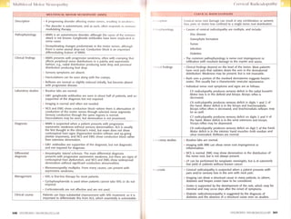
![Differential diagnosis
Management
( t't'vit rtl l(trrlit rtlolrrrtlry tt,ru*tt
CERVIC.AL RADICUTOT,ATHY.-<onI,d
Brachial plexopothy. Can produce motor and sensory [irrtlirrgs rrr
one or both arms. However, the symptoms span dermatonr,rl
distribution.
Peripherol mononeuropothy Especially the radial nerve, periplrt'r,rl
mononeuropathy can mimic cervical radiculopathy, e.9., C7 lesion
However, examination of proximal and distal musculature helps to
differentiate a peripheral from a nerve root lesion. Also, most
peripheral nerve lesions span root distributions.
Nerve root compression from disc or bony element usually can
usually be treated conservatively. Treatment options include:
Physicol therapy is extremely helpful for reducing pain and
restoring function. This should be offered to almost all patienls
Muscle reloxonfs are used if there is paraspinal muscle spasnr
that contributes to the pain.
Anti-inflommotory agents are given to most patients and can
reduce pain.
Epidurol corticosteroid injections are considered if conservativt.
measures are not helpful.
Surgicol decompression is considered especially if there is
weakness or refractory pain.
Herpes zoster is treated with analgesics and antiviral medications.
Anticonvulsants often are often used as an adjunct for control of tht,
neuropathic pain.
Tumor infiltrotion is treated with local or neuraxis radiation.
lntrathecal and systemic chemotherapy also often are given.
Corticosteroids also are helpful for the pain of tumor infiltration.
Diobetic rodiculoneuropothy produces neuropathic pain, which can
be treated not only with analgesics but also by anticonvulsants suclr
as gabapentin. These agents are less helpful for the mechanical pairr
than the neuropathic pain.
Most patients improve with treatment, although not all. lf the cause n
compressive, there is a high remission rate with medications and
therapy, although there is the possibility of recurrence of the pain at ,r
later time.
542 DISORDERS-NEUROMUSCULAR DISORDERS-NEUROMUSCULAR 54I
( r,tvit rrl l{rrrli< trloprrllry
l)t.rrtt.rlorrrr.s art<l Myololncs of Uppcr Limb
,,t,.',, lr.It,rtt, rlcrrr,rrr,rlrorr (,1 tl(,illl,tlr)[t(.
,,,,,rrlrrrri lo [r,r,r].rrr.ur(l (;,rt('ll) sltorln.ts distinct
rlr ri,rrt,, I lrlt rs ,rr lrr,rill,r onsirk'r.rltle ovcrlap
tr1r,r,rr ,rlj,rr cnl (l('ll.ll()ntcs. An alternative
rr,rt()rr( nr,r1r is Ih,rl providcd by Foerster.
( .l lo I I Scnsory Representation Anterior view
Posterior view
au
{, *t l-dffi
cz,-nl, -:]
t *+*
t' . -. ,t
c4 '.l.t**-,r
C5
C5
I
C8
.,'
i
C6
r;,-n qr-:-r'.'-
' ".-ca lr,-';: '
I Motor lmpairment Related to Level of Cervical Root Lesion
Hcrniated disc
comprcssing
nerve root](data:image/gif;base64,R0lGODlhAQABAIAAAAAAAP///yH5BAEAAAAALAAAAAABAAEAAAIBRAA7)