SlideShare a Scribd company logo
1 of 75
Radiological imaging of cord Myelopathy. 
Dr/ ABD ALLAH NAZEER. MD.
Introduction: 
The term myelopathy describes pathologic conditions that cause 
spinal cord, meningeal or peri meningeal space damage or 
dysfunction. Traumatic injuries, vascular diseases, infections and 
inflammatory or autoimmune processes may affect the spinal cord 
due to its confinement in a very small space. Spinal cord injuries 
usually have devastating consequences such as quadriplegia, 
paraplegia and severe sensory deficits. 
The history, an adequate neurological examination and the study of 
the cerebrospinal fluid (CSF) guide the diagnosis of spinal cord 
injuries. However, imaging is of great importance in order to home 
in on the diagnosis and classify the etiology appropriately. 
Many of the processes affecting the spinal cord may be reversible if 
recognized and treated early. 
The vast majority of spinal cord diseases may be treated medically, 
with surgical treatment reserved for compressive disorders, which 
constitute a neurological emergency.
Definition and clinical picture 
It is important not to mistake myelopathy for myelitis. Although both 
terms refer to spinal cord compromise due to a pathological event, 
myelopathy has multiple etiologies, while myelitis is used to refer to 
inflammatory or infectious processes. Acute transverse myelopathy 
(includes non-inflammatory etiologies) and transverse myelitis have been 
used as synonyms in the published literature. Findings of spinal tract 
injuries, a certain degree of sensory dysfunction, or urinary retention, 
point to a spinal cord injury. There are certain conditions that may mimic 
myelopathy, such as myopathy or disorders of the neuromuscular junction, 
but the absence of a sensory deficit rules them out. Myelopathies may 
have a variable course and may manifest as a single event or as a multi-phasic 
or recurrent disease. The latter is rare and is usually secondary to 
demyelinating diseases, vascular malformations of the spinal cord, or 
systemic diseases. The central nervous system (CNS) damage may be 
monofocal as in transverse myelitis and optic neuritis, or multifocal as in 
acute disseminated encephalomyelitis (ADEM) (brain and spinal cord), 
neuromyelitis optica (optic nerve and spinal cord) and multiple sclerosis 
(MS) (any area of the neural axis).
Spinal cord pathologies may be classified as acute, subacute/ 
intermittent or chronic, depending on the time course, the extent of 
the involvement, the clinical picture or syndrome, or the etiology. 
Patients with myelopathies but no evident lesions, or who present 
with multiple lesions of chronic appearance on magnetic resonance 
imaging, must be questioned about prior subtle symptoms. Acute 
onset that worsens within hours or days points to a spinal cord infarct 
or hemorrhage. When symptoms are recent, it is of paramount 
importance to rule out a surgical emergency. This requires immediate 
imaging work-up, ideally total spine magnetic resonance (MR). If 
there is evidence of spinal cord compression due to an acute lesion 
(epidural metastasis or abscess), definitive management is required 
in order to avoid damage or to adequately manage all other 
potential diagnoses. If the symptoms progress for more than three 
weeks, transverse myelitis is improbable, and other conditions must 
be considered, such as a spinal tumor, chronic compressive disease, 
dural arterio-venous fistula, metabolic disorder, sarcoidosis, or a 
degenerative process.
Spinal cord syndromes present with typical signs and symptoms caused 
by a lesion of a specific tract in a specific location that may lead to the 
etiological diagnosis. They are classified as follows: 
• Complete spinal cord: involvement of all the tracts (trauma, compression 
or acute transverse myelitis). 
• Brown Séquard or hemi-spinal cord syndrome: ipsilateral cortico-spinal 
tract, posterior columns and contralateral spinothalamic tract (multiple 
sclerosis and compression). 
• Anterior spinal cord syndrome: anterior horns, corticospinal, 
spinothalamic and autonomic tracts (anterior spinal artery infarct and 
multiple sclerosis). Posterior spinal cord syndrome: posterior columns 
(vitamin B12 or copper deficiency). 
• Central syndrome: spino-thalamic crossing, cortico-spinal and autonomic 
tracts (syringomyelia, neuromyelitis optica). 
• Medullary cone: sacral emerging fibers (post-viral myelitis). 
• Cauda equina: cauda equina nerves (acute cytomegalovirus infection, 
polyradiculits and compression) 
• Tractopathies: selective disorders (vitamin B12 deficiency, paraneoplastic 
myelopathy and multiple sclerosis).
Spinal cord abnormalities. Common causes in white and uncommon causes in yellow.
Degenerative compressive myelopathy: 
Degenerative compressive myelopathy may be classified 
according to the compression site, as follows: 
• Anterior (disc protrusion or posterior osteophytes). 
• Anterolateral (Luschka joints). 
• Lateral (facet joints). 
• Posterior (ligamentum flavum). 
It may be caused by atlanto-axial instability, spinal canal 
stenosis due to cervical spondylolysis, cervical spinal fusion, 
myelomeningocele or epidural masses. Atlanto-axial instability 
is the primary cause of degenerative compressive myelopathy. 
It is found mainly in rheumatoid arthritis, followed by Down’s 
syndrome, Morquio’s syndrome or type IV 
mucopolysaccharidoses, skeletal dysplasia, ankylosing 
spondylitis and Lesh-Nyhan syndrome.
Increased intensity of the spinal cord in C2 in the T2 weighted sequence 
due to compressive myelopathy secondary to rheumatoid arthritis.
Increased intensity and 
thickening of the spinal 
cord from the bulbo-medullary 
junction down 
to C4 in the sagittal T2 
sequence, due to 
compressive myelopathy 
in Morquio’s syndrome.
MRI showing cervical spondylotic myelopathy.
Post-traumatic compressive myelopathy: 
Post-traumatic myelopathy is four times more frequent in males, 
in particular between 16 and 30 years of age. Motor vehicle 
accidents are the most common cause, accounting for 50% of the 
events, followed by violence (firearm or stab wounds), falls from 
heights, and sports injuries (diving, American football and 
horseback-riding). The most mobile segments are more often 
affected, in particularC5-C7 and T10-L2. Clinically, quadriplegia 
predominates in 30-40% of cases, and paraplegia occurs in 6-10%. 
MR imaging is of vital importance in approaching spinal cord 
trauma because it shows location, extension and severity very 
clearly, and also reveals edema and intramedullary bleeding. 
Some studies have shown that hemorrhage and longer 
hematomas are associated with a lower rate of motor recovery. 
Over the long term, CSF leaks, infections, cysts and syringomyelia 
may develop.
a) Axial sequence with T2 gradient echo information. B) Sagittal section with T2 
information in C7 showing diminished height and signal intensity with annulus protrusion 
in C5-C6 and C6-C7; there is also central and left subarticular protrusion of the annulus 
associated with annulus and ligament tear in C7, giving rise to central spinal hyperintensity 
due to compressive myelopathy resulting from nucleus pulposus herniation.
Myelopathy due to 
traumatic cord compression. 
Myelopathy as a result of 
compression by a dorsally 
located epidural hemorrhage
Myelopathy result of a metastasis.
T2 weighted 
image with 
annulus 
protrusion in C4 
and C5, giving 
rise to spinal cord 
hyperintensity 
due to traumatic 
compressive 
myelopathy.
Abscess-related compressive myelopathy: 
Epidural abscesses are uncommon but they constitute a surgical 
emergency because they may progress rapidly within days and early 
diagnosis is difficult, leading to delayed treatment. They affect mainly 
men, with no specific age range, and the incidence has been shown to 
have increased in recent years. Risk factors are similar to those for 
spondylodiscitis, including diabetes mellitus, use of intravenous drugs, 
chronic renal failure, alcohol abuse, and immune deficiency. Human 
immunodeficiency virus has not been shown to be the cause of the 
increased incidence. It usually presents as subacute pain, fever (may be 
absent in subacute and chronic stages), increased local tenderness, 
progressive radiculopathy or myelopathy. The second phase of 
radicular irritation is followed by neurologic deficit (muscle weakness, 
abnormal sensation and incontinence) and then by paralysis in 34% of 
cases, and even death. Symptoms result from mechanical compression 
and, in some cases, from ischemia. Any segment of the spinal cord may 
be affected, but the most frequent are the thoracic and lumbar 
segments.
a) Sagittal image with T1 information showing signal-intensity changes of the lower 
T10 endplate and upper T11 endplate, and of the corresponding disc, associated with 
hyperintensity and spinal cord thickening in that segment. b) Post-gadolinium STIR 
image showing thickening of the prevertebral soft tissues, the vertebral bodies and the 
disc from T10 to T11. These enhance with contrast, together with the thickened spinal 
cord due to myelopathy resulting from an epidural abscess.
Cervical Pott’s and retropharyngeal abscess with cord myelopathy.
Tumoral compressive myelopathy: 
Myelopathy may be the initial manifestation of a malignancy in up to 
20% of cases where the only systemic symptom is weight loss. Tumors 
compressing the spinal cord may be divided into extradural and intradural. 
Extradural tumors may be classified as follows: 
• Benign: synovial cyst, osteoma, osteoblastoma, giant cell tumor, 
hemangioma, eosinophilic granuloma, schwannoma and meningioma. 
• Malignant: bone metastasis (are the cause of the most common 
myelopathy due to extradural spinal cord compression), multiple 
myeloma, lymphoma and chondrosarcoma. 
Intradural tumors are classified as follows: 
• Extra-spinal: neurofibroma, meningioma, lipoma, schwannoma and 
arachnoid cyst. 
• Intraspinal: astrocytoma, ependymoma, hemangioblastoma and 
metastasis. 
Forty per cent of patients present with radiculopathy and myelopathy 
associated with subacute dorsal pain that worsens in decubitus position. 
MRI may reveal the cause of the myelopathy and help guide the approach 
to the primary tumor.
Post-gadolinium sagittal STIR 
image showing abnormal 
signal intensity of the bodies 
of T3 and T4, with a 
pathological fracture of T3 
involving the epidural 
component and which 
compresses the spinal cord. 
There is gadolinium 
enhancement of T1, T3 and 
T4 and of the spinous 
processes, but no 
enhancement of the spinal 
cord due to metastatic 
disease. This causes tumor 
compression myelopathy.
Postgadolinium sagittal 
STIR image showing 
spinal widening in C5 
and C6, with 
enhancement of the 
spinal cord and the 
bodies of C7 and T1. 
There is also 
enhancement of the 
prevertebral soft tissues 
and of the cervical 
muscles due to 
myelopathy secondary 
to a high-grade glioma.
Astrocytoma simulating Transverse myelitis.
Spinal astrocytoma. Multiple ependymomas.
Myelopathy of vascular origin: 
The spinal cord may be affected by compressive and non-compressive vascular 
diseases, of which the most common are malformations of the dural arteriovenous 
fistula type. In cases of vascular malformation, patients present with non-specific 
clinical findings, usually distal to the site of the disease. These diseases were classified 
by Riche in 1985 as follows: 
• Intraspinal arteriovenous malformations. 
• Perispinal arteriovenous malformations. 
• Spinodural arteriovenous fistulas. 
• Epidural arteriovenous malformations. 
• Paravertebral vascular malformations. 
• Vertebral hemangiomas. 
• Complex angiomatosis (Cobb’s syndrome, Osler-Weber-Rendu syndrome). 
• Cavernomas, telangiectasias and spinal venous angiomas (do not require 
endovascular treatment). 
In 2002, Spetzler proposed the following new classification 
• Neoplastic vascular lesions: hemangioblastoma and cavernous malformation. 
• Spinal aneurism. 
• Arteriovenous fistula: extradural and intradural. The latter includes ventral (small, 
medium and large) and dorsal (one or several feeding vessels) fistulas. 
• Arteriovenous malformations: extra-intradural and intradural (intraspinal, compact, 
diffuse and of the medullary cone).
Sagittal sequence with T2 information of the medullary cone, showing serpent 
like, tortuous intra and extra-spinal images with absence of flow signal, 
associated with dorsal spine hyperintensity. b) Arteriogram confirming the 
presence of dural arteriovenous malformation with myelopathy of vascular origin.
Fifteen-year old patient with neurologic deficit of sudden onset and normal 
laboratory tests. The sagittal sequence with T2 information shows a high-intensity signal 
anterior to the spinal cord suggesting a diagnosis of myelopathy due to ischemia.
Spinal chord ischemia with DWI.
Myelopathy and dilated veins as a result of an AVF.
Compressive myelopathy due to syringomyelia: 
Syringomyelia is a rare neurologic disorder, characterized by 
the slow development of fluid-filled areas extending along the 
spinal cord, and causing symptoms such as pain, weakness 
and stiffness of the back, shoulders and limbs. In the United 
States, it is more common among African-Americans. It may 
be related to congenital or acquired malformations. Chiari’s 
malformation is a congenital abnormality in which cerebellar 
amygdalas herniate through the foramen magnum in the 
spinal canal, with altered CSF flow. This causes headache, 
double vision, dizziness and muscle weakness of the upper 
limbs. Most non-traumatic forms of syringomyelia are due to 
Chiari malformation. The acquired causes of syringomyelia 
include trauma, tuberculosis-associated chronic arachnoiditis, 
and intraspinal tumors .
a) Sagittal sequence with T2 information of the cervical spine showing Chiari 
malformation type II, absent corpus callosum and a tubular zone of high signal 
intensity in the spinal cord down from the bulbo-medullary junction, involving all 
the segments. b) Sagittal sequence with T2 information of the dorso-lumbar spine 
with myelopathy secondary to syringomyelia.
Transverse myelitis: 
Acute transverse myelitis is a spinal disorder characterized by bilateral motor, 
sensory and autonomic abnormalities because it involves the spinothalamic 
and pyramidal tracts, the posterior columns and the anterior funiculus of one 
or more levels. Middle-aged adults are most frequently affected. 
A publication established the following criteria for transverse myelopathy: 
bilateral spinal cord dysfunction during a four-week period with a well-defined 
sensory level and no history of disease, where compression has been 
ruled out. Other criteria are proposed later for the differentiation between 
inflammatory and non-inflammatory transverse myelitis, and between 
idiopathic transverse myelitis and myelitis associated with a systemic or 
nervous system disease. 
These criteria are the following (5): 
• Sensory, motor or autonomic dysfunction of spinal origin. 
• Bilateral signs and symptoms. 
• Clearly defined sensory level. 
• Spinal inflammation (CSF pleocytosis or high immunoglobulin G levels, 
or gadolinium enhancement). 
• Maximum progression during a period ranging between four hours and 
four weeks.
Twenty-year old patient with suspected multiple sclerosis. a) The sagittal 
sequence with T1 information shows widening of the spinal cord from C3 
down to C7, with no other abnormal findings. b) Central high-signal area in 
T2 sequences due to acute transverse myelopathy.
ATM on T2WI, CE-T1WI and STIR.
Acute Transverse Myelitis.
Sixty-one-year-old female patient 
with neurological abnormalities 
over the past three days, but no 
significant history. Laboratory tests 
and the medullary biopsy were all 
normal. The sagittal sequence with 
T2 information showed discal and 
osteophytic changes of the 
vertebral bodies associated with 
bulging of the inferior annulus and 
thickening and hypersensitivity of 
the cervical spinal cord from the 
craniocervical junction down to C7. 
The clinical findings and the 
additional studies established the 
diagnosis of idiopathic myelopathy.
MRI of 
representative 
cases of acute 
and subacute 
myelopathies
Para-infectious myelopathy: 
Neurological damage in parainfectious myelopathy is caused directly 
by the infection, the immune reaction against the agent, and the 
reaction of the immune system. It is usually due to a blood-borne 
infection originating in the lungs, the skin, the skeletal, genitourinary 
or digestive systems. It presents with severe motor and sphincter 
dysfunction associated with fever, meningism and skin exanthema. The 
time period for the onset of myelitis after the infection is no different 
between infectious and post-infectious myelitis: five days for small-pox 
myelitis, ten days for mycoplasm and twelve days for herpes zoster 
myelitis. Possible etiologies include the following: 
• Viral: herpes, varicella zoster, EBV, CMV, HIV, dengue, influenza, 
measles, mumps, HTLV-1, enterovirus, Coxsackie B, hepatitis A and 
C, and polio. 
• Bacterial: mycoplasm, treponema pallidum, brucella, mycobacterium 
tuberculosis and borrelia. 
• Fungi: actinomyces, blastomyces, coccydiodes and aspergillus. 
• Parasites: Schistosoma, Cysticercosis, echinococcus and toxoplasma.
a) Sagittal sequence with T1 information showing thickening of the spinal cord 
from the craniocervical junction down to the thoracic region. b) Sagittal sequence 
with T2 weighted image showing high central spinal signal. HTLV-1 infection is 
confirmed later by positive serology.
Twenty-four-year old patient 
with congenital HIV and 
tuberculosis. The image 
shows alteration in the 
shape and signal intensity of 
the vertebral bodies of T10 
and T11, of the disc and of 
the prevertebral soft tissues. 
It is associated with 
widening of the spinal cord 
with edema and contrast 
enhancement, and with fluid 
accumulations near the 
spinal canal in the post-gadolinium 
sagittal STIR 
sequence due to tuberculous 
myelopathy.
Spinal Cord Schistosomiasis with cord myelopathy.
Acute inflammatory demyelinating polyneuropathy.
Acute disseminated encephalomyelitis 
Acute disseminated encephalomyelitis (ADEM) is an uncommon inflammatory 
disease of the central nervous system, characterized by diffuse demyelination of 
the cerebral white matter and the spinal cord. It is more frequent in children and 
young adults. It has been associated with infection or vaccination, but this is not 
considered a criterion in clinical consensus. It is believed to be a single-phase 
disease with a good prognosis; however, recurrent forms make differentiation 
from MS difficult. 
ADEM has clinical manifestations that usually include encephalopathy but may 
also include focal or multifocal demyelinating inflammatory syndromes of the CNS 
such as optic neuritis and myelitis. For that reason, ADEM is a differential 
diagnosis for isolated demyelinating syndrome, which is a more common 
precursor of MS in adults. ADEM symptoms include rapidly progressing 
encephalopathy associated with seizures or multiple neurologic deficits. The spinal 
cord is affected in 11% to 28% of patients, generally in the thoracic and cervical 
segments. CSF findings are non-specific and oligoclonal bands are found in 65% of 
patients MRI shows bilateral symmetric multifocal white matter lesions, with or 
without damage of the grey matter, and extensive disease of several spinal 
segments with expansion. These lesions appear with low signal in T1 sequences, 
and well defined with a high signal in T2 sequences; gadolinium enhancement is 
variable. All patients with spinal involvement have brain damage
Twenty-eight-year old patient presenting with sudden neurologic decline. a) Coronal 
FLAIR sequence of the brain showing hyperintensity in the bulbo-medullary junction. b) 
Axial image with T2 information in the bulbo-medullary junction with anterior 
hyperintensity related to myelopathy due to acute disseminated encephalopathy.
Typical ADEM.
ADEM seen in children.
Multiple sclerosis: 
MS is a chronic demyelinating inflammatory CNS disease. It is common in 
Europe, the United States, Canada, New Zealand and Australia, but it is 
rare in Asia, the tropics and the subtropical regions. In high-risk 
populations, the incidence is one out of every 200 women. The female-to-male 
ratio varies between 1.5 and 2.5. The age of onset of symptoms 
varies by region; however the incidence is low in children, increases in 
adolescence and peaks between 25 and 35 years of age, after which it 
starts to decline. The etiology remains unknown, although environmental, 
viral and immune-mediated factors in genetically susceptible patients are 
thought to be the culprits . The strongest risk factor is family history. 
Approximately 80-85% of patients present with a relapsing picture, with 
symptoms that last for several days and improve over the course of weeks. 
In 15% of patients, the disease is progressive from the start. It is the most 
studied of all acute myelopathies, and its effects range from irreversible 
tissue loss to partial demyelination where there can be remyelination and 
repair. There is spinal cord involvement in more than 90% of patients. It 
may present in the form a cervicodorsal asymmetric transverse myelitis 
with sensory symptoms.
Twenty-eight-year old patient diagnosed with multiple sclerosis in December 
2010 with progression in time and space. a) Sagittal sequence with T2 
information showing high spinal signal in C4. b) High-signal area in C4 shown on 
a T2 gradient echo axial view due to myelopathy secondary to multiple sclerosis.
MS in the cord.
Pediatric Acute Transverse Myelitis.
Acute exacerbation of spinal MS.
Neuromyelitis optica or Devic’s syndrome: 
Neuromyelitis optica is defined as the concomitant presentation of 
myelitis and optic neuritis. This combination occurs in MS, ADEM, 
systemic lupus erythematosus and Sjögren’s syndrome. It is also found 
in association with viral and bacterial infections. 
Neuromyelitis optica is an immune CNS demyelinating condition that 
affects the spinal cord and the optic nerves. It is often mistaken with 
MS, although clinical, radiological and immunopathological tests 
suggest that they are different. The identification of the specific 
antigen of the neuromyelitis optica immunoglobulin G/aquaporin 4 
antibody implies humoral immunity, which makes it different from MS. 
It is an uncommon disorder among the Western population, with an 
incidence of 0.4 per million people per year, representing one out of 
every 200 patients with demyelinating disease. In Asia, the Caribbean 
and South America, the incidence is higher, pointing to genetic 
mechanisms. In all populations, females with a mean age of 40 are 
predominantly affected, in a 3:1 ratio.
Forty-four-year old patient with demyelinating disease and proven neurological 
decline. a) Diminished signal intensity in the upper segments of the cervical spinal 
cord in the sagittal T1 sequence. b) Sagittal sequence with T2 information showing 
hyperintensity from the bulbo-medullary junction down to C6 and C7, due to 
neuromyelitis optica myelopathy.
NMO presenting with neuritis optica.
Neuromyelitis optica with cord myelopathy.
Neuromyelitis 
Optica.
Myelopathy due to systemic disease: 
Myelitis associated with a systemic disease has been rarely described in the 
literature. It has been associated with systemic lupus erythematosus (SLE), 
Sjögren’s syndrome, scleroderma, Behçet disease, and sarcoidosis. It has been 
estimated that the frequency of myelitis in patients with SLE is 3%, but it is 
unknown in Sjögren’s syndrome. Myelitis usually occurs in the first year of the 
disease and may be its first manifestation. The hypothesis about the 
pathophysiology is still a subject for debate, and the most accepted is a 
vascular mechanism secondary to ischemic lesions. Women are more 
frequently affected than men, in an 8:1 ratio. CNS involvement in SLE is often 
found in relation with the antiphospholipid syndrome with anticardiolipin 
antibodies. The clinical symptoms often include transverse myelitis with 
severe motor and sensory dysfunction. CSF may be normal or show plecytosis, 
and although oligoclonal bands are rare, they may be present. MRI findings 
have been studied more in SLE than in Sjögren’s syndrome. Moreover, it has 
been found that the central high-signal spinal lesion in T2 sequences, CNS 
sarcoidosis may occur in isolation in the form of a myelopathy. The definitive 
diagnosis requires biopsy evidence of non-caseifying granulomatous 
inflammation of the CNS or any other compromised organ.
a) Sagittal sequence with T2 information showing spinal cord thickening 
and hyperintensity from C4 down to T2. b) Post-gadolinium STIR image 
showing enhancement due to SLE-associated myelopathy.
Diabetic myelopathy.
Spinal cord MRI. 
T2-weighted 
sequences. 
Extended 
hypersignal in a 
patient with a 
Sjögren's 
syndrome (A) 
localized in the 
centromedullary 
territory (B).
Post-radiation or electric damage: 
Neurotoxicity is a known complication of high-dose radiation. The deep white 
matter is the most affected since it comprises the cortex and the subcortical 
arcuate fibers. There are three forms of lesions: acute (weeks or months), early 
late and late (six months to two years). The latter may be irreversible, 
progressive and, on occasions, fatal; however, it may resolve spontaneously in 
some cases. Radiation myelopathy is a devastating complication of radiation 
therapy. It is a rare cause of acute myelopathy, accounting for only 2% of 
complications, and it is suggested in cases where there is a history of exposure 
to head and neck radiation (even longer than ten years), with a dose greater 
than 4,000 rads. It is an irreversible process with no effective treatment. It may 
have an early manifestation ten to sixteen weeks into radiotherapy, or a late 
manifestation, and may resolve spontaneously between two and nine months 
after onset. In the early stages, there is evidence of edema or spinal 
enhancement and, in late cases, spinal atrophy is observed It manifests as a 
transient sensory loss, progressive chronic myelopathy, acute transverse 
myelitis, or local amyotrophy. The transient sensory loss gives an electric-shock 
sensation when the neck is flexed forward (Lhermitte sign) and it resolves 
within two and thirty-six weeks. In chronic progressive myelopathy, it presents 
like a Brown Séquard syndrome lasting between three months and five years.
Patient with a history of 
radiotherapy due to 
esophageal cancer who 
complains of 
paresthesias 
and discreet loss of 
strength in the lower 
limbs, and Lhermitte 
sign. a) Sagittal T2 image 
of the dorsal spine 
with discreet fusiform 
thickening of the spinal 
cord showing high 
intensity signal and post-radiotherapy 
bone 
marrow changes. b) 
Follow-up MRI after 18 
months with evidence of 
partial regression of post- 
-radiation myelopathy.
Spinal cord MRI. Extended hypersignal in T2-weighted sequences in the 
cervical cord following laryngeal radiation with a swelling of the cord (A). 
MRI after 4 months of follow-up shows normal cord signal in T2-weighted 
sequences, but an atrophic cord in T1-weighted sequences (B).
Radiation myelopathy. 
Paraneoplastic tractopathy. Axial T2 
sections through the cord of a 69-year-old 
woman with melanoma and high 
titers of amphiphysin-immunoglobulin 
(Ig)G. Arrow points to hyperintensity in 
the region of the corticospinal tracts.
Subacute combined degeneration: 
Combined subacute degeneration is a complication of vitamin B12 deficiency, 
associated with pernicious anemia. This deficiency may be related to 
parietal-cell auto-antibodies or the intrinsic factor required for vitamin B12 
binding. There is a genetic deficiency of transcobalamin 2 (cobalamin 
transporter protein). The complete transcobalamin 2 deficiency is a recessive 
autosomal condition characterized by normal concentrations of vitamin B12 
with severe infantile megaloblastic anemia associated with neurologic 
damage . The clinical picture presents as a slowly-progressing spastic 
paraparesis with distal proprioceptive loss and symmetrical dysesthesias. 
This clinical picture is due to axonal loss and inferior cervical and thoracic 
spinal demyelination, generally in the posterior and anterolateral columns, 
that cannot be explained by compression or inflammation on MRI. The 
absence of anemia with or without macrocytosis does not rule out a vitamin 
B12 deficiency diagnosis. MRI shows high signal intensity of the posterior 
columns in T2 sequences, without contrast enhancement. Imaging 
improvement correlates with clinical improvement . The mean time to 
diagnosis since the onset of neurological symptoms due to vitamin B12 
deficiency is approximately on year, with a range that extends to four years.
a) Sagittal T2 sequence showing hyperintense signal in the dorsal spine. b) Axial 
T2 image confirming spinal damage predominantly in the posterior horns. Low 
concentrations of vitamin B12 were found, pointing to the diagnosis of 
myelopathy due to vitamin B12 deficiency. c) and d) Follow-up MRI performed 
one year after the diagnosis, showing no evidence of abnormalities.
Subacute combined degeneration with hyperintensity at the posterior column.
Acute paraneoplastic or necrotizing myelitis: 
Paraneoplastic myelopathy is a rare disease. It may occur before 
the cancer is detected. Several antibodies have been associated 
with subacute myelopathies, in general with lung, breast, thyroid, 
ovarian and prostate cancer, and Hodgkin’s lymphoma. 
The lesion often involves the thoracic spinal cord that shows a 
high-intensity signal in T2 sequences and gadolinium 
enhancement. There is increased protein concentration in the CSF. 
Myelopathy with normal MRI: 
In some cases of acute myelopathy, the MR image is normal. There 
are several explanations for this: 1) the syndrome is not a 
myelopathy (Guillain-Barré, inflammatory radiculopathy); 2) the 
picture may not be acute but rather a decompensated existing 
myelopathy (Friedreich’s ataxia, motor neuron disease, vitamin B12 
deficiency, HIV or HTLV-1 myelopathy); and 3) MRI performed during 
the convalescence period.
Large B- cell lymphoma showing cord edema progressing cranially to involve the 
cervical spine. (b) MRI demonstrated edema involving the conus medullaris.
Large B- cell lymphoma showing cord edema progressing 
cranially to involve the cervical and thoracic cord.
Conclusion. 
Myelopathy is a broad term that refers to spinal cord 
involvement of multiple etiologies. 
Spinal cord diseases often have devastating consequences, 
ranging from quadriplegia and paraplegia to severe 
sensory deficits due to its confinement in a very small 
area. 
Many of these diseases are potentially reversible if they 
are recognized on time, hence the importance of 
recognizing the significance of magnetic resonance 
imaging when approaching a multifactorial disease 
considered as one of the most critical neurological 
emergencies, where prognosis depends on an early and 
accurate diagnosis.
Thank You.

More Related Content

What's hot

Presentation1.pptx, radiological imaging of spinal cord tumour.
Presentation1.pptx, radiological imaging of spinal cord tumour.Presentation1.pptx, radiological imaging of spinal cord tumour.
Presentation1.pptx, radiological imaging of spinal cord tumour.Abdellah Nazeer
 
Neuroimaging Lecture
Neuroimaging LectureNeuroimaging Lecture
Neuroimaging Lecturetest
 
Presentation1.pptx, radiological imaging of congenital anomalies of the spine...
Presentation1.pptx, radiological imaging of congenital anomalies of the spine...Presentation1.pptx, radiological imaging of congenital anomalies of the spine...
Presentation1.pptx, radiological imaging of congenital anomalies of the spine...Abdellah Nazeer
 
imaging in neurology - demyelinating diseases
 imaging in neurology - demyelinating diseases imaging in neurology - demyelinating diseases
imaging in neurology - demyelinating diseasesNeurologyKota
 
Imaging in degenerative diseases of spine
Imaging in degenerative diseases of spineImaging in degenerative diseases of spine
Imaging in degenerative diseases of spineGirendra Shankar
 
MRI KNEE JOINT ANATOMY
MRI KNEE JOINT ANATOMYMRI KNEE JOINT ANATOMY
MRI KNEE JOINT ANATOMYNikhil Bansal
 
Imaging of white matter diseases
Imaging of white matter diseasesImaging of white matter diseases
Imaging of white matter diseasesNavni Garg
 
Mri evaluation of spine myelopathy
Mri evaluation of spine myelopathyMri evaluation of spine myelopathy
Mri evaluation of spine myelopathyDrBhishm Sevendra
 
Muscle MRI in Neuromuscular Disease
Muscle MRI in Neuromuscular DiseaseMuscle MRI in Neuromuscular Disease
Muscle MRI in Neuromuscular DiseaseDr. Arghya Deb
 
Radiologic anatomy of the cranial nerves
Radiologic anatomy of the cranial nervesRadiologic anatomy of the cranial nerves
Radiologic anatomy of the cranial nerveshazem youssef
 
Radiological findings of congenital anomalies of the spine and spinal cord
Radiological findings of congenital anomalies of the spine and spinal cordRadiological findings of congenital anomalies of the spine and spinal cord
Radiological findings of congenital anomalies of the spine and spinal cordDr. Armaan Singh
 
Craniovertebral junction
Craniovertebral junctionCraniovertebral junction
Craniovertebral junctionNeurologyKota
 
Presentation1, radiological imaging of pediatric leukodystrophy.
Presentation1, radiological imaging of pediatric leukodystrophy.Presentation1, radiological imaging of pediatric leukodystrophy.
Presentation1, radiological imaging of pediatric leukodystrophy.Abdellah Nazeer
 
Radiology of demyelinating diseases
Radiology of demyelinating diseases Radiology of demyelinating diseases
Radiology of demyelinating diseases NeurologyKota
 
Spinal neoplasms
Spinal neoplasmsSpinal neoplasms
Spinal neoplasmsfahad shafi
 
MR spectroscopy
MR spectroscopyMR spectroscopy
MR spectroscopyairwave12
 
Presentation1.pptx, radiological imaging of dementia.
Presentation1.pptx, radiological imaging of dementia.Presentation1.pptx, radiological imaging of dementia.
Presentation1.pptx, radiological imaging of dementia.Abdellah Nazeer
 
Cervical Spine Trauma Imaging
Cervical Spine Trauma ImagingCervical Spine Trauma Imaging
Cervical Spine Trauma ImagingSCGH ED CME
 

What's hot (20)

Presentation1.pptx, radiological imaging of spinal cord tumour.
Presentation1.pptx, radiological imaging of spinal cord tumour.Presentation1.pptx, radiological imaging of spinal cord tumour.
Presentation1.pptx, radiological imaging of spinal cord tumour.
 
MR Spectroscopy
MR SpectroscopyMR Spectroscopy
MR Spectroscopy
 
Neuroimaging Lecture
Neuroimaging LectureNeuroimaging Lecture
Neuroimaging Lecture
 
Presentation1.pptx, radiological imaging of congenital anomalies of the spine...
Presentation1.pptx, radiological imaging of congenital anomalies of the spine...Presentation1.pptx, radiological imaging of congenital anomalies of the spine...
Presentation1.pptx, radiological imaging of congenital anomalies of the spine...
 
imaging in neurology - demyelinating diseases
 imaging in neurology - demyelinating diseases imaging in neurology - demyelinating diseases
imaging in neurology - demyelinating diseases
 
Imaging in degenerative diseases of spine
Imaging in degenerative diseases of spineImaging in degenerative diseases of spine
Imaging in degenerative diseases of spine
 
MRI KNEE JOINT ANATOMY
MRI KNEE JOINT ANATOMYMRI KNEE JOINT ANATOMY
MRI KNEE JOINT ANATOMY
 
Imaging of white matter diseases
Imaging of white matter diseasesImaging of white matter diseases
Imaging of white matter diseases
 
Mri evaluation of spine myelopathy
Mri evaluation of spine myelopathyMri evaluation of spine myelopathy
Mri evaluation of spine myelopathy
 
Muscle MRI in Neuromuscular Disease
Muscle MRI in Neuromuscular DiseaseMuscle MRI in Neuromuscular Disease
Muscle MRI in Neuromuscular Disease
 
Radiologic anatomy of the cranial nerves
Radiologic anatomy of the cranial nervesRadiologic anatomy of the cranial nerves
Radiologic anatomy of the cranial nerves
 
Radiological findings of congenital anomalies of the spine and spinal cord
Radiological findings of congenital anomalies of the spine and spinal cordRadiological findings of congenital anomalies of the spine and spinal cord
Radiological findings of congenital anomalies of the spine and spinal cord
 
Craniovertebral junction
Craniovertebral junctionCraniovertebral junction
Craniovertebral junction
 
Presentation1, radiological imaging of pediatric leukodystrophy.
Presentation1, radiological imaging of pediatric leukodystrophy.Presentation1, radiological imaging of pediatric leukodystrophy.
Presentation1, radiological imaging of pediatric leukodystrophy.
 
Myelination disorders
Myelination disordersMyelination disorders
Myelination disorders
 
Radiology of demyelinating diseases
Radiology of demyelinating diseases Radiology of demyelinating diseases
Radiology of demyelinating diseases
 
Spinal neoplasms
Spinal neoplasmsSpinal neoplasms
Spinal neoplasms
 
MR spectroscopy
MR spectroscopyMR spectroscopy
MR spectroscopy
 
Presentation1.pptx, radiological imaging of dementia.
Presentation1.pptx, radiological imaging of dementia.Presentation1.pptx, radiological imaging of dementia.
Presentation1.pptx, radiological imaging of dementia.
 
Cervical Spine Trauma Imaging
Cervical Spine Trauma ImagingCervical Spine Trauma Imaging
Cervical Spine Trauma Imaging
 

Viewers also liked

Diagnostic Imaging of Central Nervous System Infections
Diagnostic Imaging of Central Nervous System InfectionsDiagnostic Imaging of Central Nervous System Infections
Diagnostic Imaging of Central Nervous System InfectionsMohamed M.A. Zaitoun
 
Presentation1.pptx, radiological imaging of spinal trauma and spinal cord inj...
Presentation1.pptx, radiological imaging of spinal trauma and spinal cord inj...Presentation1.pptx, radiological imaging of spinal trauma and spinal cord inj...
Presentation1.pptx, radiological imaging of spinal trauma and spinal cord inj...Abdellah Nazeer
 
Compressive spine disease
Compressive spine diseaseCompressive spine disease
Compressive spine diseaseNabil Khalil
 
Presentation1.pptx, radiological imaging of aids diseases
Presentation1.pptx, radiological imaging of aids diseasesPresentation1.pptx, radiological imaging of aids diseases
Presentation1.pptx, radiological imaging of aids diseasesAbdellah Nazeer
 
Central and Peripheral Nerve Lesions - Neel Golwala
Central and Peripheral Nerve Lesions - Neel GolwalaCentral and Peripheral Nerve Lesions - Neel Golwala
Central and Peripheral Nerve Lesions - Neel Golwalabcooper876
 
Neuromyelitis Optica
Neuromyelitis OpticaNeuromyelitis Optica
Neuromyelitis Opticasm171181
 
Presentation1.pptx, lecture for md oral examination.
Presentation1.pptx, lecture for md oral examination.Presentation1.pptx, lecture for md oral examination.
Presentation1.pptx, lecture for md oral examination.Abdellah Nazeer
 
Non compressive myelopathy
Non compressive myelopathyNon compressive myelopathy
Non compressive myelopathyHirdesh Chawla
 
Radiological assessment - Part 1
Radiological assessment - Part 1Radiological assessment - Part 1
Radiological assessment - Part 1SpinePlus
 
Presentation1.pptx, radiological imaging of adult neck masses.
Presentation1.pptx, radiological imaging of adult neck masses.Presentation1.pptx, radiological imaging of adult neck masses.
Presentation1.pptx, radiological imaging of adult neck masses.Abdellah Nazeer
 
Presentation1.pptx, radiological anatomy of the orbits, pns and petrous bone.
Presentation1.pptx, radiological anatomy of the orbits, pns and petrous bone.Presentation1.pptx, radiological anatomy of the orbits, pns and petrous bone.
Presentation1.pptx, radiological anatomy of the orbits, pns and petrous bone.Abdellah Nazeer
 
Presentation2.pptx wrist joint.
Presentation2.pptx wrist joint.Presentation2.pptx wrist joint.
Presentation2.pptx wrist joint.Abdellah Nazeer
 
Presentation1.pptx, imaging of genetic diseases. (3)
Presentation1.pptx, imaging of genetic diseases. (3)Presentation1.pptx, imaging of genetic diseases. (3)
Presentation1.pptx, imaging of genetic diseases. (3)Abdellah Nazeer
 
Imaging of cns viral infection
Imaging of cns viral infectionImaging of cns viral infection
Imaging of cns viral infectionsks200166
 
Presentation1.pptx, diagnostic pitfalls mimicking meniscal tear and post oper...
Presentation1.pptx, diagnostic pitfalls mimicking meniscal tear and post oper...Presentation1.pptx, diagnostic pitfalls mimicking meniscal tear and post oper...
Presentation1.pptx, diagnostic pitfalls mimicking meniscal tear and post oper...Abdellah Nazeer
 
spinal cord anatomy and spinal cord syndromes
spinal cord anatomy and spinal cord syndromesspinal cord anatomy and spinal cord syndromes
spinal cord anatomy and spinal cord syndromesSachin Adukia
 
Presentation1.pptx, radiological imaging of spinal dysraphism.
Presentation1.pptx, radiological imaging of spinal dysraphism.Presentation1.pptx, radiological imaging of spinal dysraphism.
Presentation1.pptx, radiological imaging of spinal dysraphism.Abdellah Nazeer
 
Optic neuritis
Optic neuritisOptic neuritis
Optic neuritisSSSIHMS-PG
 

Viewers also liked (20)

Diagnostic Imaging of Central Nervous System Infections
Diagnostic Imaging of Central Nervous System InfectionsDiagnostic Imaging of Central Nervous System Infections
Diagnostic Imaging of Central Nervous System Infections
 
Presentation1.pptx, radiological imaging of spinal trauma and spinal cord inj...
Presentation1.pptx, radiological imaging of spinal trauma and spinal cord inj...Presentation1.pptx, radiological imaging of spinal trauma and spinal cord inj...
Presentation1.pptx, radiological imaging of spinal trauma and spinal cord inj...
 
CASO 4
CASO 4CASO 4
CASO 4
 
Compressive spine disease
Compressive spine diseaseCompressive spine disease
Compressive spine disease
 
Optic neuritis
Optic neuritisOptic neuritis
Optic neuritis
 
Presentation1.pptx, radiological imaging of aids diseases
Presentation1.pptx, radiological imaging of aids diseasesPresentation1.pptx, radiological imaging of aids diseases
Presentation1.pptx, radiological imaging of aids diseases
 
Central and Peripheral Nerve Lesions - Neel Golwala
Central and Peripheral Nerve Lesions - Neel GolwalaCentral and Peripheral Nerve Lesions - Neel Golwala
Central and Peripheral Nerve Lesions - Neel Golwala
 
Neuromyelitis Optica
Neuromyelitis OpticaNeuromyelitis Optica
Neuromyelitis Optica
 
Presentation1.pptx, lecture for md oral examination.
Presentation1.pptx, lecture for md oral examination.Presentation1.pptx, lecture for md oral examination.
Presentation1.pptx, lecture for md oral examination.
 
Non compressive myelopathy
Non compressive myelopathyNon compressive myelopathy
Non compressive myelopathy
 
Radiological assessment - Part 1
Radiological assessment - Part 1Radiological assessment - Part 1
Radiological assessment - Part 1
 
Presentation1.pptx, radiological imaging of adult neck masses.
Presentation1.pptx, radiological imaging of adult neck masses.Presentation1.pptx, radiological imaging of adult neck masses.
Presentation1.pptx, radiological imaging of adult neck masses.
 
Presentation1.pptx, radiological anatomy of the orbits, pns and petrous bone.
Presentation1.pptx, radiological anatomy of the orbits, pns and petrous bone.Presentation1.pptx, radiological anatomy of the orbits, pns and petrous bone.
Presentation1.pptx, radiological anatomy of the orbits, pns and petrous bone.
 
Presentation2.pptx wrist joint.
Presentation2.pptx wrist joint.Presentation2.pptx wrist joint.
Presentation2.pptx wrist joint.
 
Presentation1.pptx, imaging of genetic diseases. (3)
Presentation1.pptx, imaging of genetic diseases. (3)Presentation1.pptx, imaging of genetic diseases. (3)
Presentation1.pptx, imaging of genetic diseases. (3)
 
Imaging of cns viral infection
Imaging of cns viral infectionImaging of cns viral infection
Imaging of cns viral infection
 
Presentation1.pptx, diagnostic pitfalls mimicking meniscal tear and post oper...
Presentation1.pptx, diagnostic pitfalls mimicking meniscal tear and post oper...Presentation1.pptx, diagnostic pitfalls mimicking meniscal tear and post oper...
Presentation1.pptx, diagnostic pitfalls mimicking meniscal tear and post oper...
 
spinal cord anatomy and spinal cord syndromes
spinal cord anatomy and spinal cord syndromesspinal cord anatomy and spinal cord syndromes
spinal cord anatomy and spinal cord syndromes
 
Presentation1.pptx, radiological imaging of spinal dysraphism.
Presentation1.pptx, radiological imaging of spinal dysraphism.Presentation1.pptx, radiological imaging of spinal dysraphism.
Presentation1.pptx, radiological imaging of spinal dysraphism.
 
Optic neuritis
Optic neuritisOptic neuritis
Optic neuritis
 

Similar to Radiological Imaging of Spinal Cord Myelopathy Causes

Spinal Cord Disease - Clinical Presentations and Management
Spinal Cord Disease - Clinical Presentations and ManagementSpinal Cord Disease - Clinical Presentations and Management
Spinal Cord Disease - Clinical Presentations and ManagementJoseph Paul, MD
 
lecture for physiothrapy.pdf
lecture for physiothrapy.pdflecture for physiothrapy.pdf
lecture for physiothrapy.pdfeyobkaseye
 
Approach to myelopathy
Approach to myelopathyApproach to myelopathy
Approach to myelopathychandan kumar
 
SPINAL CORD DISORDERS.pptx
SPINAL CORD DISORDERS.pptxSPINAL CORD DISORDERS.pptx
SPINAL CORD DISORDERS.pptxtebaradio
 
Non Traumatic Spinal cord injuries
Non Traumatic Spinal cord injuries   Non Traumatic Spinal cord injuries
Non Traumatic Spinal cord injuries Dr. Muzahid
 
Classification of spinal cord disorders
Classification of spinal cord disorders Classification of spinal cord disorders
Classification of spinal cord disorders Yapa
 
Medical student lectures hydrocephalus ,chiari ,congenital ,truma ,nerve injury
Medical student lectures hydrocephalus ,chiari ,congenital ,truma ,nerve injury Medical student lectures hydrocephalus ,chiari ,congenital ,truma ,nerve injury
Medical student lectures hydrocephalus ,chiari ,congenital ,truma ,nerve injury Mohammed Homoud
 
Surgery 5th year, 1st lecture/part two (Dr. Khalid Shokor Mahmood)
Surgery 5th year, 1st lecture/part two (Dr. Khalid Shokor Mahmood)Surgery 5th year, 1st lecture/part two (Dr. Khalid Shokor Mahmood)
Surgery 5th year, 1st lecture/part two (Dr. Khalid Shokor Mahmood)College of Medicine, Sulaymaniyah
 
Multiple sclerosis: Introduction, Risk Factors, Diagnosis and Treatment
Multiple sclerosis: Introduction, Risk Factors, Diagnosis and TreatmentMultiple sclerosis: Introduction, Risk Factors, Diagnosis and Treatment
Multiple sclerosis: Introduction, Risk Factors, Diagnosis and TreatmentEnriqueAlvarez93
 
Cervical myelopathy cme
Cervical myelopathy cmeCervical myelopathy cme
Cervical myelopathy cmegroup7usmkk
 
Spine myelopathy
Spine   myelopathySpine   myelopathy
Spine myelopathySidra Afzal
 
Final case presentation sci (kimberly walsh)
Final case presentation sci (kimberly walsh)Final case presentation sci (kimberly walsh)
Final case presentation sci (kimberly walsh)Kimberly Walsh
 
Case record...Idiopathic postinfectious transverse myelitis
Case record...Idiopathic postinfectious transverse myelitisCase record...Idiopathic postinfectious transverse myelitis
Case record...Idiopathic postinfectious transverse myelitisProfessor Yasser Metwally
 
Case record...Acute postinfectious transverse myelitis (Transverse myelitis s...
Case record...Acute postinfectious transverse myelitis (Transverse myelitis s...Case record...Acute postinfectious transverse myelitis (Transverse myelitis s...
Case record...Acute postinfectious transverse myelitis (Transverse myelitis s...Professor Yasser Metwally
 

Similar to Radiological Imaging of Spinal Cord Myelopathy Causes (20)

Spinal Cord Disease - Clinical Presentations and Management
Spinal Cord Disease - Clinical Presentations and ManagementSpinal Cord Disease - Clinical Presentations and Management
Spinal Cord Disease - Clinical Presentations and Management
 
lecture for physiothrapy.pdf
lecture for physiothrapy.pdflecture for physiothrapy.pdf
lecture for physiothrapy.pdf
 
DDX Paraplegia
DDX ParaplegiaDDX Paraplegia
DDX Paraplegia
 
Approach to myelopathy
Approach to myelopathyApproach to myelopathy
Approach to myelopathy
 
SPINAL CORD DISORDERS.pptx
SPINAL CORD DISORDERS.pptxSPINAL CORD DISORDERS.pptx
SPINAL CORD DISORDERS.pptx
 
Non Traumatic Spinal cord injuries
Non Traumatic Spinal cord injuries   Non Traumatic Spinal cord injuries
Non Traumatic Spinal cord injuries
 
Classification of spinal cord disorders
Classification of spinal cord disorders Classification of spinal cord disorders
Classification of spinal cord disorders
 
Medical student lectures hydrocephalus ,chiari ,congenital ,truma ,nerve injury
Medical student lectures hydrocephalus ,chiari ,congenital ,truma ,nerve injury Medical student lectures hydrocephalus ,chiari ,congenital ,truma ,nerve injury
Medical student lectures hydrocephalus ,chiari ,congenital ,truma ,nerve injury
 
Spinal myelopathy
Spinal myelopathySpinal myelopathy
Spinal myelopathy
 
Surgery 5th year, 1st lecture/part two (Dr. Khalid Shokor Mahmood)
Surgery 5th year, 1st lecture/part two (Dr. Khalid Shokor Mahmood)Surgery 5th year, 1st lecture/part two (Dr. Khalid Shokor Mahmood)
Surgery 5th year, 1st lecture/part two (Dr. Khalid Shokor Mahmood)
 
Multiple sclerosis: Introduction, Risk Factors, Diagnosis and Treatment
Multiple sclerosis: Introduction, Risk Factors, Diagnosis and TreatmentMultiple sclerosis: Introduction, Risk Factors, Diagnosis and Treatment
Multiple sclerosis: Introduction, Risk Factors, Diagnosis and Treatment
 
Cervical myelopathy cme
Cervical myelopathy cmeCervical myelopathy cme
Cervical myelopathy cme
 
Spine myelopathy
Spine   myelopathySpine   myelopathy
Spine myelopathy
 
Case record...Neuromyelitis optica
Case record...Neuromyelitis opticaCase record...Neuromyelitis optica
Case record...Neuromyelitis optica
 
Final case presentation sci (kimberly walsh)
Final case presentation sci (kimberly walsh)Final case presentation sci (kimberly walsh)
Final case presentation sci (kimberly walsh)
 
Spinal nerve root entrapment.pptx
Spinal nerve root entrapment.pptxSpinal nerve root entrapment.pptx
Spinal nerve root entrapment.pptx
 
Case record...Idiopathic postinfectious transverse myelitis
Case record...Idiopathic postinfectious transverse myelitisCase record...Idiopathic postinfectious transverse myelitis
Case record...Idiopathic postinfectious transverse myelitis
 
Case record...Acute postinfectious transverse myelitis (Transverse myelitis s...
Case record...Acute postinfectious transverse myelitis (Transverse myelitis s...Case record...Acute postinfectious transverse myelitis (Transverse myelitis s...
Case record...Acute postinfectious transverse myelitis (Transverse myelitis s...
 
Spinal cord injury rehabilitation
Spinal cord injury rehabilitationSpinal cord injury rehabilitation
Spinal cord injury rehabilitation
 
Orthopedics 5th year, 4th lecture (Dr. Hamid)
Orthopedics 5th year, 4th lecture (Dr. Hamid)Orthopedics 5th year, 4th lecture (Dr. Hamid)
Orthopedics 5th year, 4th lecture (Dr. Hamid)
 

More from Abdellah Nazeer

Muculoskeletal Pediatic Imaging..pptx
Muculoskeletal Pediatic Imaging..pptxMuculoskeletal Pediatic Imaging..pptx
Muculoskeletal Pediatic Imaging..pptxAbdellah Nazeer
 
Presentation1, Ultrasound of the bowel loops and the lymph nodes..pptx
Presentation1, Ultrasound of the bowel loops and the lymph nodes..pptxPresentation1, Ultrasound of the bowel loops and the lymph nodes..pptx
Presentation1, Ultrasound of the bowel loops and the lymph nodes..pptxAbdellah Nazeer
 
Presentation1 Short cases MD..pptx
Presentation1 Short cases MD..pptxPresentation1 Short cases MD..pptx
Presentation1 Short cases MD..pptxAbdellah Nazeer
 
Presentation1, MD MCQ Cases..pptx
Presentation1, MD MCQ Cases..pptxPresentation1, MD MCQ Cases..pptx
Presentation1, MD MCQ Cases..pptxAbdellah Nazeer
 
Presentation1, Short Cases Quiz..pptx
Presentation1, Short Cases Quiz..pptxPresentation1, Short Cases Quiz..pptx
Presentation1, Short Cases Quiz..pptxAbdellah Nazeer
 
Presentation1, radiological imaging of lateral hindfoot impingement.
Presentation1, radiological imaging of lateral hindfoot impingement.Presentation1, radiological imaging of lateral hindfoot impingement.
Presentation1, radiological imaging of lateral hindfoot impingement.Abdellah Nazeer
 
Presentation2, radiological anatomy of the liver and spleen.
Presentation2, radiological anatomy of the liver and spleen.Presentation2, radiological anatomy of the liver and spleen.
Presentation2, radiological anatomy of the liver and spleen.Abdellah Nazeer
 
Presentation1, artifacts and pitfalls of the wrist and elbow joints.
Presentation1, artifacts and pitfalls of the wrist and elbow joints.Presentation1, artifacts and pitfalls of the wrist and elbow joints.
Presentation1, artifacts and pitfalls of the wrist and elbow joints.Abdellah Nazeer
 
Presentation1, artifact and pitfalls of the knee, hip and ankle joints.
Presentation1, artifact and pitfalls of the knee, hip and ankle joints.Presentation1, artifact and pitfalls of the knee, hip and ankle joints.
Presentation1, artifact and pitfalls of the knee, hip and ankle joints.Abdellah Nazeer
 
Presentation1, radiological imaging of artifact and pitfalls in shoulder join...
Presentation1, radiological imaging of artifact and pitfalls in shoulder join...Presentation1, radiological imaging of artifact and pitfalls in shoulder join...
Presentation1, radiological imaging of artifact and pitfalls in shoulder join...Abdellah Nazeer
 
Presentation1, radiological imaging of internal abdominal hernia.
Presentation1, radiological imaging of internal abdominal hernia.Presentation1, radiological imaging of internal abdominal hernia.
Presentation1, radiological imaging of internal abdominal hernia.Abdellah Nazeer
 
Presentation11, radiological imaging of ovarian torsion.
Presentation11, radiological imaging of ovarian torsion.Presentation11, radiological imaging of ovarian torsion.
Presentation11, radiological imaging of ovarian torsion.Abdellah Nazeer
 
Presentation1, musculoskeletal anatomy.
Presentation1, musculoskeletal anatomy.Presentation1, musculoskeletal anatomy.
Presentation1, musculoskeletal anatomy.Abdellah Nazeer
 
Presentation1, new mri techniques in the diagnosis and monitoring of multiple...
Presentation1, new mri techniques in the diagnosis and monitoring of multiple...Presentation1, new mri techniques in the diagnosis and monitoring of multiple...
Presentation1, new mri techniques in the diagnosis and monitoring of multiple...Abdellah Nazeer
 
Presentation1, radiological application of diffusion weighted mri in neck mas...
Presentation1, radiological application of diffusion weighted mri in neck mas...Presentation1, radiological application of diffusion weighted mri in neck mas...
Presentation1, radiological application of diffusion weighted mri in neck mas...Abdellah Nazeer
 
Presentation1, radiological application of diffusion weighted images in breas...
Presentation1, radiological application of diffusion weighted images in breas...Presentation1, radiological application of diffusion weighted images in breas...
Presentation1, radiological application of diffusion weighted images in breas...Abdellah Nazeer
 
Presentation1, radiological application of diffusion weighted images in abdom...
Presentation1, radiological application of diffusion weighted images in abdom...Presentation1, radiological application of diffusion weighted images in abdom...
Presentation1, radiological application of diffusion weighted images in abdom...Abdellah Nazeer
 
Presentation1, radiological application of diffusion weighted imges in neuror...
Presentation1, radiological application of diffusion weighted imges in neuror...Presentation1, radiological application of diffusion weighted imges in neuror...
Presentation1, radiological application of diffusion weighted imges in neuror...Abdellah Nazeer
 
Presentation1, mr physics.
Presentation1, mr physics.Presentation1, mr physics.
Presentation1, mr physics.Abdellah Nazeer
 
Presentation1. ct physics.
Presentation1. ct physics.Presentation1. ct physics.
Presentation1. ct physics.Abdellah Nazeer
 

More from Abdellah Nazeer (20)

Muculoskeletal Pediatic Imaging..pptx
Muculoskeletal Pediatic Imaging..pptxMuculoskeletal Pediatic Imaging..pptx
Muculoskeletal Pediatic Imaging..pptx
 
Presentation1, Ultrasound of the bowel loops and the lymph nodes..pptx
Presentation1, Ultrasound of the bowel loops and the lymph nodes..pptxPresentation1, Ultrasound of the bowel loops and the lymph nodes..pptx
Presentation1, Ultrasound of the bowel loops and the lymph nodes..pptx
 
Presentation1 Short cases MD..pptx
Presentation1 Short cases MD..pptxPresentation1 Short cases MD..pptx
Presentation1 Short cases MD..pptx
 
Presentation1, MD MCQ Cases..pptx
Presentation1, MD MCQ Cases..pptxPresentation1, MD MCQ Cases..pptx
Presentation1, MD MCQ Cases..pptx
 
Presentation1, Short Cases Quiz..pptx
Presentation1, Short Cases Quiz..pptxPresentation1, Short Cases Quiz..pptx
Presentation1, Short Cases Quiz..pptx
 
Presentation1, radiological imaging of lateral hindfoot impingement.
Presentation1, radiological imaging of lateral hindfoot impingement.Presentation1, radiological imaging of lateral hindfoot impingement.
Presentation1, radiological imaging of lateral hindfoot impingement.
 
Presentation2, radiological anatomy of the liver and spleen.
Presentation2, radiological anatomy of the liver and spleen.Presentation2, radiological anatomy of the liver and spleen.
Presentation2, radiological anatomy of the liver and spleen.
 
Presentation1, artifacts and pitfalls of the wrist and elbow joints.
Presentation1, artifacts and pitfalls of the wrist and elbow joints.Presentation1, artifacts and pitfalls of the wrist and elbow joints.
Presentation1, artifacts and pitfalls of the wrist and elbow joints.
 
Presentation1, artifact and pitfalls of the knee, hip and ankle joints.
Presentation1, artifact and pitfalls of the knee, hip and ankle joints.Presentation1, artifact and pitfalls of the knee, hip and ankle joints.
Presentation1, artifact and pitfalls of the knee, hip and ankle joints.
 
Presentation1, radiological imaging of artifact and pitfalls in shoulder join...
Presentation1, radiological imaging of artifact and pitfalls in shoulder join...Presentation1, radiological imaging of artifact and pitfalls in shoulder join...
Presentation1, radiological imaging of artifact and pitfalls in shoulder join...
 
Presentation1, radiological imaging of internal abdominal hernia.
Presentation1, radiological imaging of internal abdominal hernia.Presentation1, radiological imaging of internal abdominal hernia.
Presentation1, radiological imaging of internal abdominal hernia.
 
Presentation11, radiological imaging of ovarian torsion.
Presentation11, radiological imaging of ovarian torsion.Presentation11, radiological imaging of ovarian torsion.
Presentation11, radiological imaging of ovarian torsion.
 
Presentation1, musculoskeletal anatomy.
Presentation1, musculoskeletal anatomy.Presentation1, musculoskeletal anatomy.
Presentation1, musculoskeletal anatomy.
 
Presentation1, new mri techniques in the diagnosis and monitoring of multiple...
Presentation1, new mri techniques in the diagnosis and monitoring of multiple...Presentation1, new mri techniques in the diagnosis and monitoring of multiple...
Presentation1, new mri techniques in the diagnosis and monitoring of multiple...
 
Presentation1, radiological application of diffusion weighted mri in neck mas...
Presentation1, radiological application of diffusion weighted mri in neck mas...Presentation1, radiological application of diffusion weighted mri in neck mas...
Presentation1, radiological application of diffusion weighted mri in neck mas...
 
Presentation1, radiological application of diffusion weighted images in breas...
Presentation1, radiological application of diffusion weighted images in breas...Presentation1, radiological application of diffusion weighted images in breas...
Presentation1, radiological application of diffusion weighted images in breas...
 
Presentation1, radiological application of diffusion weighted images in abdom...
Presentation1, radiological application of diffusion weighted images in abdom...Presentation1, radiological application of diffusion weighted images in abdom...
Presentation1, radiological application of diffusion weighted images in abdom...
 
Presentation1, radiological application of diffusion weighted imges in neuror...
Presentation1, radiological application of diffusion weighted imges in neuror...Presentation1, radiological application of diffusion weighted imges in neuror...
Presentation1, radiological application of diffusion weighted imges in neuror...
 
Presentation1, mr physics.
Presentation1, mr physics.Presentation1, mr physics.
Presentation1, mr physics.
 
Presentation1. ct physics.
Presentation1. ct physics.Presentation1. ct physics.
Presentation1. ct physics.
 

Radiological Imaging of Spinal Cord Myelopathy Causes

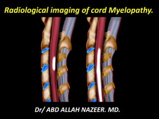
  • 1. Radiological imaging of cord Myelopathy. Dr/ ABD ALLAH NAZEER. MD.
  • 2. Introduction: The term myelopathy describes pathologic conditions that cause spinal cord, meningeal or peri meningeal space damage or dysfunction. Traumatic injuries, vascular diseases, infections and inflammatory or autoimmune processes may affect the spinal cord due to its confinement in a very small space. Spinal cord injuries usually have devastating consequences such as quadriplegia, paraplegia and severe sensory deficits. The history, an adequate neurological examination and the study of the cerebrospinal fluid (CSF) guide the diagnosis of spinal cord injuries. However, imaging is of great importance in order to home in on the diagnosis and classify the etiology appropriately. Many of the processes affecting the spinal cord may be reversible if recognized and treated early. The vast majority of spinal cord diseases may be treated medically, with surgical treatment reserved for compressive disorders, which constitute a neurological emergency.
  • 3. Definition and clinical picture It is important not to mistake myelopathy for myelitis. Although both terms refer to spinal cord compromise due to a pathological event, myelopathy has multiple etiologies, while myelitis is used to refer to inflammatory or infectious processes. Acute transverse myelopathy (includes non-inflammatory etiologies) and transverse myelitis have been used as synonyms in the published literature. Findings of spinal tract injuries, a certain degree of sensory dysfunction, or urinary retention, point to a spinal cord injury. There are certain conditions that may mimic myelopathy, such as myopathy or disorders of the neuromuscular junction, but the absence of a sensory deficit rules them out. Myelopathies may have a variable course and may manifest as a single event or as a multi-phasic or recurrent disease. The latter is rare and is usually secondary to demyelinating diseases, vascular malformations of the spinal cord, or systemic diseases. The central nervous system (CNS) damage may be monofocal as in transverse myelitis and optic neuritis, or multifocal as in acute disseminated encephalomyelitis (ADEM) (brain and spinal cord), neuromyelitis optica (optic nerve and spinal cord) and multiple sclerosis (MS) (any area of the neural axis).
  • 4. Spinal cord pathologies may be classified as acute, subacute/ intermittent or chronic, depending on the time course, the extent of the involvement, the clinical picture or syndrome, or the etiology. Patients with myelopathies but no evident lesions, or who present with multiple lesions of chronic appearance on magnetic resonance imaging, must be questioned about prior subtle symptoms. Acute onset that worsens within hours or days points to a spinal cord infarct or hemorrhage. When symptoms are recent, it is of paramount importance to rule out a surgical emergency. This requires immediate imaging work-up, ideally total spine magnetic resonance (MR). If there is evidence of spinal cord compression due to an acute lesion (epidural metastasis or abscess), definitive management is required in order to avoid damage or to adequately manage all other potential diagnoses. If the symptoms progress for more than three weeks, transverse myelitis is improbable, and other conditions must be considered, such as a spinal tumor, chronic compressive disease, dural arterio-venous fistula, metabolic disorder, sarcoidosis, or a degenerative process.
  • 5. Spinal cord syndromes present with typical signs and symptoms caused by a lesion of a specific tract in a specific location that may lead to the etiological diagnosis. They are classified as follows: • Complete spinal cord: involvement of all the tracts (trauma, compression or acute transverse myelitis). • Brown Séquard or hemi-spinal cord syndrome: ipsilateral cortico-spinal tract, posterior columns and contralateral spinothalamic tract (multiple sclerosis and compression). • Anterior spinal cord syndrome: anterior horns, corticospinal, spinothalamic and autonomic tracts (anterior spinal artery infarct and multiple sclerosis). Posterior spinal cord syndrome: posterior columns (vitamin B12 or copper deficiency). • Central syndrome: spino-thalamic crossing, cortico-spinal and autonomic tracts (syringomyelia, neuromyelitis optica). • Medullary cone: sacral emerging fibers (post-viral myelitis). • Cauda equina: cauda equina nerves (acute cytomegalovirus infection, polyradiculits and compression) • Tractopathies: selective disorders (vitamin B12 deficiency, paraneoplastic myelopathy and multiple sclerosis).
  • 6.
  • 7. Spinal cord abnormalities. Common causes in white and uncommon causes in yellow.
  • 8. Degenerative compressive myelopathy: Degenerative compressive myelopathy may be classified according to the compression site, as follows: • Anterior (disc protrusion or posterior osteophytes). • Anterolateral (Luschka joints). • Lateral (facet joints). • Posterior (ligamentum flavum). It may be caused by atlanto-axial instability, spinal canal stenosis due to cervical spondylolysis, cervical spinal fusion, myelomeningocele or epidural masses. Atlanto-axial instability is the primary cause of degenerative compressive myelopathy. It is found mainly in rheumatoid arthritis, followed by Down’s syndrome, Morquio’s syndrome or type IV mucopolysaccharidoses, skeletal dysplasia, ankylosing spondylitis and Lesh-Nyhan syndrome.
  • 9. Increased intensity of the spinal cord in C2 in the T2 weighted sequence due to compressive myelopathy secondary to rheumatoid arthritis.
  • 10. Increased intensity and thickening of the spinal cord from the bulbo-medullary junction down to C4 in the sagittal T2 sequence, due to compressive myelopathy in Morquio’s syndrome.
  • 11. MRI showing cervical spondylotic myelopathy.
  • 12. MRI showing cervical spondylotic myelopathy.
  • 13. Post-traumatic compressive myelopathy: Post-traumatic myelopathy is four times more frequent in males, in particular between 16 and 30 years of age. Motor vehicle accidents are the most common cause, accounting for 50% of the events, followed by violence (firearm or stab wounds), falls from heights, and sports injuries (diving, American football and horseback-riding). The most mobile segments are more often affected, in particularC5-C7 and T10-L2. Clinically, quadriplegia predominates in 30-40% of cases, and paraplegia occurs in 6-10%. MR imaging is of vital importance in approaching spinal cord trauma because it shows location, extension and severity very clearly, and also reveals edema and intramedullary bleeding. Some studies have shown that hemorrhage and longer hematomas are associated with a lower rate of motor recovery. Over the long term, CSF leaks, infections, cysts and syringomyelia may develop.
  • 14. a) Axial sequence with T2 gradient echo information. B) Sagittal section with T2 information in C7 showing diminished height and signal intensity with annulus protrusion in C5-C6 and C6-C7; there is also central and left subarticular protrusion of the annulus associated with annulus and ligament tear in C7, giving rise to central spinal hyperintensity due to compressive myelopathy resulting from nucleus pulposus herniation.
  • 15. Myelopathy due to traumatic cord compression. Myelopathy as a result of compression by a dorsally located epidural hemorrhage
  • 16. Myelopathy result of a metastasis.
  • 17. T2 weighted image with annulus protrusion in C4 and C5, giving rise to spinal cord hyperintensity due to traumatic compressive myelopathy.
  • 18. Abscess-related compressive myelopathy: Epidural abscesses are uncommon but they constitute a surgical emergency because they may progress rapidly within days and early diagnosis is difficult, leading to delayed treatment. They affect mainly men, with no specific age range, and the incidence has been shown to have increased in recent years. Risk factors are similar to those for spondylodiscitis, including diabetes mellitus, use of intravenous drugs, chronic renal failure, alcohol abuse, and immune deficiency. Human immunodeficiency virus has not been shown to be the cause of the increased incidence. It usually presents as subacute pain, fever (may be absent in subacute and chronic stages), increased local tenderness, progressive radiculopathy or myelopathy. The second phase of radicular irritation is followed by neurologic deficit (muscle weakness, abnormal sensation and incontinence) and then by paralysis in 34% of cases, and even death. Symptoms result from mechanical compression and, in some cases, from ischemia. Any segment of the spinal cord may be affected, but the most frequent are the thoracic and lumbar segments.
  • 19. a) Sagittal image with T1 information showing signal-intensity changes of the lower T10 endplate and upper T11 endplate, and of the corresponding disc, associated with hyperintensity and spinal cord thickening in that segment. b) Post-gadolinium STIR image showing thickening of the prevertebral soft tissues, the vertebral bodies and the disc from T10 to T11. These enhance with contrast, together with the thickened spinal cord due to myelopathy resulting from an epidural abscess.
  • 20. Cervical Pott’s and retropharyngeal abscess with cord myelopathy.
  • 21. Tumoral compressive myelopathy: Myelopathy may be the initial manifestation of a malignancy in up to 20% of cases where the only systemic symptom is weight loss. Tumors compressing the spinal cord may be divided into extradural and intradural. Extradural tumors may be classified as follows: • Benign: synovial cyst, osteoma, osteoblastoma, giant cell tumor, hemangioma, eosinophilic granuloma, schwannoma and meningioma. • Malignant: bone metastasis (are the cause of the most common myelopathy due to extradural spinal cord compression), multiple myeloma, lymphoma and chondrosarcoma. Intradural tumors are classified as follows: • Extra-spinal: neurofibroma, meningioma, lipoma, schwannoma and arachnoid cyst. • Intraspinal: astrocytoma, ependymoma, hemangioblastoma and metastasis. Forty per cent of patients present with radiculopathy and myelopathy associated with subacute dorsal pain that worsens in decubitus position. MRI may reveal the cause of the myelopathy and help guide the approach to the primary tumor.
  • 22. Post-gadolinium sagittal STIR image showing abnormal signal intensity of the bodies of T3 and T4, with a pathological fracture of T3 involving the epidural component and which compresses the spinal cord. There is gadolinium enhancement of T1, T3 and T4 and of the spinous processes, but no enhancement of the spinal cord due to metastatic disease. This causes tumor compression myelopathy.
  • 23. Postgadolinium sagittal STIR image showing spinal widening in C5 and C6, with enhancement of the spinal cord and the bodies of C7 and T1. There is also enhancement of the prevertebral soft tissues and of the cervical muscles due to myelopathy secondary to a high-grade glioma.
  • 26. Myelopathy of vascular origin: The spinal cord may be affected by compressive and non-compressive vascular diseases, of which the most common are malformations of the dural arteriovenous fistula type. In cases of vascular malformation, patients present with non-specific clinical findings, usually distal to the site of the disease. These diseases were classified by Riche in 1985 as follows: • Intraspinal arteriovenous malformations. • Perispinal arteriovenous malformations. • Spinodural arteriovenous fistulas. • Epidural arteriovenous malformations. • Paravertebral vascular malformations. • Vertebral hemangiomas. • Complex angiomatosis (Cobb’s syndrome, Osler-Weber-Rendu syndrome). • Cavernomas, telangiectasias and spinal venous angiomas (do not require endovascular treatment). In 2002, Spetzler proposed the following new classification • Neoplastic vascular lesions: hemangioblastoma and cavernous malformation. • Spinal aneurism. • Arteriovenous fistula: extradural and intradural. The latter includes ventral (small, medium and large) and dorsal (one or several feeding vessels) fistulas. • Arteriovenous malformations: extra-intradural and intradural (intraspinal, compact, diffuse and of the medullary cone).
  • 27. Sagittal sequence with T2 information of the medullary cone, showing serpent like, tortuous intra and extra-spinal images with absence of flow signal, associated with dorsal spine hyperintensity. b) Arteriogram confirming the presence of dural arteriovenous malformation with myelopathy of vascular origin.
  • 28. Fifteen-year old patient with neurologic deficit of sudden onset and normal laboratory tests. The sagittal sequence with T2 information shows a high-intensity signal anterior to the spinal cord suggesting a diagnosis of myelopathy due to ischemia.
  • 30. Myelopathy and dilated veins as a result of an AVF.
  • 31. Compressive myelopathy due to syringomyelia: Syringomyelia is a rare neurologic disorder, characterized by the slow development of fluid-filled areas extending along the spinal cord, and causing symptoms such as pain, weakness and stiffness of the back, shoulders and limbs. In the United States, it is more common among African-Americans. It may be related to congenital or acquired malformations. Chiari’s malformation is a congenital abnormality in which cerebellar amygdalas herniate through the foramen magnum in the spinal canal, with altered CSF flow. This causes headache, double vision, dizziness and muscle weakness of the upper limbs. Most non-traumatic forms of syringomyelia are due to Chiari malformation. The acquired causes of syringomyelia include trauma, tuberculosis-associated chronic arachnoiditis, and intraspinal tumors .
  • 32. a) Sagittal sequence with T2 information of the cervical spine showing Chiari malformation type II, absent corpus callosum and a tubular zone of high signal intensity in the spinal cord down from the bulbo-medullary junction, involving all the segments. b) Sagittal sequence with T2 information of the dorso-lumbar spine with myelopathy secondary to syringomyelia.
  • 33. Transverse myelitis: Acute transverse myelitis is a spinal disorder characterized by bilateral motor, sensory and autonomic abnormalities because it involves the spinothalamic and pyramidal tracts, the posterior columns and the anterior funiculus of one or more levels. Middle-aged adults are most frequently affected. A publication established the following criteria for transverse myelopathy: bilateral spinal cord dysfunction during a four-week period with a well-defined sensory level and no history of disease, where compression has been ruled out. Other criteria are proposed later for the differentiation between inflammatory and non-inflammatory transverse myelitis, and between idiopathic transverse myelitis and myelitis associated with a systemic or nervous system disease. These criteria are the following (5): • Sensory, motor or autonomic dysfunction of spinal origin. • Bilateral signs and symptoms. • Clearly defined sensory level. • Spinal inflammation (CSF pleocytosis or high immunoglobulin G levels, or gadolinium enhancement). • Maximum progression during a period ranging between four hours and four weeks.
  • 34. Twenty-year old patient with suspected multiple sclerosis. a) The sagittal sequence with T1 information shows widening of the spinal cord from C3 down to C7, with no other abnormal findings. b) Central high-signal area in T2 sequences due to acute transverse myelopathy.
  • 35. ATM on T2WI, CE-T1WI and STIR.
  • 37. Sixty-one-year-old female patient with neurological abnormalities over the past three days, but no significant history. Laboratory tests and the medullary biopsy were all normal. The sagittal sequence with T2 information showed discal and osteophytic changes of the vertebral bodies associated with bulging of the inferior annulus and thickening and hypersensitivity of the cervical spinal cord from the craniocervical junction down to C7. The clinical findings and the additional studies established the diagnosis of idiopathic myelopathy.
  • 38. MRI of representative cases of acute and subacute myelopathies
  • 39. Para-infectious myelopathy: Neurological damage in parainfectious myelopathy is caused directly by the infection, the immune reaction against the agent, and the reaction of the immune system. It is usually due to a blood-borne infection originating in the lungs, the skin, the skeletal, genitourinary or digestive systems. It presents with severe motor and sphincter dysfunction associated with fever, meningism and skin exanthema. The time period for the onset of myelitis after the infection is no different between infectious and post-infectious myelitis: five days for small-pox myelitis, ten days for mycoplasm and twelve days for herpes zoster myelitis. Possible etiologies include the following: • Viral: herpes, varicella zoster, EBV, CMV, HIV, dengue, influenza, measles, mumps, HTLV-1, enterovirus, Coxsackie B, hepatitis A and C, and polio. • Bacterial: mycoplasm, treponema pallidum, brucella, mycobacterium tuberculosis and borrelia. • Fungi: actinomyces, blastomyces, coccydiodes and aspergillus. • Parasites: Schistosoma, Cysticercosis, echinococcus and toxoplasma.
  • 40. a) Sagittal sequence with T1 information showing thickening of the spinal cord from the craniocervical junction down to the thoracic region. b) Sagittal sequence with T2 weighted image showing high central spinal signal. HTLV-1 infection is confirmed later by positive serology.
  • 41. Twenty-four-year old patient with congenital HIV and tuberculosis. The image shows alteration in the shape and signal intensity of the vertebral bodies of T10 and T11, of the disc and of the prevertebral soft tissues. It is associated with widening of the spinal cord with edema and contrast enhancement, and with fluid accumulations near the spinal canal in the post-gadolinium sagittal STIR sequence due to tuberculous myelopathy.
  • 42. Spinal Cord Schistosomiasis with cord myelopathy.
  • 44. Acute disseminated encephalomyelitis Acute disseminated encephalomyelitis (ADEM) is an uncommon inflammatory disease of the central nervous system, characterized by diffuse demyelination of the cerebral white matter and the spinal cord. It is more frequent in children and young adults. It has been associated with infection or vaccination, but this is not considered a criterion in clinical consensus. It is believed to be a single-phase disease with a good prognosis; however, recurrent forms make differentiation from MS difficult. ADEM has clinical manifestations that usually include encephalopathy but may also include focal or multifocal demyelinating inflammatory syndromes of the CNS such as optic neuritis and myelitis. For that reason, ADEM is a differential diagnosis for isolated demyelinating syndrome, which is a more common precursor of MS in adults. ADEM symptoms include rapidly progressing encephalopathy associated with seizures or multiple neurologic deficits. The spinal cord is affected in 11% to 28% of patients, generally in the thoracic and cervical segments. CSF findings are non-specific and oligoclonal bands are found in 65% of patients MRI shows bilateral symmetric multifocal white matter lesions, with or without damage of the grey matter, and extensive disease of several spinal segments with expansion. These lesions appear with low signal in T1 sequences, and well defined with a high signal in T2 sequences; gadolinium enhancement is variable. All patients with spinal involvement have brain damage
  • 45. Twenty-eight-year old patient presenting with sudden neurologic decline. a) Coronal FLAIR sequence of the brain showing hyperintensity in the bulbo-medullary junction. b) Axial image with T2 information in the bulbo-medullary junction with anterior hyperintensity related to myelopathy due to acute disseminated encephalopathy.
  • 47. ADEM seen in children.
  • 48. Multiple sclerosis: MS is a chronic demyelinating inflammatory CNS disease. It is common in Europe, the United States, Canada, New Zealand and Australia, but it is rare in Asia, the tropics and the subtropical regions. In high-risk populations, the incidence is one out of every 200 women. The female-to-male ratio varies between 1.5 and 2.5. The age of onset of symptoms varies by region; however the incidence is low in children, increases in adolescence and peaks between 25 and 35 years of age, after which it starts to decline. The etiology remains unknown, although environmental, viral and immune-mediated factors in genetically susceptible patients are thought to be the culprits . The strongest risk factor is family history. Approximately 80-85% of patients present with a relapsing picture, with symptoms that last for several days and improve over the course of weeks. In 15% of patients, the disease is progressive from the start. It is the most studied of all acute myelopathies, and its effects range from irreversible tissue loss to partial demyelination where there can be remyelination and repair. There is spinal cord involvement in more than 90% of patients. It may present in the form a cervicodorsal asymmetric transverse myelitis with sensory symptoms.
  • 49. Twenty-eight-year old patient diagnosed with multiple sclerosis in December 2010 with progression in time and space. a) Sagittal sequence with T2 information showing high spinal signal in C4. b) High-signal area in C4 shown on a T2 gradient echo axial view due to myelopathy secondary to multiple sclerosis.
  • 50.
  • 51. MS in the cord.
  • 53. Acute exacerbation of spinal MS.
  • 54. Neuromyelitis optica or Devic’s syndrome: Neuromyelitis optica is defined as the concomitant presentation of myelitis and optic neuritis. This combination occurs in MS, ADEM, systemic lupus erythematosus and Sjögren’s syndrome. It is also found in association with viral and bacterial infections. Neuromyelitis optica is an immune CNS demyelinating condition that affects the spinal cord and the optic nerves. It is often mistaken with MS, although clinical, radiological and immunopathological tests suggest that they are different. The identification of the specific antigen of the neuromyelitis optica immunoglobulin G/aquaporin 4 antibody implies humoral immunity, which makes it different from MS. It is an uncommon disorder among the Western population, with an incidence of 0.4 per million people per year, representing one out of every 200 patients with demyelinating disease. In Asia, the Caribbean and South America, the incidence is higher, pointing to genetic mechanisms. In all populations, females with a mean age of 40 are predominantly affected, in a 3:1 ratio.
  • 55. Forty-four-year old patient with demyelinating disease and proven neurological decline. a) Diminished signal intensity in the upper segments of the cervical spinal cord in the sagittal T1 sequence. b) Sagittal sequence with T2 information showing hyperintensity from the bulbo-medullary junction down to C6 and C7, due to neuromyelitis optica myelopathy.
  • 56. NMO presenting with neuritis optica.
  • 57. Neuromyelitis optica with cord myelopathy.
  • 59. Myelopathy due to systemic disease: Myelitis associated with a systemic disease has been rarely described in the literature. It has been associated with systemic lupus erythematosus (SLE), Sjögren’s syndrome, scleroderma, Behçet disease, and sarcoidosis. It has been estimated that the frequency of myelitis in patients with SLE is 3%, but it is unknown in Sjögren’s syndrome. Myelitis usually occurs in the first year of the disease and may be its first manifestation. The hypothesis about the pathophysiology is still a subject for debate, and the most accepted is a vascular mechanism secondary to ischemic lesions. Women are more frequently affected than men, in an 8:1 ratio. CNS involvement in SLE is often found in relation with the antiphospholipid syndrome with anticardiolipin antibodies. The clinical symptoms often include transverse myelitis with severe motor and sensory dysfunction. CSF may be normal or show plecytosis, and although oligoclonal bands are rare, they may be present. MRI findings have been studied more in SLE than in Sjögren’s syndrome. Moreover, it has been found that the central high-signal spinal lesion in T2 sequences, CNS sarcoidosis may occur in isolation in the form of a myelopathy. The definitive diagnosis requires biopsy evidence of non-caseifying granulomatous inflammation of the CNS or any other compromised organ.
  • 60. a) Sagittal sequence with T2 information showing spinal cord thickening and hyperintensity from C4 down to T2. b) Post-gadolinium STIR image showing enhancement due to SLE-associated myelopathy.
  • 62. Spinal cord MRI. T2-weighted sequences. Extended hypersignal in a patient with a Sjögren's syndrome (A) localized in the centromedullary territory (B).
  • 63. Post-radiation or electric damage: Neurotoxicity is a known complication of high-dose radiation. The deep white matter is the most affected since it comprises the cortex and the subcortical arcuate fibers. There are three forms of lesions: acute (weeks or months), early late and late (six months to two years). The latter may be irreversible, progressive and, on occasions, fatal; however, it may resolve spontaneously in some cases. Radiation myelopathy is a devastating complication of radiation therapy. It is a rare cause of acute myelopathy, accounting for only 2% of complications, and it is suggested in cases where there is a history of exposure to head and neck radiation (even longer than ten years), with a dose greater than 4,000 rads. It is an irreversible process with no effective treatment. It may have an early manifestation ten to sixteen weeks into radiotherapy, or a late manifestation, and may resolve spontaneously between two and nine months after onset. In the early stages, there is evidence of edema or spinal enhancement and, in late cases, spinal atrophy is observed It manifests as a transient sensory loss, progressive chronic myelopathy, acute transverse myelitis, or local amyotrophy. The transient sensory loss gives an electric-shock sensation when the neck is flexed forward (Lhermitte sign) and it resolves within two and thirty-six weeks. In chronic progressive myelopathy, it presents like a Brown Séquard syndrome lasting between three months and five years.
  • 64. Patient with a history of radiotherapy due to esophageal cancer who complains of paresthesias and discreet loss of strength in the lower limbs, and Lhermitte sign. a) Sagittal T2 image of the dorsal spine with discreet fusiform thickening of the spinal cord showing high intensity signal and post-radiotherapy bone marrow changes. b) Follow-up MRI after 18 months with evidence of partial regression of post- -radiation myelopathy.
  • 65. Spinal cord MRI. Extended hypersignal in T2-weighted sequences in the cervical cord following laryngeal radiation with a swelling of the cord (A). MRI after 4 months of follow-up shows normal cord signal in T2-weighted sequences, but an atrophic cord in T1-weighted sequences (B).
  • 66. Radiation myelopathy. Paraneoplastic tractopathy. Axial T2 sections through the cord of a 69-year-old woman with melanoma and high titers of amphiphysin-immunoglobulin (Ig)G. Arrow points to hyperintensity in the region of the corticospinal tracts.
  • 67. Subacute combined degeneration: Combined subacute degeneration is a complication of vitamin B12 deficiency, associated with pernicious anemia. This deficiency may be related to parietal-cell auto-antibodies or the intrinsic factor required for vitamin B12 binding. There is a genetic deficiency of transcobalamin 2 (cobalamin transporter protein). The complete transcobalamin 2 deficiency is a recessive autosomal condition characterized by normal concentrations of vitamin B12 with severe infantile megaloblastic anemia associated with neurologic damage . The clinical picture presents as a slowly-progressing spastic paraparesis with distal proprioceptive loss and symmetrical dysesthesias. This clinical picture is due to axonal loss and inferior cervical and thoracic spinal demyelination, generally in the posterior and anterolateral columns, that cannot be explained by compression or inflammation on MRI. The absence of anemia with or without macrocytosis does not rule out a vitamin B12 deficiency diagnosis. MRI shows high signal intensity of the posterior columns in T2 sequences, without contrast enhancement. Imaging improvement correlates with clinical improvement . The mean time to diagnosis since the onset of neurological symptoms due to vitamin B12 deficiency is approximately on year, with a range that extends to four years.
  • 68. a) Sagittal T2 sequence showing hyperintense signal in the dorsal spine. b) Axial T2 image confirming spinal damage predominantly in the posterior horns. Low concentrations of vitamin B12 were found, pointing to the diagnosis of myelopathy due to vitamin B12 deficiency. c) and d) Follow-up MRI performed one year after the diagnosis, showing no evidence of abnormalities.
  • 69. Subacute combined degeneration with hyperintensity at the posterior column.
  • 70. Acute paraneoplastic or necrotizing myelitis: Paraneoplastic myelopathy is a rare disease. It may occur before the cancer is detected. Several antibodies have been associated with subacute myelopathies, in general with lung, breast, thyroid, ovarian and prostate cancer, and Hodgkin’s lymphoma. The lesion often involves the thoracic spinal cord that shows a high-intensity signal in T2 sequences and gadolinium enhancement. There is increased protein concentration in the CSF. Myelopathy with normal MRI: In some cases of acute myelopathy, the MR image is normal. There are several explanations for this: 1) the syndrome is not a myelopathy (Guillain-Barré, inflammatory radiculopathy); 2) the picture may not be acute but rather a decompensated existing myelopathy (Friedreich’s ataxia, motor neuron disease, vitamin B12 deficiency, HIV or HTLV-1 myelopathy); and 3) MRI performed during the convalescence period.
  • 71. Large B- cell lymphoma showing cord edema progressing cranially to involve the cervical spine. (b) MRI demonstrated edema involving the conus medullaris.
  • 72. Large B- cell lymphoma showing cord edema progressing cranially to involve the cervical and thoracic cord.
  • 73.
  • 74. Conclusion. Myelopathy is a broad term that refers to spinal cord involvement of multiple etiologies. Spinal cord diseases often have devastating consequences, ranging from quadriplegia and paraplegia to severe sensory deficits due to its confinement in a very small area. Many of these diseases are potentially reversible if they are recognized on time, hence the importance of recognizing the significance of magnetic resonance imaging when approaching a multifactorial disease considered as one of the most critical neurological emergencies, where prognosis depends on an early and accurate diagnosis.