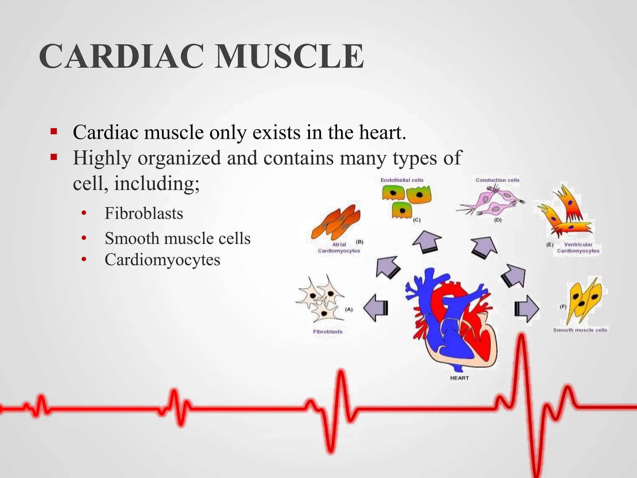
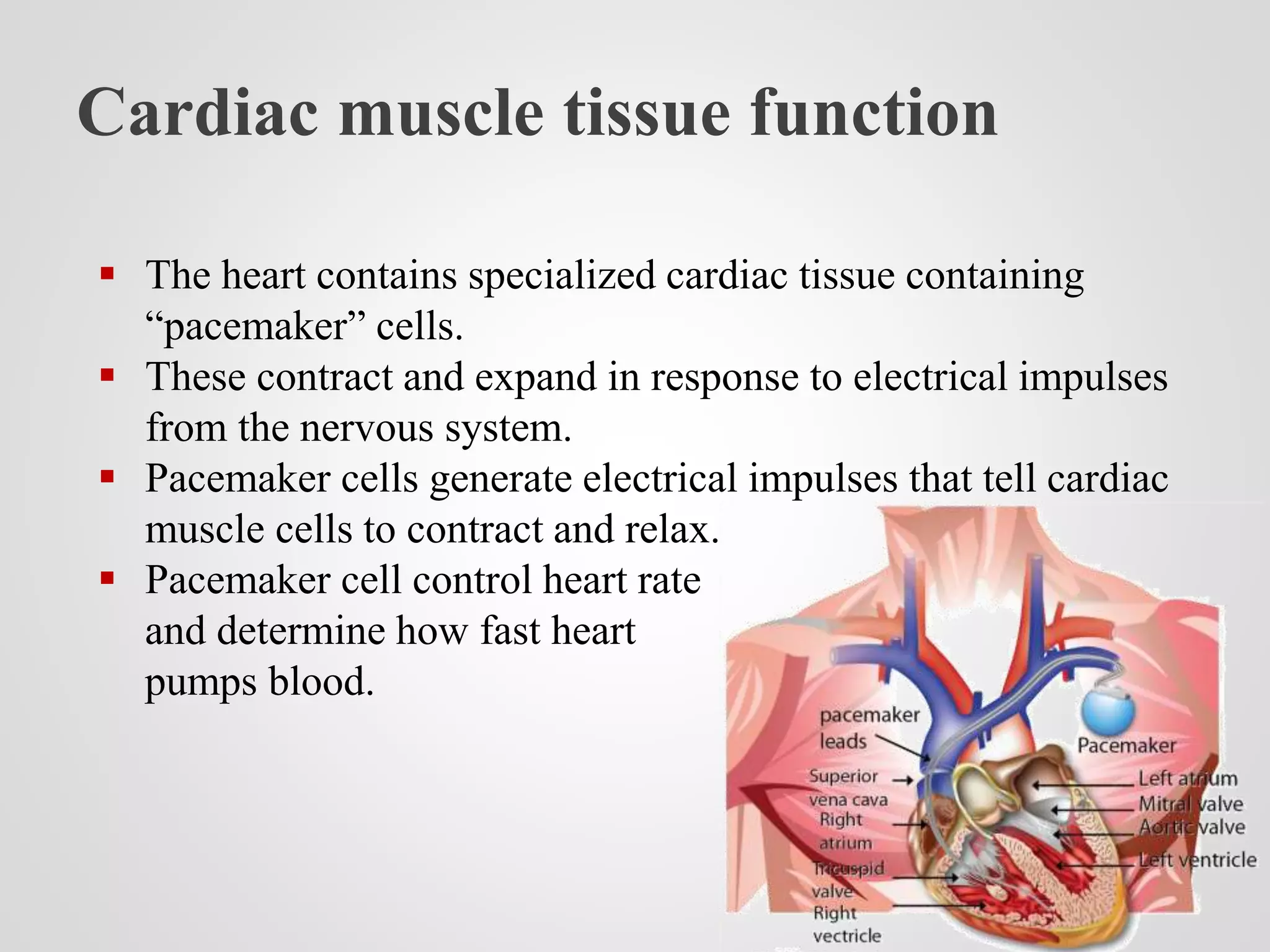
Submitted By:
Fatima Adil
Roll Number
18371514-106
Submitted To:
Dr. Syed Zakir Hussain Shah
Topic:
Cardiac Muscles
MUSCLE TISSUES
 The human body contains three different kinds of
muscle tissue:
• Skeletal muscle tissue
• smooth muscle tissue
• Cardiac muscle tissue
CARDIAC MUSCLE
 Cardiac muscle only exists in the heart.
 Highly organized and contains many types of
cell, including;
• Fibroblasts
• Smooth muscle cells
• Cardiomyocytes
What are heart muscles made of?
 Intercalated discs
• Small connections that join cardiac
muscle cells to each other.
 Gap junctions
• Part of the intercalated discs.
• Allows the muscle to contract in a
coordinated way.
What are heart muscles made of?
 Desmosomes
• Found within intercalated discs.
• Hold the cardiac muscle fibers together during a contraction.
 Nucleus
• Control center of a cell.
• It contains all of the cell’s genetic material.
STRUCTURE
 Appear striated or striped due to myosin and actin filaments.
 Myosin filament connects to two actin filaments forming
sarcomere.
 Cardiac muscle cells contain
• One nucleus, but some have two.
• Mitochondria
Pathway of Cardiac Muscle Contraction
 Pacemaker cells initiate the action potential.
 This activates calcium channels in the T-tubules, resulting in
an influx of calcium ions into the cardiomyocyte.
 Calcium binds to troponin-C which moves the troponin
complex away.
 Removal of the troponin complex frees the actin to be
bound by myosin and initiates contraction.
Pathway of Cardiac Muscle Contraction
 Myosin head binds to ATP and pulls the actin filaments
toward the center of the sarcomere, contracting the muscle.
 Intracellular calcium is then removed dropping intracellular
calcium concentration.
 troponin complex returns to its inhibiting, ending
contraction.
HEART LAYERS
 Heart consist of three layers:
• Epicardium- The outer layer of the heart wall.
• Myocardium- The middle layer.
• Endocardium- The inner layer.
Cardiac muscle tissue function
 The heart contains specialized cardiac tissue containing
“pacemaker” cells.
 These contract and expand in response to electrical impulses
from the nervous system.
 Pacemaker cells generate electrical impulses that tell cardiac
muscle cells to contract and relax.
 Pacemaker cell control heart rate
and determine how fast heart
pumps blood.
Cardiomyopathy
 Cardiomyopathy is one of the main conditions that can affect
cardiac muscle tissue.
 It’s a disease that makes it harder for your heart to pump
blood.
 TYPES:
 Hypertrophic cardiomyopathy-
• The cardiac muscles enlarge and thicken
 Restrictive cardiomyopathy-
• The ventricles become stiff.
 Dilated cardiomyopathy-
• Ventricles become larger & weaker
 Arrhythmogenic right ventricular dysplasia-
• The cardiac muscle tissue of right ventricle is replaced with fatty or
fiber-rich tissue.
• This can lead to arrhythmia, which refers to an abnormal heart rate or
rhythm.
 Symptoms of cardiomyopathy:
• Difficulty breathing or shortness of breath
• Fatigue
• Swelling of the legs, ankles, and feet
• Inflammation in the abdomen or neck
• Irregular heartbeat
• Heart murmurs
• Dizziness or lightheadedness
 Causes:
• A family history of cardiomyopathy or heart failure
• High blood pressure
• Obesity
• Heavy alcohol consumption
• Use of certain recreational drugs
• Past heart attacks or heart infections
 Factors that can increase a person’s risk of
cardiomyopathy include:
• Diabetes
• Thyroid disease
• Coronary heart disease
• Heart attack
• Chronic high blood pressure
• Viral infections that affect the heart muscle
• Valvular disease of the heart
• Heavy alcohol consumption
• A family history of cardiomyopathy
How does exercise impact cardiac muscle
tissue?
 Exercise can strengthen your cardiac muscle.
 Exercise can also help reduce risk of developing
cardiomyopathy and make your heart work more efficiently.
 Some examples of aerobic exercises include:
• Running or jogging
• Walking or hiking
• Cycling
• Swimming
The bottom line
 Cardiac muscle tissue is one of the three types of muscle in
your body.
 It’s only found in your heart.
 Involved in coordinated contractions that keep your heart
beating.
 To keep your cardiac muscle working efficiently try to get in
some sort of exercise more days of the week than not.
THANK YOU

Cardiac muscle

  • 1.
    Submitted By: Fatima Adil RollNumber 18371514-106 Submitted To: Dr. Syed Zakir Hussain Shah Topic: Cardiac Muscles
  • 2.
    MUSCLE TISSUES  Thehuman body contains three different kinds of muscle tissue: • Skeletal muscle tissue • smooth muscle tissue • Cardiac muscle tissue
  • 3.
    CARDIAC MUSCLE  Cardiacmuscle only exists in the heart.  Highly organized and contains many types of cell, including; • Fibroblasts • Smooth muscle cells • Cardiomyocytes
  • 4.
    What are heartmuscles made of?  Intercalated discs • Small connections that join cardiac muscle cells to each other.  Gap junctions • Part of the intercalated discs. • Allows the muscle to contract in a coordinated way.
  • 5.
    What are heartmuscles made of?  Desmosomes • Found within intercalated discs. • Hold the cardiac muscle fibers together during a contraction.  Nucleus • Control center of a cell. • It contains all of the cell’s genetic material.
  • 6.
    STRUCTURE  Appear striatedor striped due to myosin and actin filaments.  Myosin filament connects to two actin filaments forming sarcomere.  Cardiac muscle cells contain • One nucleus, but some have two. • Mitochondria
  • 7.
    Pathway of CardiacMuscle Contraction  Pacemaker cells initiate the action potential.  This activates calcium channels in the T-tubules, resulting in an influx of calcium ions into the cardiomyocyte.  Calcium binds to troponin-C which moves the troponin complex away.  Removal of the troponin complex frees the actin to be bound by myosin and initiates contraction.
  • 8.
    Pathway of CardiacMuscle Contraction  Myosin head binds to ATP and pulls the actin filaments toward the center of the sarcomere, contracting the muscle.  Intracellular calcium is then removed dropping intracellular calcium concentration.  troponin complex returns to its inhibiting, ending contraction.
  • 9.
    HEART LAYERS  Heartconsist of three layers: • Epicardium- The outer layer of the heart wall. • Myocardium- The middle layer. • Endocardium- The inner layer.
  • 10.
    Cardiac muscle tissuefunction  The heart contains specialized cardiac tissue containing “pacemaker” cells.  These contract and expand in response to electrical impulses from the nervous system.  Pacemaker cells generate electrical impulses that tell cardiac muscle cells to contract and relax.  Pacemaker cell control heart rate and determine how fast heart pumps blood.
  • 11.
    Cardiomyopathy  Cardiomyopathy isone of the main conditions that can affect cardiac muscle tissue.  It’s a disease that makes it harder for your heart to pump blood.  TYPES:  Hypertrophic cardiomyopathy- • The cardiac muscles enlarge and thicken  Restrictive cardiomyopathy- • The ventricles become stiff.
  • 12.
     Dilated cardiomyopathy- •Ventricles become larger & weaker  Arrhythmogenic right ventricular dysplasia- • The cardiac muscle tissue of right ventricle is replaced with fatty or fiber-rich tissue. • This can lead to arrhythmia, which refers to an abnormal heart rate or rhythm.
  • 13.
     Symptoms ofcardiomyopathy: • Difficulty breathing or shortness of breath • Fatigue • Swelling of the legs, ankles, and feet • Inflammation in the abdomen or neck • Irregular heartbeat • Heart murmurs • Dizziness or lightheadedness
  • 14.
     Causes: • Afamily history of cardiomyopathy or heart failure • High blood pressure • Obesity • Heavy alcohol consumption • Use of certain recreational drugs • Past heart attacks or heart infections
  • 15.
     Factors thatcan increase a person’s risk of cardiomyopathy include: • Diabetes • Thyroid disease • Coronary heart disease • Heart attack • Chronic high blood pressure • Viral infections that affect the heart muscle • Valvular disease of the heart • Heavy alcohol consumption • A family history of cardiomyopathy
  • 16.
    How does exerciseimpact cardiac muscle tissue?  Exercise can strengthen your cardiac muscle.  Exercise can also help reduce risk of developing cardiomyopathy and make your heart work more efficiently.  Some examples of aerobic exercises include: • Running or jogging • Walking or hiking • Cycling • Swimming
  • 17.
    The bottom line Cardiac muscle tissue is one of the three types of muscle in your body.  It’s only found in your heart.  Involved in coordinated contractions that keep your heart beating.  To keep your cardiac muscle working efficiently try to get in some sort of exercise more days of the week than not.
  • 18.