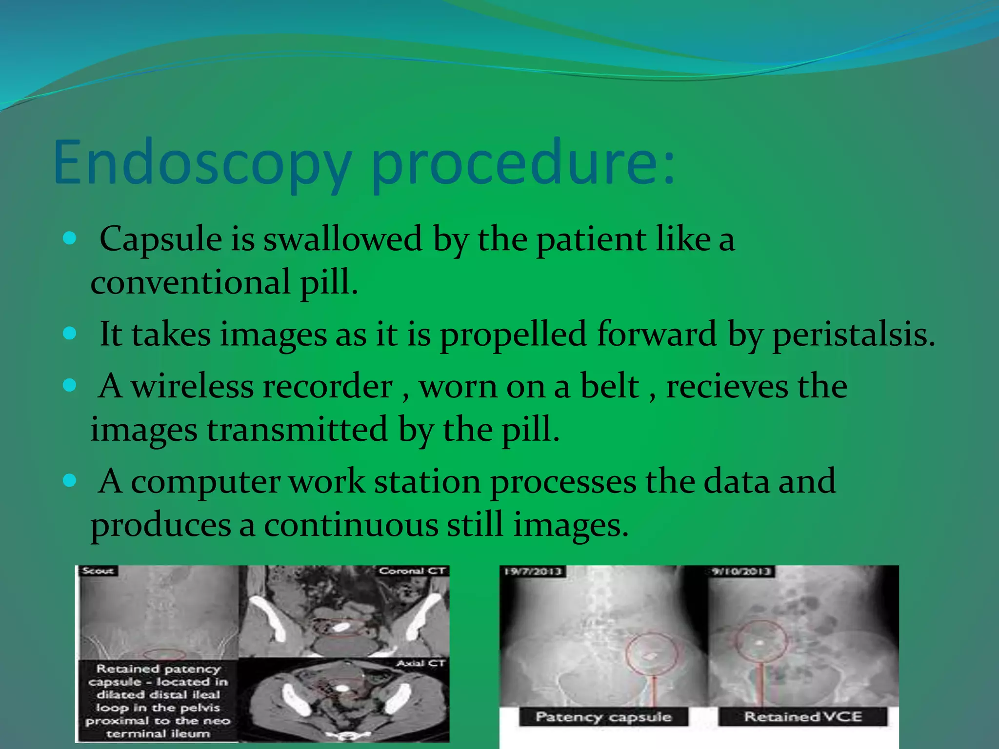
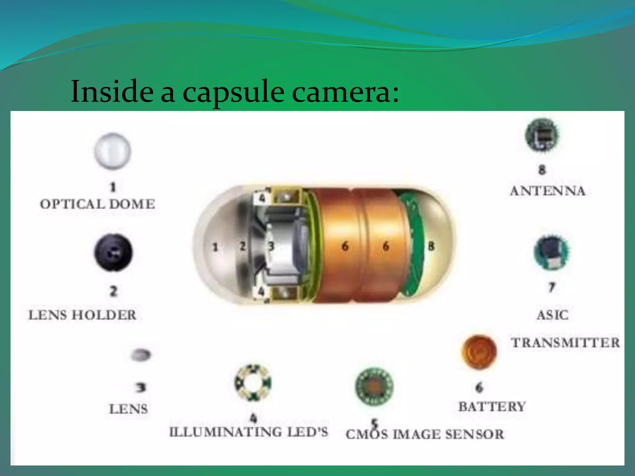
By
SATHISHKUMAR G
(sathishsak111@gmail.com)
What is capsule endoscopy?
 Capsule endoscopy leads the doctors examine the lining of the middle part
of one’s gastrointestinal tract,which include the three portion of the small
intestine(duodenum , jejunum , ileum).
 Doctor will give a pill sized video camera for a patient to swallow.
 This camera has its own light source and takes picture of small intestine as it
passess through .These pictures are send to a small recording device patient
have to wear on this body.
About:
 Capsule endoscopy was first used in humans in 1999.
 The first publication on capsule endoscopy was
published in nature in 2000.
How does capsule endoscopy work:
 Eight aerial leads that are attached around the patients
abdomen collecting data.
 Capsule ingested as any other capsule.
 Patient can drink clears immediately,but no solid food
for three hours .
 Attached to the leads is the recorder and the patient
should report back if it stops recording for any reason.
 Belt and aerial should be worn for 8 hours after
swallowing or until the recorder stops recording.
 Recorder and aerials are returned , but the capsule is
disposable!
 Images are downloaded and processed prior to
interpretation.
Endoscopy procedure:
 Capsule is swallowed by the patient like a
conventional pill.
 It takes images as it is propelled forward by peristalsis.
 A wireless recorder , worn on a belt , recieves the
images transmitted by the pill.
 A computer work station processes the data and
produces a continuous still images.
Description:
 The capsule consists of seven optical fibres
 One for elimination and the rest six for collecting
light.
 The tip will illuminate red , green & blue laser light
helping in visuality.
 All this processing together combined will give us
two-dimensional picture helping in diagnosis.
 The images can be retrieved from the recording device
worn around patient waist as a belt.
Types of Endoscopic capsules:
Inside a capsule camera:
Uses:
 Crohn’s Disease.
 Malabsorption Disorders.
 Tumors of the small intestine & Vascular Disorders.
 Ulcerative colitis.
Limitations:
 Slow Gastric/Intestinal motility.
 Poorer quality of images as compared to Fiberoptic
scopes.
 The position of the capsule can not be accurately
controlled.
 Potentially obstructed views.
 Morbidly obese patients.
 Interpretation of results are very observer dependent.
 Findings may be of unknown significance or relevence.
CAPSULE ENDOSCOPY
CAPSULE ENDOSCOPY

CAPSULE ENDOSCOPY

  • 1.
  • 2.
    What is capsuleendoscopy?  Capsule endoscopy leads the doctors examine the lining of the middle part of one’s gastrointestinal tract,which include the three portion of the small intestine(duodenum , jejunum , ileum).  Doctor will give a pill sized video camera for a patient to swallow.  This camera has its own light source and takes picture of small intestine as it passess through .These pictures are send to a small recording device patient have to wear on this body.
  • 3.
    About:  Capsule endoscopywas first used in humans in 1999.  The first publication on capsule endoscopy was published in nature in 2000.
  • 4.
    How does capsuleendoscopy work:  Eight aerial leads that are attached around the patients abdomen collecting data.  Capsule ingested as any other capsule.  Patient can drink clears immediately,but no solid food for three hours .  Attached to the leads is the recorder and the patient should report back if it stops recording for any reason.
  • 5.
     Belt andaerial should be worn for 8 hours after swallowing or until the recorder stops recording.  Recorder and aerials are returned , but the capsule is disposable!  Images are downloaded and processed prior to interpretation.
  • 6.
    Endoscopy procedure:  Capsuleis swallowed by the patient like a conventional pill.  It takes images as it is propelled forward by peristalsis.  A wireless recorder , worn on a belt , recieves the images transmitted by the pill.  A computer work station processes the data and produces a continuous still images.
  • 7.
    Description:  The capsuleconsists of seven optical fibres  One for elimination and the rest six for collecting light.  The tip will illuminate red , green & blue laser light helping in visuality.  All this processing together combined will give us two-dimensional picture helping in diagnosis.  The images can be retrieved from the recording device worn around patient waist as a belt.
  • 8.
  • 9.
  • 19.
    Uses:  Crohn’s Disease. Malabsorption Disorders.  Tumors of the small intestine & Vascular Disorders.  Ulcerative colitis.
  • 21.
    Limitations:  Slow Gastric/Intestinalmotility.  Poorer quality of images as compared to Fiberoptic scopes.  The position of the capsule can not be accurately controlled.  Potentially obstructed views.  Morbidly obese patients.  Interpretation of results are very observer dependent.  Findings may be of unknown significance or relevence.