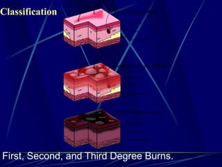
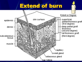
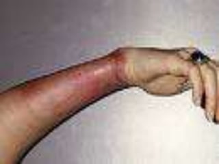
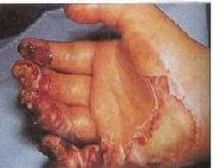
Burn
Burns are skin injuries resulting
from heat, Flame, electric current,
chemicals, friction, or excessive
sunlight exposure. Based on the
standard depth of injury.
skin
Anatomy of skin
1. Human skin is divided into two layers
a. epidermis (outermost)
b. dermis ( innermost)
2. separated by basement membrane (BM)
3. A layer of loose connective tissue
beneath the dermis called
hypodermis, or subcutis
The Skin Overview
The Human Skin
1. Average about 3000 square inches
or 2 square meters.
2. Weighs about 6 pounds (15% of BW)
3. The largest organ
4. Receives 1/3 of the body’s blood volume
5.Varies in thickness (0.5mm~6mm)
6. constantly being renewed (26~42 days).
The most important function of the
skin is to act as a barrier against
infection. The skin prevents loss of
body fluids, thus preventing
dehydration. The skin also regulates
the body temperature by controlling
the amount of evaporation of fluids
from the sweat glands. The skin serves
a cosmetic effect by giving the body
shape.
When the skin is burned, these functions
are impaired or lost completely. The severity
of the skin injury depends upon the size of
the injury, depth of the wound, part of the
body injured, age of the patient, and past
medical history. Because of the importance
of the skin, it becomes clear that injury can
be traumatic and life threatening. Recovery
from burn injury involves four major
aspects: burn wound management, physical
therapy, nutrition, and emotional support.
Pathophysiology
Pathophysiologic changes result from
major burns during the initial burn-
shock period. Changes include tissue
hypo perfusion and organ hypo
function secondary to decreased
cardiac output, followed by a
hyperdynamic and hyper metabolic
phase.
Assessment
Rule of nine
First, Second, and Third Degree Burns.
Classification
Assessment
with all trauma victims, a primary and
secondary trauma survey, including
assessment of airway, breathing, and
circulation as well as vital signs, is done.
Other assessment parameters specific to
the burn injury focus on extent and
severity of burn injury.
Extend of burn
First-degree (superficial)
burns
Superficial tissue destruction involving the
epidermis only.
Local pain and erythema; blisters are absent
for about 24 hours.
Mild to absent systemic response.
Rapid healing (normally 3 to 5 days) without
scarring.
First-degree burns generally do not require
treatment, except in the case of large burns
of infants or elderly persons.
Second-degree (partial-thickness)
burns
Tissue destruction involving the epidermis
and part of the dermis.
Skin appearing red to pale and moist
Formation of wet, thin-walled blisters
immediately after injury.
Intact tactile and pain sensors
Healing in 21 to 28 days with variable
amount of scarring.
Third-degree (full-thickness)
burns
Tissue destruction involving the epidermis,
dermis, and underlying subcutaneous
tissue.
Injury appearing white, cherry red, or black;
the injury may or may not contain deep
blisters or visible thrombosed veins.
Dry, hard, leathery appearance due to loss
of epidermal elasticity
Painless to touch because of destruction of
all superficial nerve ending in skin.
Nursing diagnosis
Impaired Gas Exchange related to
inhalation injury
Ineffective Breathing Pattern related to
circumferential chest burn, upper airway
obstruction.
Risk for Infection related to loss of skin
barrier and altered immune response
Body Image Disturbance related to
cosmetic and functional sequelae of burn
wound.
Nursing interventions
Stop the burning process if possible
Remove all clothing and jewelry
Ensure patent airway
Prepare for incubation
Cannulate two veins
Cool the burn for several minutes. Do not use
ice.
Remove restrictive objects.
Cover the wound with sterile dressing or a
clean, dry cloth.
Prevent shock by initiating I.V. fluid
therapy immediately.
Ensure that the client avoids oral
intake and is placed in an upright
position t prevent aspiration of
vomitus.
Transport the client to the nearest
emergency medical center. Note the
time of burn (needed for
resuscitation).
Administer 100% Oxygen until the arterial blood gas
determination demonstrates adequate oxygenation,
and perform frequent neuralgic assessment until
hypoxia resolves.
Support pulmonary function through early intubation
and volume ventilator-assisted respiration with optimal
positive end expiratory pressure, large tidal volume,
and lowest possible inspired oxygen concentration.
Provide pain relief.
Monitor acid-base balance and electrolyte levels.
Provide appropriate fluid resuscitation, based on the
parkland formula.
Thanks for your
keen concentration!

Burns

  • 3.
    Burn Burns are skininjuries resulting from heat, Flame, electric current, chemicals, friction, or excessive sunlight exposure. Based on the standard depth of injury.
  • 4.
  • 5.
    Anatomy of skin 1.Human skin is divided into two layers a. epidermis (outermost) b. dermis ( innermost) 2. separated by basement membrane (BM) 3. A layer of loose connective tissue beneath the dermis called hypodermis, or subcutis
  • 6.
  • 7.
    The Human Skin 1.Average about 3000 square inches or 2 square meters. 2. Weighs about 6 pounds (15% of BW) 3. The largest organ 4. Receives 1/3 of the body’s blood volume 5.Varies in thickness (0.5mm~6mm) 6. constantly being renewed (26~42 days).
  • 10.
    The most importantfunction of the skin is to act as a barrier against infection. The skin prevents loss of body fluids, thus preventing dehydration. The skin also regulates the body temperature by controlling the amount of evaporation of fluids from the sweat glands. The skin serves a cosmetic effect by giving the body shape.
  • 11.
    When the skinis burned, these functions are impaired or lost completely. The severity of the skin injury depends upon the size of the injury, depth of the wound, part of the body injured, age of the patient, and past medical history. Because of the importance of the skin, it becomes clear that injury can be traumatic and life threatening. Recovery from burn injury involves four major aspects: burn wound management, physical therapy, nutrition, and emotional support.
  • 12.
    Pathophysiology Pathophysiologic changes resultfrom major burns during the initial burn- shock period. Changes include tissue hypo perfusion and organ hypo function secondary to decreased cardiac output, followed by a hyperdynamic and hyper metabolic phase.
  • 14.
  • 15.
  • 16.
    First, Second, andThird Degree Burns. Classification
  • 17.
    Assessment with all traumavictims, a primary and secondary trauma survey, including assessment of airway, breathing, and circulation as well as vital signs, is done. Other assessment parameters specific to the burn injury focus on extent and severity of burn injury.
  • 18.
  • 19.
    First-degree (superficial) burns Superficial tissuedestruction involving the epidermis only. Local pain and erythema; blisters are absent for about 24 hours. Mild to absent systemic response. Rapid healing (normally 3 to 5 days) without scarring. First-degree burns generally do not require treatment, except in the case of large burns of infants or elderly persons.
  • 21.
    Second-degree (partial-thickness) burns Tissue destructioninvolving the epidermis and part of the dermis. Skin appearing red to pale and moist Formation of wet, thin-walled blisters immediately after injury. Intact tactile and pain sensors Healing in 21 to 28 days with variable amount of scarring.
  • 23.
    Third-degree (full-thickness) burns Tissue destructioninvolving the epidermis, dermis, and underlying subcutaneous tissue. Injury appearing white, cherry red, or black; the injury may or may not contain deep blisters or visible thrombosed veins. Dry, hard, leathery appearance due to loss of epidermal elasticity Painless to touch because of destruction of all superficial nerve ending in skin.
  • 25.
    Nursing diagnosis Impaired GasExchange related to inhalation injury Ineffective Breathing Pattern related to circumferential chest burn, upper airway obstruction. Risk for Infection related to loss of skin barrier and altered immune response Body Image Disturbance related to cosmetic and functional sequelae of burn wound.
  • 26.
    Nursing interventions Stop theburning process if possible Remove all clothing and jewelry Ensure patent airway Prepare for incubation Cannulate two veins Cool the burn for several minutes. Do not use ice. Remove restrictive objects. Cover the wound with sterile dressing or a clean, dry cloth.
  • 27.
    Prevent shock byinitiating I.V. fluid therapy immediately. Ensure that the client avoids oral intake and is placed in an upright position t prevent aspiration of vomitus. Transport the client to the nearest emergency medical center. Note the time of burn (needed for resuscitation).
  • 29.
    Administer 100% Oxygenuntil the arterial blood gas determination demonstrates adequate oxygenation, and perform frequent neuralgic assessment until hypoxia resolves. Support pulmonary function through early intubation and volume ventilator-assisted respiration with optimal positive end expiratory pressure, large tidal volume, and lowest possible inspired oxygen concentration. Provide pain relief. Monitor acid-base balance and electrolyte levels. Provide appropriate fluid resuscitation, based on the parkland formula.
  • 30.
    Thanks for your keenconcentration!