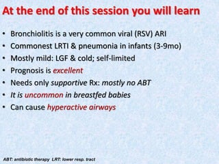
Call Girls Coimbatore Just Call 9907093804 Top Class Call Girl Service Available
Bronchiolitis
1.
2.
3.
4.
5.
6.
7.
8.
9. Bronchiole: A tiny continuation of the bronchi & connects to
the alveoli. Resp. bronchioles the final branches
10.
11.
12.
13. A clinical scenario
• Age: 5mo, formula fed, with UR catarrh (rhinitis, -/+
cough, LGF x3d). Then suddenly developed fast br.,
tachycardia, chest indrawing, wheezing. Still
playful, ~normal feeding
What is the Dx?
15. At the end of this session you will learn
• Bronchiolitis is a very common viral (RSV) ARI
• Commonest LRTI & pneumonia in infants (3-9mo)
• Mostly mild: LGF & cold; self-limited
• Prognosis is excellent
• Needs only supportive Rx: mostly no ABT
• It is uncommon in breastfed babies
• Can cause hyperactive airways
ABT: antibiotic therapy LRT: lower resp. tract
16. INTRODUCTION
Bronchiolitis: inflammation of bronchioles with excessive
mucus & bronchospasm
• Common c/of hospitalization
• May be associated with significant MM
• Can cause Hyperactive/Reactive Airway:
– Bronchospasm from allergy/infection
– Mostly temporary
– Not BA; but asthma-like syn.
– May later become BA (child is old enough for bronchial
challenge test)
17. MICROBIOLOGY
• Typically viral: RSV: 80%
– Influenza, parainfluenza A B C
– Rhinovirus
– Metapneumovirus
– Coronavirus (SARS. MERS)
– Bocavirus
• Occasionally M pneumoniae
18. RSV
Ubiquitous
• Seasonal
– Temperate Southern H: May-Sep, peak May-July
– ,, Northern H: Nov.- April, peak Jan-Feb
– Tropical: rainy season
• 90% by 2y age have exposure
METAPNEUMOVIRUS
• Paramyxovirus; may co-infect with other viruses
• May cause pneumonia
PARAINFLUENZA: Usually type 3
20. INFLUENZA VIRUS
• Very similar to RSV or parainfluenza v. in CF
• Similar in distribution to RSV
BOCAVIRUS: May cause pertussis-like illness
RHINOVIRUS
• >160 serotypes! Mainly ‘common cold’
• Affects LRT in children with chr. LD
• Often co-infects with other viruses
CORONAVIRUS
• 2nd commonest c/of common cold
• Non-SARS types cause bronchiolitis
• SARS, MERS
21. EPIDEMIOLOGY
• Typically 3-9 mo
– 60% <6 mo; 80% <1y. Not beyond 5y
• 60% of all LRTI in infants. 3% admission
Incidence: 31/1,000 infants
RSV in admitted pts.
• 40% of all LRTI in 1st y of life
• 20% of all LRTI in 5 y ..
23. FACTORS FOR SEVERE B.
• Preterm, LBW, age: <6w
• Cystic fibrosis
• BA, Chr. LD or anatomical defects of airways
• Cardiac disease, kidney disease
• Immunodeficiency
• ENVIRONMENTAL: active/passive smoking, overcrowding,
child care centre, high altitude
24. CLASSICAL PRESENTATION
• Starts as URT catarrh: rhinitis, -/+ cough, LGF: 1-3d then:
– fast br., tachycardia, chest indrawing, wheezing
• Mostly mild
• Full recovery: 2-8w
• No recurrence in RSV. If any, look for HD, BA, CF, etc.
25. PHYSICAL SIGNS
• Tachypnea: cut-offs
– <60d 60 bpm (Preterm 70)
– 2mo-12mo 50 ,,
– 1y- 5y 40 ,,
• Vesicular br. with prolonged expiration, diffuse wheezes
and crackles
• ~Dehydration
• ~conjunctivitis or AOM
• ~cyanosis or apnea in young infants
26. S/of SEVERE DISEASE
Indication of admission: average stay: 3 d
• Age <3 mo.
• Toxic look, dehydrated, apneic spells
• RR: >70
• O2 <90% on room air; CO2 trapping
• Nasal flare (pneumonia)
• Atelectasis
• Poor feeding
• S/o HF
• 2y infx.
Parent unable to home care
27. Apnea in bronchiolitis
• 20% of admitted RSV Bronchiolitis in < 2-3 mo, prematurity
• May be the presenting symptom
• Recurrence rate 50%
• Mortality <2%
28. Dehydration in ARI
• Fever
• Fast br.
• Poor intake
• Vomiting
• Runny nose
• Parenteral diarrhea
29. DIAGNOSIS: Clinical
• Based on: h/o & PE
• Supported by CXR
• hyperinflation, flat diaphragm, air bronchograms,
peribronchial cuffing, patchy infiltrates, atelectasis
30. Air trapping. Peribronchial cuffing due to wall thickening. Minimal
focal atelectasis. Tubular heart. Flat low-set D
33. DX: VIRAL ISOLATION
• Generally not warranted. It rarely affects Rx/outcomes
– it may decrease AB use; may stop spread
– may help guide antiviral Rx
• Nasal aspirate: Ag detection; EM; FAB tests; culture & PCR
OTHER TESTS
Mostly in complications
• CBC: 2y infx.
• CRP: …
• ABG to evaluate respiratory failure
• CXR for pneumonia, heart disease
34. DD
• BA
• Bronchitis/pneumonia
• Chr. lung D
• FB, GERD or aspiration
• Cong. HD or HF
• Vascular rings, bronchomalacia, complete tracheal rings or
other anomalies
COURSE
Depends on co-morbidities. Usually self-limited
– wheezing may continue >1w
– Anorexia & disturbed sleep may persist for 2-4w
35. DD: Bronchiolitis & Bronchitis
• Anatomical: bronchioles are very small & delicate airways
that lead to alveoli ("cul de sacs“ for gas exchange).
Bronchi are much larger "pipes" immediately after the
trachea
• Bronchiolitis is an infant to early childhood illness
• Bronchitis is more seen in teens & adults
• Cigarette smoke is a predisposing factor for both d.
• Both are viral inf. & do not require ABT
• Bronchitis has more productive cough
36. TREATMENT: SUPPORTIVE CARE
• Clean airway with saline, correct dehydration
• Antipyretics SOS
• Humidified O2
• Mechanical ventilation for pCO2 >55 or apnea
• Monitor:
– apnea, hypoxia
– hydration, respiratory
failure, HF
37. CHEST PHYSIOTHERAPY
• Not recommended: no cl. improvement, nor reduces O2
need or shorten hospital stay
• May increase distress & irritability
FLUID ADMINISTRATION
Oral & IVF in dehydration
Monitor for fluid overload as SIADH may occur
BRONCHODILATORS: Not recommended
Consider in severe wheezing
Albuterol/racemic epinephrine may work; but SE
common: tachycardia, hypoxemia, tremor,
constipation, insomnia, etc.
Anticholinergics: No benefit
38. CORTICOSTERIODS
Not recommended
• May help in chr. LD or recurrent wheezing
• Prednisone, dexamethasone
• Inhaler: not helpful
RIBAVIRIN
• Not routinely recommended
• Very costly. May be useful in severe RSV
• Must be used early
ANTIBIOTICS: No routine ABT. Used in 2y bacterial infx.
(positive culture, AOM, Consolidation on CXR)
39. NON-STANDARD THERAPIES
• Heliox
– helium & O2 decreases breathing work
– only small benefit in limited patients
• RSV-IG or Palivizumab
– no improvement in routine cases
• Surfactant
– may decrease duration of mechanical ventilation or ICU
stay
40. DISCHARGE CRITERIA
• Normal RR
• Adequate feeding
• No supplemental O2
• Caretaker educated & confident; capable of bulb suctioning
PROGNOSIS: Excellent
• MR <1% in admitted children
• 80% deaths in infants
41. COMPLICATIONS
Highest in high-risks
• Apnea: in young infants
• Respiratory failure: 15%
• 2y bacterial inf.: 1%
• Collapse, consolidation
• Otitis media
Prognosis
• Excellent
• Death: <2% (2y bacterial pn. & interstitial pn.)
42. CXR: hyperinflation with flattened diaphragm & bilateral atelectasis in
the R apical & L basal regions in a 16d-old with severe bronchiolitis
43. PREVENTION
• Hand washing; avoid viral contact, smoking
• New vaccine under trials
• Hyperimmune Ig
• MAB: palivizumab
– 55% less admission for preterm/chr. LD & 45% for cong.
HD. Given monthly through RSV season
• Influenza vaccine
44. BRONCHIOLITIS & B.A.
• RSV is a risk for recurrent wheeze (40%) & reduced FEV1
up to age 11y
• Association of RSV with later BA
– may be a predisposition factor
45. Bronchiolitis obliterans (BO)
• Or obliterative/constrictive B. usually adenovirus.
Rare but fatal (irreversible): fibrosis +/or
inflammation: block
B. obliterans organizing pn. (BOOP) or cryptogenic
organizing pn. (COP)
• Non-infx.; often in Rh A, or with amiodarone
• CF & CXR show pn. But no response to ABT
48. DH: Asymmetry of L hemidiaphragm. Gas-filled organs or a NGT within the chest
confirm DX. confused in diaphragmatic paralysis or after lung reduction surgery
50. Pneumoperitoneum: mostly perforation. Erect XR: air crescent under D. Sometimes, a double-
wall, or Rigler's, sign can be seen which refers to internal & external air outlining the intestinal
51.
52. A tension PT: air under pressure: one-way valve. tracheal shift, hyperresonance & no
BS, distended neck veins, hypoxia. affected lung collapses; widened IC spaces
53. Pneumomediastinum. Most commonly follows injury to esophagus/adjacent alveoli. CXR: free air may
outline anatomic structures. Common findings are a thin line of radiolucency that outlines the cardiac
outline, vertically oriented streaks of air in the mediastinum, a double bronchial wall sign, or lucency
around the right pulmonary artery, the "ring around the artery" sign. Air is most easily detected
retrosternally on lateral CXR. Air is fixed in a pneumomediastinum & does not rise to the highest point
54. Airway FB, most often in children; the
commonest site is R main B due to its being
posterior, straighter, & wider. Indirect s/of
ingestion include focal overinflation with partial
obstruction or collapse in complete obstruction.
See a radiopaque object lodged in the R main
bronchus of a child
55.
56. Aspiration pn.: aspirated flora or gastric contents. A. pneumonitis: from chemical
insult. CXR: bilateral opacities in the middle or lower zones. Acute: infiltrates or lobar
57. Rt. middle lobe collapse with obliteration of R heart border
59. MCQs
• RSV is the commonest c/of bronchiolitis
• ABT is usually required in B
• Most B are later associated with BA
• In EBF babies B is rare
• Anticholingergic nebulization is beneficial in B
• B is usually a killer D
• SARS/MERS is caused by RSV
• Antiviral Rx is beneficial in all B cases