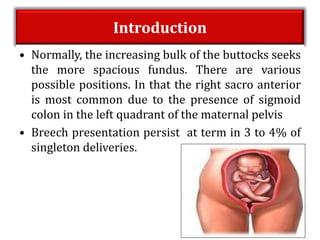
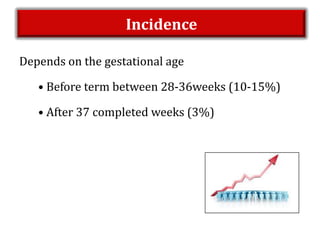
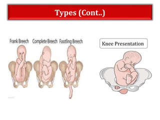
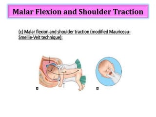
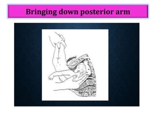
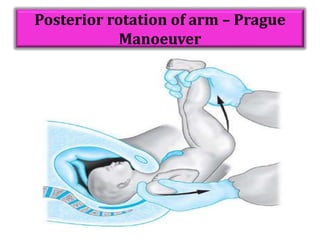
MRS. SONY SARA P.J
ASSO. PROFESSOR
MSC (N),OBSTETRICS AND GYNECOLOGICAL NURSING
GANGA COLLEGE OF NURSING
COIMBATORE
BREECH PRESENTATION
Content Overview
• Introduction
• Definition
• Incidence
• Types of breech
• Causes of breech
• Diagnosis
• Management of breech
• Mechanism of breech delivery
• Assisted breech delivery
• Prognosis
Introduction
• Normally, the increasing bulk of the buttocks seeks
the more spacious fundus. There are various
possible positions. In that the right sacro anterior
is most common due to the presence of sigmoid
colon in the left quadrant of the maternal pelvis
• Breech presentation persist at term in 3 to 4% of
singleton deliveries.
Definition – Breech Presentation
Breech presentation is when the lie of the baby
is longitudinal and pelvic or Podalic pole of foetus
presents at the brim and cephalic pole at the fundus.
Incidence
Depends on the gestational age
• Before term between 28-36weeks (10-15%)
• After 37 completed weeks (3%)
Types
Complete
Breech/Flexed Breech
Presentation Frank Breech
Footling Presentation
Knee Presentation
Incomplete Breech
Types (Cont..)
Knee Presentation
Aetiology
Maternal Factors
Fetal Factors
Placental and Amniotic Fluid
Factors
Maternal Factors
Multiparity
Uterine Anomalies
Fibroid Factors
Fetal Factors
Multiple Pregnancy
Short cord Intrauterine death
Fetal Anomalies
Placental And Amniotic Fluid Factors
Placenta praevia
Cornufundal
placenta
Prematurity Polyhydramnios
Diagnosis
• Abdominal examination
• Ultrasonography
• Pelvic examination
Abdominal Examination
Ultrasonography
• It confirms the clinical diagnosis
• It helps to detect fetal congenital anomalies
• To find out type of breech
• It helps to localise the placenta
• Assessment of liquor
• Attitude of the head
Vaginal Examination
S.No Types Breech Cephalic
1. Frank breech Ischial Tuberosity, Scarum,
Anus,External Genitalia
Fetal sutures and
Fontanalles
Muscular resistance of anus
with or without Meconium
Less yielding jaws
Straight line : Ischial
Tuberosity & Anus
Triangular : Malar
Eminences & Mouth
2. Complete
breech
Feet along side the buttocks
3. Footling One or both feet Inferior t the
buttocks
Clinical Examination
Positions
Mechanism of Labour
• Lie : Longitudinal
• Presentation : Breech
• Presenting parts : Anterior part of the Left
buttocks.
• Attitude : Complete Flexion (Complete Breech),
Flexion of hip and Extension of knee(Frank Breech)
• Denominator : Sacrum
• Positions : Left & right sacro anterior (LSA & RSA),
right & left sacro posterior (RSP and LSP)
Cont..
Delivery of Buttocks & lower limbs
• Engagement
• Descent
• Internal rotation of breech
• More descent with Lateral flexion
• Birth of buttocks
• Restitution
Delivery of Shoulders & arms
• Engagement
• Descent
• Internal rotation of shoulders
• Birth of shoulders by lateral flexion
• Restitution
• External rotation
Cont..
Delivery of After coming Head
• Engagement
• Descent
• Flexion
• Internal rotation
• Birth of head by flexion
Cont..
Cont..
Delivery of the buttocks :
 Engagement :
• The engaging diameter is “bi-trochanteric diameter”
(10cm)
• It is passes the pelvic brim, the breech is engaged.
 Descent :
• It occurs until the buttocks reaches the pelvic floor.
 Internal Rotation :
• It occurs 1/8th of the circle until it comes under the
Symphysis Pubis.
Cont..
 Further descent with lateral flexion of the trunk.
 Delivery of the trunk and the lower limbs follows.
 Restitution occurs.
Delivery of the shoulders :
 The engaging diameter of the shoulder is “Bi-
Sacromial diameter” is 12cm.
 Internal rotation of the shoulder occurs 1/8th of
the circle.
 Delivery of the posterior shoulder followed by
anterior one.
 Restitution and External Rotation.
Cont..
Delivery of the head:
 The engaging diameter is either through the
opposite oblique diameter is “Sub Occipito
Frontal”(10cm)
 Descent with Increasing flexion.
 Internal rotation of the occiput occurs 1/8th of the
circle.
 Further descent takes place.
 The head is born by lateral flexion.
Management
Antenatal management
 External cephalic version
* Performed at 36-37weeks
* Follow prerequisites
* It has higher success rates in
- Multiparous
- Adequate AF
- Average Fetal Weight
- Complete Breech
External Cephalic Version
ECV- Complications
• Fetal bradycardia
• Placental abruption
• Fetomaternal haemorrhage
• Uterine rupture.
Management of Breech at Term
• Mode of delivery
• Elective caesarean section
– Estimated fetal weight <3.5kg
– Footling breech
– Hyperextended head
– Complicated breech
– Placenta Praevia
– Contracted pelvis.
Vaginal Breech Delivery
 Spontaneous Breech: No manipulation of infant
is necessary
 Assisted Breech delivery: Fetus descend
spontaneously up to the umbilicus then, the rest of
the fetus is extracted using additional manoeuvres.
 Total Breech Extraction: The entire body is
extracted. This is indicated only if there is evidence
of fetal distress unresponsiveness to routine
manoeuvers and a caesarean delivery is not
possible.
Cont..
• Criteria for patient selection
Complete/Frank breech
EFW : 2-3.5kg
Flexed head
Uncomplicated Breech
Assisted Breech Delivery
Principles :
1. Never to rush
2. Never pull from below but push from above
3. Always keep the fetus with the back anteriorly.
Prerequisites:
1. A skilled Obstetrician
2. Anaesthetist
3. An assistant- for fundal pressure
4. Instruments, suture materials for episiotomy
5. Resuscitation articles – baby if asphyxiated
6. Neonatologist
Cont..
General guidelines :
• Patient and attenders to be counselled
• Informed written consent
• IOL-not recommended
• Late ARM
• Oxytocin augmentation – usually avoided
• Maintain Partogram
Maneuvers used in Delivery of After
Coming Head
Burns Marshall Method
Forceps Delivery
Malar Flexion and Shoulder Traction
Management of Complicated Breech
• Delay in descent of the breech
– At the outlet
– In the cavity
– At the brim
• Causes may be :
 Pelvic contraction
 Big baby
 Weak uterine contraction
Frank Breech Contraction (Pinard’s
Maneuver)
Extended Arms - Lovset’s Maneuver
Bringing down posterior arm
Nuchal arm
Posterior rotation of arm – Prague
Manoeuver
Complications
Fetal complications Maternal complications
• Intrauterine fetal death
• Intracranial haemorrhage
• Birth asphyxia
• Birth injuries
 Haematoma
 Fractures
 Visceral injuries
 Nerve injury
 Long term
neurological damage
• Increased Caesarean
delivery
• Genital tract injuries
Conclusion
• In view of insignificant difference in the
fetomaternal outcome balanced decision about
mode of delivery on case by case basis will go a
long way in improving both fetal and maternal
outcome.
Reference
• D.C.Dutta,”Textbook of Obstetrics including Perinatology and
Contraception". Seventh Edition.
• Jacob, Annamma (2009). A Comprehensive Textbook of Midwifer
y. Second Edition. New Delhi: Jaypee Brothers
Medical Publishers.
• J.B. Sharma, “Midwifery & Gynaecological Nursing” Avichal
Publishing company:1st edition
• www.ncbl.nlm.nih.gov breech presentation: overview
BREECH PRESENTATION
BREECH PRESENTATION

BREECH PRESENTATION

  • 1.
    MRS. SONY SARAP.J ASSO. PROFESSOR MSC (N),OBSTETRICS AND GYNECOLOGICAL NURSING GANGA COLLEGE OF NURSING COIMBATORE
  • 2.
  • 3.
    Content Overview • Introduction •Definition • Incidence • Types of breech • Causes of breech • Diagnosis • Management of breech • Mechanism of breech delivery • Assisted breech delivery • Prognosis
  • 4.
    Introduction • Normally, theincreasing bulk of the buttocks seeks the more spacious fundus. There are various possible positions. In that the right sacro anterior is most common due to the presence of sigmoid colon in the left quadrant of the maternal pelvis • Breech presentation persist at term in 3 to 4% of singleton deliveries.
  • 5.
    Definition – BreechPresentation Breech presentation is when the lie of the baby is longitudinal and pelvic or Podalic pole of foetus presents at the brim and cephalic pole at the fundus.
  • 6.
    Incidence Depends on thegestational age • Before term between 28-36weeks (10-15%) • After 37 completed weeks (3%)
  • 7.
    Types Complete Breech/Flexed Breech Presentation FrankBreech Footling Presentation Knee Presentation Incomplete Breech
  • 8.
  • 9.
  • 10.
  • 11.
    Fetal Factors Multiple Pregnancy Shortcord Intrauterine death Fetal Anomalies
  • 12.
    Placental And AmnioticFluid Factors Placenta praevia Cornufundal placenta Prematurity Polyhydramnios
  • 13.
    Diagnosis • Abdominal examination •Ultrasonography • Pelvic examination
  • 14.
  • 15.
    Ultrasonography • It confirmsthe clinical diagnosis • It helps to detect fetal congenital anomalies • To find out type of breech • It helps to localise the placenta • Assessment of liquor • Attitude of the head
  • 16.
    Vaginal Examination S.No TypesBreech Cephalic 1. Frank breech Ischial Tuberosity, Scarum, Anus,External Genitalia Fetal sutures and Fontanalles Muscular resistance of anus with or without Meconium Less yielding jaws Straight line : Ischial Tuberosity & Anus Triangular : Malar Eminences & Mouth 2. Complete breech Feet along side the buttocks 3. Footling One or both feet Inferior t the buttocks
  • 17.
  • 18.
  • 19.
    Mechanism of Labour •Lie : Longitudinal • Presentation : Breech • Presenting parts : Anterior part of the Left buttocks. • Attitude : Complete Flexion (Complete Breech), Flexion of hip and Extension of knee(Frank Breech) • Denominator : Sacrum • Positions : Left & right sacro anterior (LSA & RSA), right & left sacro posterior (RSP and LSP)
  • 20.
    Cont.. Delivery of Buttocks& lower limbs • Engagement • Descent • Internal rotation of breech • More descent with Lateral flexion • Birth of buttocks • Restitution Delivery of Shoulders & arms • Engagement • Descent • Internal rotation of shoulders • Birth of shoulders by lateral flexion • Restitution • External rotation
  • 21.
    Cont.. Delivery of Aftercoming Head • Engagement • Descent • Flexion • Internal rotation • Birth of head by flexion
  • 22.
  • 23.
    Cont.. Delivery of thebuttocks :  Engagement : • The engaging diameter is “bi-trochanteric diameter” (10cm) • It is passes the pelvic brim, the breech is engaged.  Descent : • It occurs until the buttocks reaches the pelvic floor.  Internal Rotation : • It occurs 1/8th of the circle until it comes under the Symphysis Pubis.
  • 24.
    Cont..  Further descentwith lateral flexion of the trunk.  Delivery of the trunk and the lower limbs follows.  Restitution occurs. Delivery of the shoulders :  The engaging diameter of the shoulder is “Bi- Sacromial diameter” is 12cm.  Internal rotation of the shoulder occurs 1/8th of the circle.  Delivery of the posterior shoulder followed by anterior one.  Restitution and External Rotation.
  • 25.
    Cont.. Delivery of thehead:  The engaging diameter is either through the opposite oblique diameter is “Sub Occipito Frontal”(10cm)  Descent with Increasing flexion.  Internal rotation of the occiput occurs 1/8th of the circle.  Further descent takes place.  The head is born by lateral flexion.
  • 26.
    Management Antenatal management  Externalcephalic version * Performed at 36-37weeks * Follow prerequisites * It has higher success rates in - Multiparous - Adequate AF - Average Fetal Weight - Complete Breech
  • 27.
  • 28.
    ECV- Complications • Fetalbradycardia • Placental abruption • Fetomaternal haemorrhage • Uterine rupture.
  • 29.
    Management of Breechat Term • Mode of delivery • Elective caesarean section – Estimated fetal weight <3.5kg – Footling breech – Hyperextended head – Complicated breech – Placenta Praevia – Contracted pelvis.
  • 30.
    Vaginal Breech Delivery Spontaneous Breech: No manipulation of infant is necessary  Assisted Breech delivery: Fetus descend spontaneously up to the umbilicus then, the rest of the fetus is extracted using additional manoeuvres.  Total Breech Extraction: The entire body is extracted. This is indicated only if there is evidence of fetal distress unresponsiveness to routine manoeuvers and a caesarean delivery is not possible.
  • 31.
    Cont.. • Criteria forpatient selection Complete/Frank breech EFW : 2-3.5kg Flexed head Uncomplicated Breech
  • 32.
    Assisted Breech Delivery Principles: 1. Never to rush 2. Never pull from below but push from above 3. Always keep the fetus with the back anteriorly. Prerequisites: 1. A skilled Obstetrician 2. Anaesthetist 3. An assistant- for fundal pressure 4. Instruments, suture materials for episiotomy 5. Resuscitation articles – baby if asphyxiated 6. Neonatologist
  • 33.
    Cont.. General guidelines : •Patient and attenders to be counselled • Informed written consent • IOL-not recommended • Late ARM • Oxytocin augmentation – usually avoided • Maintain Partogram
  • 34.
    Maneuvers used inDelivery of After Coming Head
  • 35.
  • 36.
  • 37.
    Malar Flexion andShoulder Traction
  • 38.
    Management of ComplicatedBreech • Delay in descent of the breech – At the outlet – In the cavity – At the brim • Causes may be :  Pelvic contraction  Big baby  Weak uterine contraction
  • 39.
    Frank Breech Contraction(Pinard’s Maneuver)
  • 40.
    Extended Arms -Lovset’s Maneuver
  • 41.
  • 42.
  • 43.
    Posterior rotation ofarm – Prague Manoeuver
  • 44.
    Complications Fetal complications Maternalcomplications • Intrauterine fetal death • Intracranial haemorrhage • Birth asphyxia • Birth injuries  Haematoma  Fractures  Visceral injuries  Nerve injury  Long term neurological damage • Increased Caesarean delivery • Genital tract injuries
  • 45.
    Conclusion • In viewof insignificant difference in the fetomaternal outcome balanced decision about mode of delivery on case by case basis will go a long way in improving both fetal and maternal outcome.
  • 46.
    Reference • D.C.Dutta,”Textbook ofObstetrics including Perinatology and Contraception". Seventh Edition. • Jacob, Annamma (2009). A Comprehensive Textbook of Midwifer y. Second Edition. New Delhi: Jaypee Brothers Medical Publishers. • J.B. Sharma, “Midwifery & Gynaecological Nursing” Avichal Publishing company:1st edition • www.ncbl.nlm.nih.gov breech presentation: overview