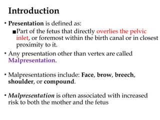
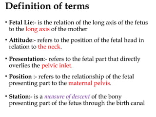
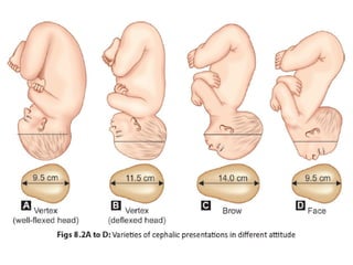
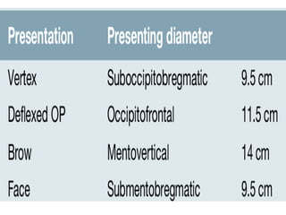
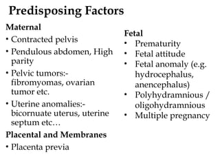
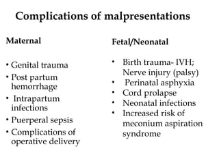
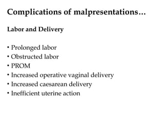
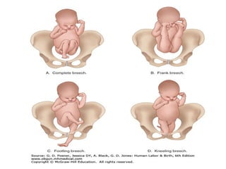
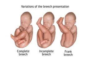
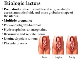
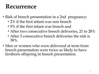
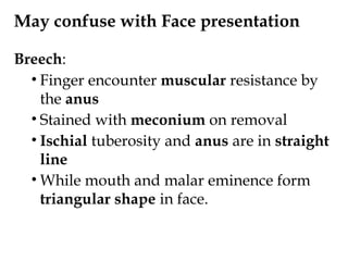
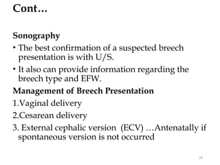
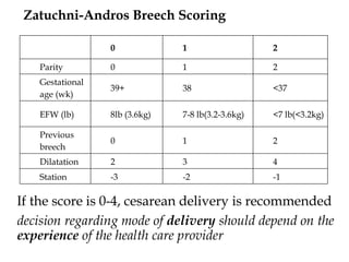
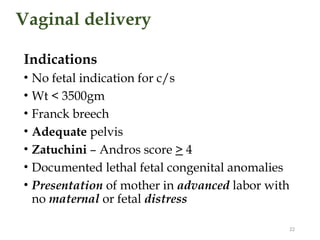
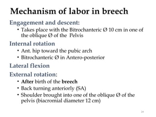
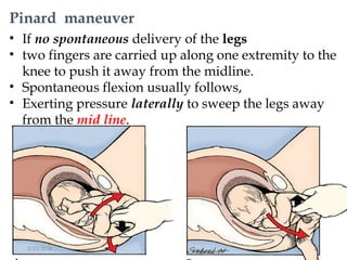
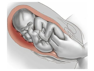
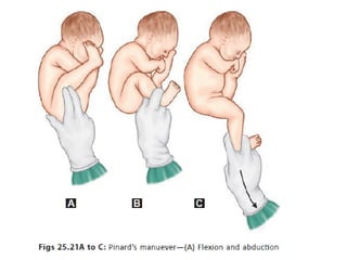
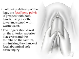
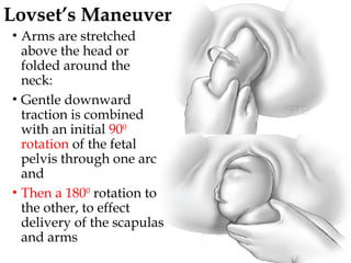
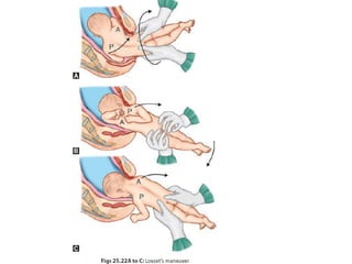
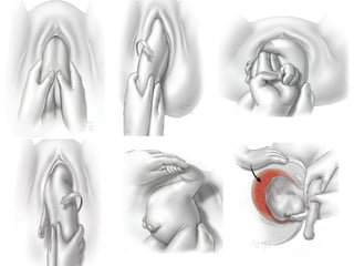
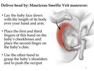
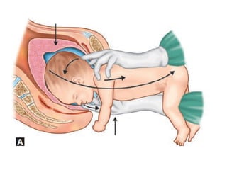
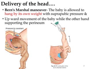
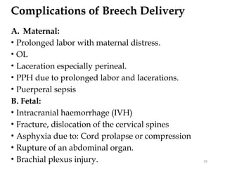
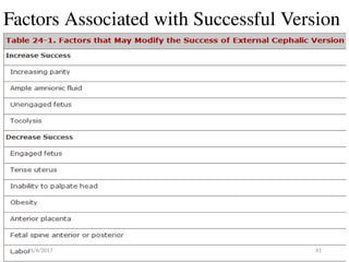
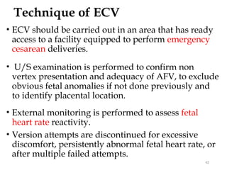
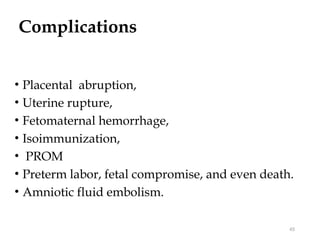
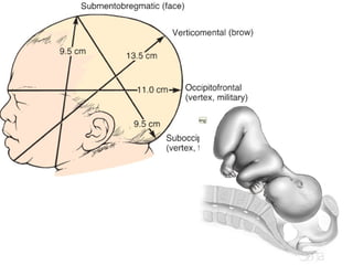
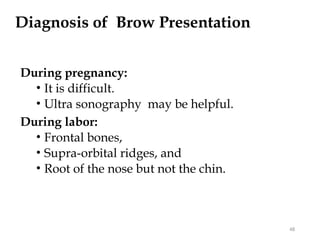
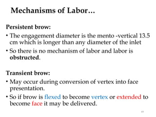
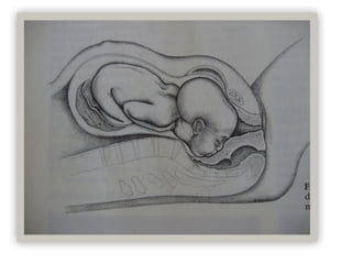
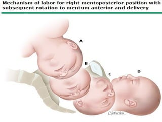
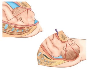
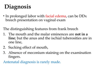
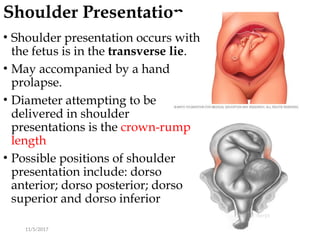
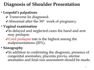
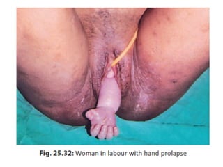
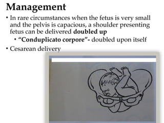
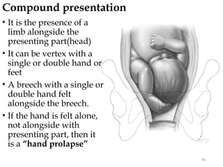
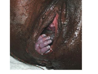
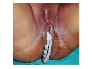
Malpresentations
Learning objectives
• Introduction
• Definition of mal presentation
• Diagnosis of mal presentation
• Management of mal presentation
• Complication of mal presentation
2
Introduction
• Presentation is defined as:
▪Part of the fetus that directly overlies the pelvic
inlet, or foremost within the birth canal or in closest
proximity to it.
• Any presentation other than vertex are called
Malpresentation.
• Malpresentations include: Face, brow, breech,
shoulder, or compound.
• Malpresentation is often associated with increased
risk to both the mother and the fetus
Definition of terms
• Fetal Lie:- is the relation of the long axis of the fetus
to the long axis of the mother
• Attitude:- refers to the position of the fetal head in
relation to the neck.
• Presentation:- refers to the fetal part that directly
overlies the pelvic inlet.
• Position :- refers to the relationship of the fetal
presenting part to the maternal pelvis.
• Station:- is a measure of descent of the bony
presenting part of the fetus through the birth canal
Predisposing Factors
Maternal
• Contracted pelvis
• Pendulous abdomen, High
parity
• Pelvic tumors:-
fibromyomas, ovarian
tumor etc.
• Uterine anomalies:-
bicornuate uterus, uterine
septum etc…
Placental and Membranes
• Placenta previa
Fetal
• Prematurity
• Fetal attitude
• Fetal anomaly (e.g.
hydrocephalus,
anencephalus)
• Polyhydramnious /
oligohydramnious
• Multiple pregnancy
Complications of malpresentations
Maternal
• Genital trauma
• Post partum
hemorrhage
• Intrapartum
infections
• Puerperal sepsis
• Complications of
operative delivery
Fetal/Neonatal
• Birth trauma- IVH;
Nerve injury (palsy)
• Perinatal asphyxia
• Cord prolapse
• Neonatal infections
• Increased risk of
meconium aspiration
syndrome
Complications of malpresentations…
Labor and Delivery
• Prolonged labor
• Obstructed labor
• PROM
• Increased operative vaginal delivery
• Increased caesarean delivery
• Inefficient uterine action
Breech presentation
• It is a longitudinal lie in which the buttocks is the
presenting part with or without the lower limbs.
• Diagnosis can be made through abdominal palpation,
vaginal examination and confirmed by ultrasound
Types
• Frank: hip flexed and knee extended commonest
• Complete: hip and knee are flexed, in multipara
• Footling: hip and leg are extended, one /both feet
present,
• Knee: the hips are extended with knees flexed
Prematurity, fetal malformation, Uterine anomalies, and polar
placentation are commonly observed causative factors
Etiologic factors
• Prematurity : due to small foetal size, relatively
excess amniotic fluid, and more globular shape of
the uterus.
• Multiple pregnancy:
• Poly-and oligohydramnios.
• Hydrocephalus, anencephalus.
• Bicornuate and septate uterus.
• Uterine & pelvic tumors.
• Placenta praevia
Recurrence
• Risk of breech presentation in a 2nd pregnancy:
• 2% if the first infant was non breech
• 9% if the first infant was breech and
• After two consecutive breech deliveries, 21 to 28%
• After 3 consecutive breech deliveries the risk is
38%.
• Men or women who were delivered at term from
breech presentation were twice as likely to have
firstborn offspring in breech presentation.
15
Diagnosis
Abdominal Examination after 34 weeks
• Leopold maneuver:
• The hard, round, readily ballotable fetal head occupy
the fundus.
• FHB will be heard more easily at or above the umbilicus
Vaginal Examination
• Frank breech presentation:
• Ischial tuberosities, sacrum, and the anus ..palpable
• After further fetal descent, the external genitalia may be
distinguished.
• The sacrum are palpated to establish the position and
presentation
16
May confuse with Face presentation
Breech:
• Finger encounter muscular resistance by
the anus
• Stained with meconium on removal
• Ischial tuberosity and anus are in straight
line
• While mouth and malar eminence form
triangular shape in face.
Cont…
Sonography
• The best confirmation of a suspected breech
presentation is with U/S.
• It also can provide information regarding the
breech type and EFW.
Management of Breech Presentation
1.Vaginal delivery
2.Cesarean delivery
3. External cephalic version (ECV) …Antenatally if
spontaneous version is not occurred
19
Management
Indications for C/S
• large fetus ( EFW ≥3500gm) or < 1.5kg
• Any degree of pelvic contracture
• Footling breech
• Ux dysfunction
• Severe IUGR
• Breech with poor obstetric performance
(previous perinatal death)
• Elderly primigravida
• Others:
▪Request for sterilization
• Zatuchini – Andros score <4
If the score is 0-4, cesarean delivery is recommended
decision regarding mode of delivery should depend on the
experience of the health care provider
Zatuchni-Andros Breech Scoring
0 1 2
Parity 0 1 2
Gestational
age (wk)
39+ 38 <37
EFW (lb) 8lb (3.6kg) 7-8 lb(3.2-3.6kg) <7 lb(<3.2kg)
Previous
breech
0 1 2
Dilatation 2 3 4
Station -3 -2 -1
Vaginal delivery
Indications
• No fetal indication for c/s
• Wt < 3500gm
• Franck breech
• Adequate pelvis
• Zatuchini – Andros score > 4
• Documented lethal fetal congenital anomalies
• Presentation of mother in advanced labor with
no maternal or fetal distress
22
Methods of Vaginal Delivery
General methods of vaginal breech delivery:
1. Spontaneous breech delivery: without any
traction or manipulation other than support of the
newborn.
2. Partial breech extraction. The fetus is delivered
spontaneously as far as the umbilicus, but the
remainder of the body is extracted will be assisted
by maneuvers.
3. Total breech extraction. The entire body of the
fetus is extracted by the obstetrician. Replaced by
C/S
23
Mechanism of labor in breech
Engagement and descent:
• Takes place with the Bitrochanteric Ø 10 cm in one of
the oblique Ø of the Pelvis
Internal rotation
• Ant. hip toward the pubic arch
• Bitrochanteric Ø in Antero-posterior
Lateral flexion
External rotation:
• After birth of the breech
• Back turning anteriorly (SA)
• Shoulder brought into one of the oblique Ø of the
pelvis (biacromial diameter 12 cm)
24
Assisted vaginal breech delivery
• Instruct the mother to bear down with every
contraction (2nd stage).
• Episiotomy – when fetal anus is visible and
perineum distended unless perineum is well relaxed.
• Allow the breech to be delivered and wait without
intervention till body born up to the level of
umbilicus.
• The golden rule is to "Keep your hands off“ to
deliver the fetal buttock and
• The body is allowed to deliver spontaneously up to
the level of the umbilicus
25
• After the umbilicus has
been reached, wait
spontaneous delivery of
the legs .
• If not delivered
spontaneously, deliver
one leg at a time:
• Flexion and abduction
of thigh, subsequent
delivery of the lower leg.
• Do not pull the baby
while the legs are being
delivered
27
Pinard maneuver
• If no spontaneous delivery of the legs
• two fingers are carried up along one extremity to the
knee to push it away from the midline.
• Spontaneous flexion usually follows,
• Exerting pressure laterally to sweep the legs away
from the mid line.
2/23/2016 28
31
• Following delivery of the
legs, the fetal bony pelvis
is grasped with both
hands, using a cloth
towel moistened with
warm water.
• The fingers should rest
on the anterior superior
iliac crests and the
thumbs on the sacrum,
minimizing the chance of
fetal abdominal soft
tissue injury
32
Lovset’s Maneuver
• Arms are stretched
above the head or
folded around the
neck:
• Gentle downward
traction is combined
with an initial 900
rotation of the fetal
pelvis through one arc
and
• Then a 1800
rotation to
the other, to effect
delivery of the scapulas
and arms
11/4/2017 34
Deliver head by: Mauriceau Smellie Veit maneuver:
35
• Lay the baby face down
with the length of its body
over your hand and arm.
• Place the first and third
fingers of this hand on the
baby’s cheekbones and
place the second finger on
the baby’s chin.
• Use the other hand to
grasp the baby’s shoulders
and to push the occiput
Delivery of the head….
• Bern's Marshal maneuver- The baby is allowed to
hang by its own weight with suprapubic pressure &
• Up ward movement of the baby while the other hand
supporting the perineum
37
• Piper forceps can be used to
deliver the after coming
head, when MSV maneuver
cannot be accomplished
easily.
• Suspension of the body of
the fetus in a towel
effectively holds the fetus
and helps keep the arms
out of the way
38
Forceps to After coming Head
Complications of Breech Delivery
A. Maternal:
• Prolonged labor with maternal distress.
• OL
• Laceration especially perineal.
• PPH due to prolonged labor and lacerations.
• Puerperal sepsis
B. Fetal:
• Intracranial haemorrhage (IVH)
• Fracture, dislocation of the cervical spines
• Asphyxia due to: Cord prolapse or compression
• Rupture of an abdominal organ.
• Brachial plexus injury. 39
External Cephalic Version(ECV)
• When a breech presentation is recognized prior to
labor in a woman who has reached 36 weeks'
gestation, ECV should be considered.
• Before this time, there is a relatively high incidence
of recurrence.(unstable)
• After 36 weeks, however, the likelihood of
spontaneous version is low.
• Version is C/I if vaginal delivery is not an option
• Anti-D immune globulin is given if indicated.
40
Factors Associated with Successful Version
11/4/2017 41
Technique of ECV
• ECV should be carried out in an area that has ready
access to a facility equipped to perform emergency
cesarean deliveries.
• U/S examination is performed to confirm non
vertex presentation and adequacy of AFV, to exclude
obvious fetal anomalies if not done previously and
to identify placental location.
• External monitoring is performed to assess fetal
heart rate reactivity.
• Version attempts are discontinued for excessive
discomfort, persistently abnormal fetal heart rate, or
after multiple failed attempts.
42
Technique…
• A forward roll of the
fetus usually is
attempted first.
• Each hand grasps one
of the fetal poles, and
• The buttocks are
elevated from the
maternal pelvis and
displaced laterally.
43
Technique…
• The buttocks are then
gently guided toward
the fundus, while the
head is directed
toward the pelvis.
• If the forward roll is
unsuccessful, then a
backward flip is
attempted.
44
Complications
• Placental abruption,
• Uterine rupture,
• Fetomaternal hemorrhage,
• Isoimmunization,
• PROM
• Preterm labor, fetal compromise, and even death.
• Amniotic fluid embolism.
45
Brow Presentation
• It is a cephalic presentation in which the head is
midway between flexion and extension.
• Diagnosed when the portion of the fetal head
between the orbital ridge and the anterior fontanel
presents at the pelvic inlet.
• Presenting diameters in brow presentation:
• Biparietal diameter – 9.5 cms
• Mentovertical diameter- 13.5 cms
• Most brow presentations at term are transitory
presentations in early labor.
Diagnosis of Brow Presentation
During pregnancy:
• It is difficult.
• Ultra sonography may be helpful.
During labor:
• Frontal bones,
• Supra-orbital ridges, and
• Root of the nose but not the chin.
48
Mechanisms of Labor…
Persistent brow:
• The engagement diameter is the mento -vertical 13.5
cm which is longer than any diameter of the inlet
• So there is no mechanism of labor and labor is
obstructed.
Transient brow:
• May occur during conversion of vertex into face
presentation.
• So if brow is flexed to become vertex or extended to
become face it may be delivered.
49
Management:
Early in the first stage:
1. Observe carefully and given a chance for
spontaneous conversion into either face or vertex.
2. Exclude contracted pelvis, if present do S/C
In the second stage:
If considered as persistent brow so:
1. Caesarean section is done if the foetus is living.
2. Craniotomy if the foetus is dead.
50
Face presentation
• It is a cephalic presentation in which the head is
completely extended, allowing the occiput to touch
the back
Boundaries:
• Superiorly; supra orbital ridges and the root of the
nose.
• Inferiorly; the mentum.
• Sub-mento bregmatic: 9.5
• Denominator : mentum
Face presentation…
• Possible positions 8 (e.g. MA, left mento transverse..)
• It is presumed to occur because of factors that:
1. Favor extension: goiter and excessive nuchal
cords
2. Prevent flexion of the fetal neck.
• Other factors includes:
▪CPD, Contracted maternal pelvis & platypelloid
pelvis
▪Prematurity/low birth weight, polyhydramnios
▪Macrosomia
▪Multiparity
▪Extreme laxity of the anterior abdominal wall
Mechanism of action
• In mento anterior positions, delivery follows by
flexion of the head towards the symphysis pubis
(face to pubis)
• In MA vertex fit into the hollow of the sacrum and
the chin fits under the symphysis.
• Persistent MP positions cannot be delivered
vaginally as the fetal brow pressing over the
symphysis precludes flexion
• Presenting diameter sterno-bregmatic(18cm)
• An open fetal mouth may act as a fulcrum
against the sacrum preventing further descent.
11/5/2017 57
Diagnosis
• In prolonged labor with facial edema, can be DDx
breech presentation on vaginal exam
The distinguishing features from frank breech
1. The mouth and the malar eminences are not in a
line; but the anus and the ischial tuberosities are in
one line,
2. Sucking effect of mouth,
3. Absence of meconium staining on the examination
fingers.
Antenatal diagnosis is rarely made.
Management
• Vaginal delivery can be tried in MA and
rotated MP
• Augmentation is controversial
• C/S if persistent MP
Shoulder Presentation
11/5/2017 62
• Shoulder presentation occurs with
the fetus is in the transverse lie.
• May accompanied by a hand
prolapse.
• Diameter attempting to be
delivered in shoulder
presentations is the crown-rump
length
• Possible positions of shoulder
presentation include: dorso
anterior; dorso posterior; dorso
superior and dorso inferior
Diagnosis of Shoulder Presentation
• Leopold’s palpations
✔ Transverse lie diagnosed.
✔Abnormal after the 34th
week of pregnancy.
• Vaginal examination
✔In delayed and neglected cases the hand and arm
may prolapse.
✔Cord prolapse rate is the highest among the
malpresentations (20%).
• Sonography
✔In addition to confirming the diagnosis, presence of
congenital anomalies, placenta previa, uterine
anomalies and fetal size assessment should be made.
63
Management
• In rare circumstances when the fetus is very small
and the pelvis is capacious, a shoulder presenting
fetus can be delivered doubled up
• “Conduplicato corpore”- doubled upon itself
• Cesarean delivery
Compound presentation
66
• It is the presence of a
limb alongside the
presenting part(head)
• It can be vertex with a
single or double hand or
feet
• A breech with a single or
double hand felt
alongside the breech.
• If the hand is felt alone,
not alongside with
presenting part, then it
is a “hand prolapse”
Mechanism of labor
• The usual progress in a compound presentation
diagnosed in early labor is the gradual regression of
the extremity upward as the vertex is pushed
downwards by uterine action.
• In cases of CPD, the brow may persist and requires
follow up with labor progress and appropriate
management.
• Induction and augmentation and instrumental
deliveries are contraindicated in compound
presentations
69
Thank You!!!

11.. Malpresentation and malpositio.pptx

  • 1.
  • 2.
    Learning objectives • Introduction •Definition of mal presentation • Diagnosis of mal presentation • Management of mal presentation • Complication of mal presentation 2
  • 3.
    Introduction • Presentation isdefined as: ▪Part of the fetus that directly overlies the pelvic inlet, or foremost within the birth canal or in closest proximity to it. • Any presentation other than vertex are called Malpresentation. • Malpresentations include: Face, brow, breech, shoulder, or compound. • Malpresentation is often associated with increased risk to both the mother and the fetus
  • 4.
    Definition of terms •Fetal Lie:- is the relation of the long axis of the fetus to the long axis of the mother • Attitude:- refers to the position of the fetal head in relation to the neck. • Presentation:- refers to the fetal part that directly overlies the pelvic inlet. • Position :- refers to the relationship of the fetal presenting part to the maternal pelvis. • Station:- is a measure of descent of the bony presenting part of the fetus through the birth canal
  • 7.
    Predisposing Factors Maternal • Contractedpelvis • Pendulous abdomen, High parity • Pelvic tumors:- fibromyomas, ovarian tumor etc. • Uterine anomalies:- bicornuate uterus, uterine septum etc… Placental and Membranes • Placenta previa Fetal • Prematurity • Fetal attitude • Fetal anomaly (e.g. hydrocephalus, anencephalus) • Polyhydramnious / oligohydramnious • Multiple pregnancy
  • 8.
    Complications of malpresentations Maternal •Genital trauma • Post partum hemorrhage • Intrapartum infections • Puerperal sepsis • Complications of operative delivery Fetal/Neonatal • Birth trauma- IVH; Nerve injury (palsy) • Perinatal asphyxia • Cord prolapse • Neonatal infections • Increased risk of meconium aspiration syndrome
  • 9.
    Complications of malpresentations… Laborand Delivery • Prolonged labor • Obstructed labor • PROM • Increased operative vaginal delivery • Increased caesarean delivery • Inefficient uterine action
  • 10.
    Breech presentation • Itis a longitudinal lie in which the buttocks is the presenting part with or without the lower limbs. • Diagnosis can be made through abdominal palpation, vaginal examination and confirmed by ultrasound Types • Frank: hip flexed and knee extended commonest • Complete: hip and knee are flexed, in multipara • Footling: hip and leg are extended, one /both feet present, • Knee: the hips are extended with knees flexed
  • 13.
    Prematurity, fetal malformation,Uterine anomalies, and polar placentation are commonly observed causative factors
  • 14.
    Etiologic factors • Prematurity: due to small foetal size, relatively excess amniotic fluid, and more globular shape of the uterus. • Multiple pregnancy: • Poly-and oligohydramnios. • Hydrocephalus, anencephalus. • Bicornuate and septate uterus. • Uterine & pelvic tumors. • Placenta praevia
  • 15.
    Recurrence • Risk ofbreech presentation in a 2nd pregnancy: • 2% if the first infant was non breech • 9% if the first infant was breech and • After two consecutive breech deliveries, 21 to 28% • After 3 consecutive breech deliveries the risk is 38%. • Men or women who were delivered at term from breech presentation were twice as likely to have firstborn offspring in breech presentation. 15
  • 16.
    Diagnosis Abdominal Examination after34 weeks • Leopold maneuver: • The hard, round, readily ballotable fetal head occupy the fundus. • FHB will be heard more easily at or above the umbilicus Vaginal Examination • Frank breech presentation: • Ischial tuberosities, sacrum, and the anus ..palpable • After further fetal descent, the external genitalia may be distinguished. • The sacrum are palpated to establish the position and presentation 16
  • 18.
    May confuse withFace presentation Breech: • Finger encounter muscular resistance by the anus • Stained with meconium on removal • Ischial tuberosity and anus are in straight line • While mouth and malar eminence form triangular shape in face.
  • 19.
    Cont… Sonography • The bestconfirmation of a suspected breech presentation is with U/S. • It also can provide information regarding the breech type and EFW. Management of Breech Presentation 1.Vaginal delivery 2.Cesarean delivery 3. External cephalic version (ECV) …Antenatally if spontaneous version is not occurred 19
  • 20.
    Management Indications for C/S •large fetus ( EFW ≥3500gm) or < 1.5kg • Any degree of pelvic contracture • Footling breech • Ux dysfunction • Severe IUGR • Breech with poor obstetric performance (previous perinatal death) • Elderly primigravida • Others: ▪Request for sterilization • Zatuchini – Andros score <4
  • 21.
    If the scoreis 0-4, cesarean delivery is recommended decision regarding mode of delivery should depend on the experience of the health care provider Zatuchni-Andros Breech Scoring 0 1 2 Parity 0 1 2 Gestational age (wk) 39+ 38 <37 EFW (lb) 8lb (3.6kg) 7-8 lb(3.2-3.6kg) <7 lb(<3.2kg) Previous breech 0 1 2 Dilatation 2 3 4 Station -3 -2 -1
  • 22.
    Vaginal delivery Indications • Nofetal indication for c/s • Wt < 3500gm • Franck breech • Adequate pelvis • Zatuchini – Andros score > 4 • Documented lethal fetal congenital anomalies • Presentation of mother in advanced labor with no maternal or fetal distress 22
  • 23.
    Methods of VaginalDelivery General methods of vaginal breech delivery: 1. Spontaneous breech delivery: without any traction or manipulation other than support of the newborn. 2. Partial breech extraction. The fetus is delivered spontaneously as far as the umbilicus, but the remainder of the body is extracted will be assisted by maneuvers. 3. Total breech extraction. The entire body of the fetus is extracted by the obstetrician. Replaced by C/S 23
  • 24.
    Mechanism of laborin breech Engagement and descent: • Takes place with the Bitrochanteric Ø 10 cm in one of the oblique Ø of the Pelvis Internal rotation • Ant. hip toward the pubic arch • Bitrochanteric Ø in Antero-posterior Lateral flexion External rotation: • After birth of the breech • Back turning anteriorly (SA) • Shoulder brought into one of the oblique Ø of the pelvis (biacromial diameter 12 cm) 24
  • 25.
    Assisted vaginal breechdelivery • Instruct the mother to bear down with every contraction (2nd stage). • Episiotomy – when fetal anus is visible and perineum distended unless perineum is well relaxed. • Allow the breech to be delivered and wait without intervention till body born up to the level of umbilicus. • The golden rule is to "Keep your hands off“ to deliver the fetal buttock and • The body is allowed to deliver spontaneously up to the level of the umbilicus 25
  • 27.
    • After theumbilicus has been reached, wait spontaneous delivery of the legs . • If not delivered spontaneously, deliver one leg at a time: • Flexion and abduction of thigh, subsequent delivery of the lower leg. • Do not pull the baby while the legs are being delivered 27
  • 28.
    Pinard maneuver • Ifno spontaneous delivery of the legs • two fingers are carried up along one extremity to the knee to push it away from the midline. • Spontaneous flexion usually follows, • Exerting pressure laterally to sweep the legs away from the mid line. 2/23/2016 28
  • 31.
    31 • Following deliveryof the legs, the fetal bony pelvis is grasped with both hands, using a cloth towel moistened with warm water. • The fingers should rest on the anterior superior iliac crests and the thumbs on the sacrum, minimizing the chance of fetal abdominal soft tissue injury
  • 32.
    32 Lovset’s Maneuver • Armsare stretched above the head or folded around the neck: • Gentle downward traction is combined with an initial 900 rotation of the fetal pelvis through one arc and • Then a 1800 rotation to the other, to effect delivery of the scapulas and arms
  • 34.
  • 35.
    Deliver head by:Mauriceau Smellie Veit maneuver: 35 • Lay the baby face down with the length of its body over your hand and arm. • Place the first and third fingers of this hand on the baby’s cheekbones and place the second finger on the baby’s chin. • Use the other hand to grasp the baby’s shoulders and to push the occiput
  • 37.
    Delivery of thehead…. • Bern's Marshal maneuver- The baby is allowed to hang by its own weight with suprapubic pressure & • Up ward movement of the baby while the other hand supporting the perineum 37
  • 38.
    • Piper forcepscan be used to deliver the after coming head, when MSV maneuver cannot be accomplished easily. • Suspension of the body of the fetus in a towel effectively holds the fetus and helps keep the arms out of the way 38 Forceps to After coming Head
  • 39.
    Complications of BreechDelivery A. Maternal: • Prolonged labor with maternal distress. • OL • Laceration especially perineal. • PPH due to prolonged labor and lacerations. • Puerperal sepsis B. Fetal: • Intracranial haemorrhage (IVH) • Fracture, dislocation of the cervical spines • Asphyxia due to: Cord prolapse or compression • Rupture of an abdominal organ. • Brachial plexus injury. 39
  • 40.
    External Cephalic Version(ECV) •When a breech presentation is recognized prior to labor in a woman who has reached 36 weeks' gestation, ECV should be considered. • Before this time, there is a relatively high incidence of recurrence.(unstable) • After 36 weeks, however, the likelihood of spontaneous version is low. • Version is C/I if vaginal delivery is not an option • Anti-D immune globulin is given if indicated. 40
  • 41.
    Factors Associated withSuccessful Version 11/4/2017 41
  • 42.
    Technique of ECV •ECV should be carried out in an area that has ready access to a facility equipped to perform emergency cesarean deliveries. • U/S examination is performed to confirm non vertex presentation and adequacy of AFV, to exclude obvious fetal anomalies if not done previously and to identify placental location. • External monitoring is performed to assess fetal heart rate reactivity. • Version attempts are discontinued for excessive discomfort, persistently abnormal fetal heart rate, or after multiple failed attempts. 42
  • 43.
    Technique… • A forwardroll of the fetus usually is attempted first. • Each hand grasps one of the fetal poles, and • The buttocks are elevated from the maternal pelvis and displaced laterally. 43
  • 44.
    Technique… • The buttocksare then gently guided toward the fundus, while the head is directed toward the pelvis. • If the forward roll is unsuccessful, then a backward flip is attempted. 44
  • 45.
    Complications • Placental abruption, •Uterine rupture, • Fetomaternal hemorrhage, • Isoimmunization, • PROM • Preterm labor, fetal compromise, and even death. • Amniotic fluid embolism. 45
  • 46.
    Brow Presentation • Itis a cephalic presentation in which the head is midway between flexion and extension. • Diagnosed when the portion of the fetal head between the orbital ridge and the anterior fontanel presents at the pelvic inlet. • Presenting diameters in brow presentation: • Biparietal diameter – 9.5 cms • Mentovertical diameter- 13.5 cms • Most brow presentations at term are transitory presentations in early labor.
  • 48.
    Diagnosis of BrowPresentation During pregnancy: • It is difficult. • Ultra sonography may be helpful. During labor: • Frontal bones, • Supra-orbital ridges, and • Root of the nose but not the chin. 48
  • 49.
    Mechanisms of Labor… Persistentbrow: • The engagement diameter is the mento -vertical 13.5 cm which is longer than any diameter of the inlet • So there is no mechanism of labor and labor is obstructed. Transient brow: • May occur during conversion of vertex into face presentation. • So if brow is flexed to become vertex or extended to become face it may be delivered. 49
  • 50.
    Management: Early in thefirst stage: 1. Observe carefully and given a chance for spontaneous conversion into either face or vertex. 2. Exclude contracted pelvis, if present do S/C In the second stage: If considered as persistent brow so: 1. Caesarean section is done if the foetus is living. 2. Craniotomy if the foetus is dead. 50
  • 51.
    Face presentation • Itis a cephalic presentation in which the head is completely extended, allowing the occiput to touch the back Boundaries: • Superiorly; supra orbital ridges and the root of the nose. • Inferiorly; the mentum. • Sub-mento bregmatic: 9.5 • Denominator : mentum
  • 52.
    Face presentation… • Possiblepositions 8 (e.g. MA, left mento transverse..) • It is presumed to occur because of factors that: 1. Favor extension: goiter and excessive nuchal cords 2. Prevent flexion of the fetal neck. • Other factors includes: ▪CPD, Contracted maternal pelvis & platypelloid pelvis ▪Prematurity/low birth weight, polyhydramnios ▪Macrosomia ▪Multiparity ▪Extreme laxity of the anterior abdominal wall
  • 54.
    Mechanism of action •In mento anterior positions, delivery follows by flexion of the head towards the symphysis pubis (face to pubis) • In MA vertex fit into the hollow of the sacrum and the chin fits under the symphysis. • Persistent MP positions cannot be delivered vaginally as the fetal brow pressing over the symphysis precludes flexion • Presenting diameter sterno-bregmatic(18cm) • An open fetal mouth may act as a fulcrum against the sacrum preventing further descent.
  • 57.
  • 59.
    Diagnosis • In prolongedlabor with facial edema, can be DDx breech presentation on vaginal exam The distinguishing features from frank breech 1. The mouth and the malar eminences are not in a line; but the anus and the ischial tuberosities are in one line, 2. Sucking effect of mouth, 3. Absence of meconium staining on the examination fingers. Antenatal diagnosis is rarely made.
  • 61.
    Management • Vaginal deliverycan be tried in MA and rotated MP • Augmentation is controversial • C/S if persistent MP
  • 62.
    Shoulder Presentation 11/5/2017 62 •Shoulder presentation occurs with the fetus is in the transverse lie. • May accompanied by a hand prolapse. • Diameter attempting to be delivered in shoulder presentations is the crown-rump length • Possible positions of shoulder presentation include: dorso anterior; dorso posterior; dorso superior and dorso inferior
  • 63.
    Diagnosis of ShoulderPresentation • Leopold’s palpations ✔ Transverse lie diagnosed. ✔Abnormal after the 34th week of pregnancy. • Vaginal examination ✔In delayed and neglected cases the hand and arm may prolapse. ✔Cord prolapse rate is the highest among the malpresentations (20%). • Sonography ✔In addition to confirming the diagnosis, presence of congenital anomalies, placenta previa, uterine anomalies and fetal size assessment should be made. 63
  • 65.
    Management • In rarecircumstances when the fetus is very small and the pelvis is capacious, a shoulder presenting fetus can be delivered doubled up • “Conduplicato corpore”- doubled upon itself • Cesarean delivery
  • 66.
    Compound presentation 66 • Itis the presence of a limb alongside the presenting part(head) • It can be vertex with a single or double hand or feet • A breech with a single or double hand felt alongside the breech. • If the hand is felt alone, not alongside with presenting part, then it is a “hand prolapse”
  • 69.
    Mechanism of labor •The usual progress in a compound presentation diagnosed in early labor is the gradual regression of the extremity upward as the vertex is pushed downwards by uterine action. • In cases of CPD, the brow may persist and requires follow up with labor progress and appropriate management. • Induction and augmentation and instrumental deliveries are contraindicated in compound presentations 69
  • 70.

Editor's Notes

  • #3 Vertex presentation is normal presentation which accounts 95-96%.
  • #6 13.5
  • #7 Most known causes of malpresentations act by preventing the natural rotation of the fetal head
  • #10 Incidence 3-4% at term Footling: in primigravida Complete: in multipara
  • #13 Footling breech(incomplete breech)
  • #18 sakling
  • #20 To decrease birth trauma
  • #21 Staff skilled in breech delivery and facilities available for safe emergency cesarean delivery
  • #22 Induction or augmentation of labor in women with a breech presentation is controversial The membranes are left intact to prevent cord prolapse due to the irregular outline of the breech.
  • #23 assisted breech extraction TBE= Replaced by c/s except in desperate conditions: Fetal distress in 2nd stage of labor, Cord prolapse or entanglement around the leg, Need for expeditious delivery 2nd twin
  • #28 Delivery of the leg
  • #31 Hip bone to prevent injury
  • #32 ARMS ARE FELT ON CHEST: Allow the arms to disengage spontaneously one by one.
  • #35 MSV= assistant may give suprapubic pressure
  • #37 The suboccipital area → if seen delivery could be by lifting the child up towards the mothers abdomen Wigand maneuver like MSV but differs, One hand put on the suprapubic area to provide suprapubic pressure
  • #46 Unstable presentation
  • #48 No adv of dx during px
  • #51 Presenting diameter
  • #54 MP position is impossible as the fetal brow pressing over the symphysis precludes flexion.
  • #55 In MP: chin in the hollow of the sacrum and the head is unable to enter the pelvis
  • #58 bregmaticosternal diameter 18 submento bregmatic, 9.5 cm.
  • #65 head and thorax then pass through the pelvic cavity at the same time
  • #66 anterior or lower to the vertex
  • #68 Cord prolapse risk