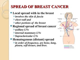
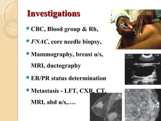
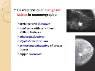
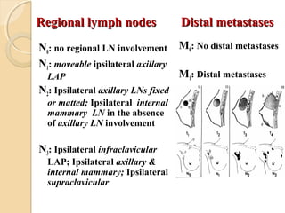
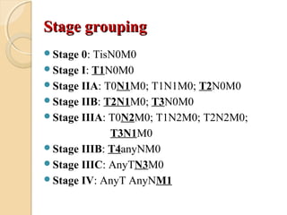
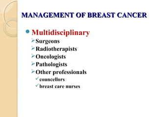
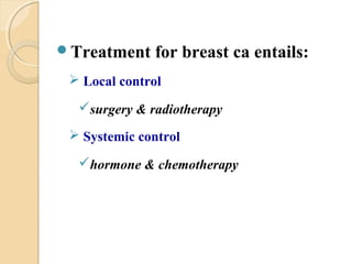
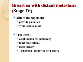
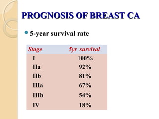
BREAST CANCERBREAST CANCER
Mizan Kidanu
March,18/2013
OUTLINEOUTLINE
Introduction
Epidemiology
Risk factors
Classification
Diagnosis
Treatment
Prognosis
INTRODUCTIONINTRODUCTION
• Two ventral bands of thickened
ectoderm (mammary ridges,
milk lines)
• These ridges disappear after a
short time, except small portions
that persist in the pectoral
region
• when normal regression fails
accessory breasts (polymastia)
or accessory nipples (polythelia)
may occur
ANATOMYANATOMY
Location
 2nd
to 6th
rib
 lateral border of
sternum to anterior
axillary line
 lies on pec. major
and seratus anterior
Contains:
 Fat, glandular tissue,
suspensory ligament
• Blood supply
internal mammary
axillary artery
intercoastal arteries
• Venous drainage
axillary vein
internal thoracic vein
lateral thoracic vein
intercoastal vein
•Lymphatic drainage
axillary LNs ~85%
internal mammary LNs
•Axillary LNs
receive approximately
85% of the drainage
grouped into:
lateral
anterior
posterior
central
apical
interpectoral
Axillary LNs
 with respect to pectoralis
minor muscle they are
grouped into:
Level-I
Level-II
Level-III
EPIDEMIOLOGYEPIDEMIOLOGY
Is the most common female cancer (26%)
2nd
common cause of cancer death in women
Main cause of death in women ages 40-59 yrs
Mortality rates have declined since the use of
screening mammography and improvements of
adjuvant therapies
Invasive ductal ca is the commonest type
Relative distributionsRelative distributions
 Upper outer quadrant ~ 60%
 Upper inner quadrant ~12%
 Lower outer quadrant ~ 10%
 Lower inner quadrant ~ 6%
 Central quadrant ~ 12%
60% 12%
6%10%
12%
RISK FACTORSRISK FACTORS
 Sex - >99% occur in females
 Early menarche, late menopause, nulliparity,
older age at first live birth
 Age - is rare below 20 yeas of age
 Radiaton exposure
 Family Hx of breast CA
 Genetic factors … BRCA-1 or BRCA-2
 Prior breast cancer
 Obesity
 Dietary factors
 Smoking & increased alcohol consumption
 Hormone replacement therapy & OCP
CLASSIFICATIONCLASSIFICATION
Carcinoma in situ, CISCarcinoma in situ, CIS
 Ductal carcinoma in situ(DIS)Ductal carcinoma in situ(DIS)
 Lobular Carcinoma in situ(LIS)Lobular Carcinoma in situ(LIS)
Invasive carcinomaInvasive carcinoma
 DuctalDuctal
scirrhous carcinoma
medullary
mucinous (colloid)
papillary
tubular
 LobularLobular
 Paget’s disease of the nipple
Carcinoma in-situCarcinoma in-situ
 Malignant cells in the duct system or lobules but
no invasion of the basement membrane
 Since the use of screening mammography there is
a 14-fold increase in the incidence
 Multicentricity - refers to the occurrence of a 2nd
breast cancer outside the breast quadrant of the
primary cancer (or at least 4 cm away)
 Multifocality - refers to the occurrence of a 2nd
cancer within the same breast quadrant as the
primary cancer (or within 4 cm of it)
LCIS
marker of increased risk for invasive
breast carcinoma, not anatomic
precursor
bilateral in 50-70%
develops only in the female breast
multicentric in 60-90%
DCIS
anatomic precursor of invasive ductal
carcinoma
multicentricity for DCIS is 40-80%
bilateral in 10-20%
CLINICAL FEATURESCLINICAL FEATURES
Lump
hard, painless swelling
Change in the skin
puckering
Peau d’orange
skin ulceration
skin nodules
Nipple changes
distortion, inversion
discharge
eczema (paget’s disease)
Metastatic disease
regional LNs
distant sites
SPREAD OF BREAST CANCERSPREAD OF BREAST CANCER
Local spread with in the breast
involves the skin & fascia
chest wall and
other portions of the breast
Regional spread of breast cancer
axillary LNs
internal mammary LNs
Supraclavicular LNs
Hematogenous (distant) spread
in order of frequency, are bone, lung,
pleura, soft tissues, and liver.
Axillary nodal metastasesAxillary nodal metastases
Common site of spread (45% at presentation)
Spread depends on the primary tumor (size)
Clinical assessment is unreliable
Axillary nodal spread Vs prognosis
number of nodes affected
level of nodal disease
DIAGNOSISDIAGNOSIS
History
duration of illness
associated symptoms (pain, headache, cough,
nipple discharge)
age at menarche and menstruation status
(pre or postmenopausal)
age at first delivery
family history of breast cancer,….
Physical examination
Inspection
arms by her side or straight
up in the air
hands on her hips
arms extended forward in a
sitting position leaning
forward to accentuate any
skin retraction
symmetry, size, and shape,
peau d'orange, nipple or skin
retraction or erythema
 Palpation
supine position
examine all quadrants
examine with the palmar
aspects of the fingers
avoiding a grasping or
pinching motion
assesses all three levels of
axillary lymphadenopathy
location, size, consistency,
shape, mobility, fixation,...
InvestigationsInvestigations
CBC, Blood group & Rh,
FNAC, core needle biopsy,
Mammography, breast u/s,
MRI, ductography
ER/PR status determination
Metastasis - LFT, CXR, CT,
MRI, abd u/s,….
Characterstics of malignant
lesions in mammography:
architectural distortion
solid mass with or without
stellate features
microcalcifications
stippled calcifications
asymmetric thickening of breast
tissues
nipple retraction
Triple AssessmentTriple Assessment
Any patient with a breast lump or other
symptoms suspicious of carcinoma, the Dx
should be made by a combination of:
1. Clinical assessment
2. Radiological imaging and
3. Tissue sample (cytological or histological)
Positive predictive value is 99.9%
TNM StagingTNM Staging
Primary tumor (T)
Tx: Primary tumor cannot be assessed
T0: No evidence of primary tumor
Tis: carcinoma in situ
T1 : ≤2 cm in greatest dimension
T2: >2 cm but not >5 cm in greatest dimension
T3: >5 cm in greatest dimension
T4: any size with direct extension to (a) chest
wall or (b) skin
Regional lymph nodesRegional lymph nodes
N0: no regional LN involvement
N1: moveable ipsilateral axillary
LAP
N2: Ipsilateral axillary LNs fixed
or matted; Ipsilateral internal
mammary LN in the absence
of axillary LN involvement
N3: Ipsilateral infraclavicular
LAP; Ipsilateral axillary &
internal mammary; Ipsilateral
supraclavicular
M0: No distal metastases
M1: Distal metastases
Distal metastasesDistal metastases
Stage groupingStage grouping
Stage 0: TisN0M0
Stage I: T1N0M0
Stage IIA: T0N1M0; T1N1M0; T2N0M0
Stage IIB: T2N1M0; T3N0M0
Stage IIIA: T0N2M0; T1N2M0; T2N2M0;
T3N1M0
Stage IIIB: T4anyNM0
Stage IIIC: AnyTN3M0
Stage IV: AnyT AnyNM1
MANAGEMENT OF BREAST CANCERMANAGEMENT OF BREAST CANCER
Multidisciplinary
Surgeons
Radiotherapists
Oncologists
Pathologists
Other professionals
councellors
breast care nurses
Treatment for breast ca entails:
 Local control
surgery & radiotherapy
 Systemic control
hormone & chemotherapy
SURGERYSURGERY
1-Wide local excision (lumpectomy)
2-Total (simple) mastectomy
 removes all breast tissue, nipple areola complex, and
skin
3-Modified Radical Mastectomy (MRM)
 preserves pectoralis major and minor muscles, allowing
removal of level I & II but not level III axillary
4-Radical mastectomy
 removes all breast tissue, skin, nipple areola complex,
pectoralis major and minor muscles, and level I, II, &
III axillary LNs
Factors affecting type of treatmentFactors affecting type of treatment
Lymph node status
 +ve node: needs adjuvant treatment
Size and extent of tumor
 large tumors recur more often
Histology
 CIS: no adjuvant treatment
Hormone receptors status
Age and/or menopausal status
Treatment of early breast cancerTreatment of early breast cancer
((Stage I & IIStage I & II))
Breast conservation - resection of the primary
breast ca with a normal margin, adjuvant
radiation therapy, and assessment of regional
lymph node status
Mastectomy with sentinel lymph node and/or
axillary LN dissection
Breast conserving surgeryBreast conserving surgery
Excision of the tumor with a rim of
normal tissue
lumpectomy
segmental mastectomy
partial mastectomy
quadrantectomy
Contraindications for breast
conserving operations(BCS)
tumor >4cm
multicentricity
centrally located tumors
poor tumor differentiation
node positive disease
positive margin after re-excision
Hx of previous radiotherapy
pregnancy
HORMONAL THERAPYHORMONAL THERAPY
Immunoassays & immunohistochemical
methods are employed to measure levels of ER
Patients with significant increase in ER
respond favourably to endocrine therapy
E.g: Tamoxifen therapy
CHEMOTHERAPYCHEMOTHERAPY
Adjuvant chemotherapy for early invasive
breast ca is indicated in all patients with:
node-positive cancers
tumor >1 cm
node-negative cancers of >0.5 cm with adverse
prognostic features (blood vessel or lymph vessel
invasion, high histologic grade, HER-2/neu
overexpression, and negative hormone receptor
status)
Locally advanced breast cancerLocally advanced breast cancer
(( Stage-IIIStage-III))
 Neoadjuvant chemotherapy
 Usually a modified radical
mastectomy (MRM)
 Followed by adjuvant
radiation therapy
Breast ca with distant metastasisBreast ca with distant metastasis
((Stage IVStage IV))
Aim of management
 provide palliation
 symptomatic relief
Treatment
 combination chemotherapy
 toilet mastectomy
 radiotherapy
 Tamoxifen therapy in ER positive
COMPLICATIONS OF MASTECTOMYCOMPLICATIONS OF MASTECTOMY
Seromas - the most common
Wound infections
Hemorrhage
Lymphedema - increased risk in:
 extensive ALND
 the delivery of radiation therapy
 the presence of pathologic lymph nodes
 obesity
Nerve injury
FOLLOW UPFOLLOW UP
Assess local recurrence, especially in BCT
Assess the contralateral breast
Detect psychiatric morbidity
Allow provision of prosthesis
Early detection & treatment of metastatic
disease
PROGNOSIS OF BREAST CAPROGNOSIS OF BREAST CA
5-year survival rate
Stage 5yr survival
I 100%
IIa 92%
IIb 81%
IIIa 67%
IIIb 54%
IV 18%
Breast  ca

Breast ca

  • 1.
  • 2.
  • 3.
    INTRODUCTIONINTRODUCTION • Two ventralbands of thickened ectoderm (mammary ridges, milk lines) • These ridges disappear after a short time, except small portions that persist in the pectoral region • when normal regression fails accessory breasts (polymastia) or accessory nipples (polythelia) may occur
  • 4.
    ANATOMYANATOMY Location  2nd to 6th rib lateral border of sternum to anterior axillary line  lies on pec. major and seratus anterior Contains:  Fat, glandular tissue, suspensory ligament
  • 5.
    • Blood supply internalmammary axillary artery intercoastal arteries • Venous drainage axillary vein internal thoracic vein lateral thoracic vein intercoastal vein •Lymphatic drainage axillary LNs ~85% internal mammary LNs
  • 6.
    •Axillary LNs receive approximately 85%of the drainage grouped into: lateral anterior posterior central apical interpectoral
  • 7.
    Axillary LNs  withrespect to pectoralis minor muscle they are grouped into: Level-I Level-II Level-III
  • 8.
    EPIDEMIOLOGYEPIDEMIOLOGY Is the mostcommon female cancer (26%) 2nd common cause of cancer death in women Main cause of death in women ages 40-59 yrs Mortality rates have declined since the use of screening mammography and improvements of adjuvant therapies Invasive ductal ca is the commonest type
  • 9.
    Relative distributionsRelative distributions Upper outer quadrant ~ 60%  Upper inner quadrant ~12%  Lower outer quadrant ~ 10%  Lower inner quadrant ~ 6%  Central quadrant ~ 12% 60% 12% 6%10% 12%
  • 10.
    RISK FACTORSRISK FACTORS Sex - >99% occur in females  Early menarche, late menopause, nulliparity, older age at first live birth  Age - is rare below 20 yeas of age  Radiaton exposure  Family Hx of breast CA  Genetic factors … BRCA-1 or BRCA-2  Prior breast cancer  Obesity  Dietary factors  Smoking & increased alcohol consumption  Hormone replacement therapy & OCP
  • 11.
    CLASSIFICATIONCLASSIFICATION Carcinoma in situ,CISCarcinoma in situ, CIS  Ductal carcinoma in situ(DIS)Ductal carcinoma in situ(DIS)  Lobular Carcinoma in situ(LIS)Lobular Carcinoma in situ(LIS) Invasive carcinomaInvasive carcinoma  DuctalDuctal scirrhous carcinoma medullary mucinous (colloid) papillary tubular  LobularLobular  Paget’s disease of the nipple
  • 12.
    Carcinoma in-situCarcinoma in-situ Malignant cells in the duct system or lobules but no invasion of the basement membrane  Since the use of screening mammography there is a 14-fold increase in the incidence  Multicentricity - refers to the occurrence of a 2nd breast cancer outside the breast quadrant of the primary cancer (or at least 4 cm away)  Multifocality - refers to the occurrence of a 2nd cancer within the same breast quadrant as the primary cancer (or within 4 cm of it)
  • 13.
    LCIS marker of increasedrisk for invasive breast carcinoma, not anatomic precursor bilateral in 50-70% develops only in the female breast multicentric in 60-90% DCIS anatomic precursor of invasive ductal carcinoma multicentricity for DCIS is 40-80% bilateral in 10-20%
  • 14.
    CLINICAL FEATURESCLINICAL FEATURES Lump hard,painless swelling Change in the skin puckering Peau d’orange skin ulceration skin nodules Nipple changes distortion, inversion discharge eczema (paget’s disease) Metastatic disease regional LNs distant sites
  • 15.
    SPREAD OF BREASTCANCERSPREAD OF BREAST CANCER Local spread with in the breast involves the skin & fascia chest wall and other portions of the breast Regional spread of breast cancer axillary LNs internal mammary LNs Supraclavicular LNs Hematogenous (distant) spread in order of frequency, are bone, lung, pleura, soft tissues, and liver.
  • 16.
    Axillary nodal metastasesAxillarynodal metastases Common site of spread (45% at presentation) Spread depends on the primary tumor (size) Clinical assessment is unreliable Axillary nodal spread Vs prognosis number of nodes affected level of nodal disease
  • 17.
    DIAGNOSISDIAGNOSIS History duration of illness associatedsymptoms (pain, headache, cough, nipple discharge) age at menarche and menstruation status (pre or postmenopausal) age at first delivery family history of breast cancer,….
  • 18.
    Physical examination Inspection arms byher side or straight up in the air hands on her hips arms extended forward in a sitting position leaning forward to accentuate any skin retraction symmetry, size, and shape, peau d'orange, nipple or skin retraction or erythema
  • 19.
     Palpation supine position examineall quadrants examine with the palmar aspects of the fingers avoiding a grasping or pinching motion assesses all three levels of axillary lymphadenopathy location, size, consistency, shape, mobility, fixation,...
  • 20.
    InvestigationsInvestigations CBC, Blood group& Rh, FNAC, core needle biopsy, Mammography, breast u/s, MRI, ductography ER/PR status determination Metastasis - LFT, CXR, CT, MRI, abd u/s,….
  • 21.
    Characterstics of malignant lesionsin mammography: architectural distortion solid mass with or without stellate features microcalcifications stippled calcifications asymmetric thickening of breast tissues nipple retraction
  • 22.
    Triple AssessmentTriple Assessment Anypatient with a breast lump or other symptoms suspicious of carcinoma, the Dx should be made by a combination of: 1. Clinical assessment 2. Radiological imaging and 3. Tissue sample (cytological or histological) Positive predictive value is 99.9%
  • 23.
    TNM StagingTNM Staging Primarytumor (T) Tx: Primary tumor cannot be assessed T0: No evidence of primary tumor Tis: carcinoma in situ T1 : ≤2 cm in greatest dimension T2: >2 cm but not >5 cm in greatest dimension T3: >5 cm in greatest dimension T4: any size with direct extension to (a) chest wall or (b) skin
  • 24.
    Regional lymph nodesRegionallymph nodes N0: no regional LN involvement N1: moveable ipsilateral axillary LAP N2: Ipsilateral axillary LNs fixed or matted; Ipsilateral internal mammary LN in the absence of axillary LN involvement N3: Ipsilateral infraclavicular LAP; Ipsilateral axillary & internal mammary; Ipsilateral supraclavicular M0: No distal metastases M1: Distal metastases Distal metastasesDistal metastases
  • 25.
    Stage groupingStage grouping Stage0: TisN0M0 Stage I: T1N0M0 Stage IIA: T0N1M0; T1N1M0; T2N0M0 Stage IIB: T2N1M0; T3N0M0 Stage IIIA: T0N2M0; T1N2M0; T2N2M0; T3N1M0 Stage IIIB: T4anyNM0 Stage IIIC: AnyTN3M0 Stage IV: AnyT AnyNM1
  • 26.
    MANAGEMENT OF BREASTCANCERMANAGEMENT OF BREAST CANCER Multidisciplinary Surgeons Radiotherapists Oncologists Pathologists Other professionals councellors breast care nurses
  • 27.
    Treatment for breastca entails:  Local control surgery & radiotherapy  Systemic control hormone & chemotherapy
  • 28.
    SURGERYSURGERY 1-Wide local excision(lumpectomy) 2-Total (simple) mastectomy  removes all breast tissue, nipple areola complex, and skin 3-Modified Radical Mastectomy (MRM)  preserves pectoralis major and minor muscles, allowing removal of level I & II but not level III axillary 4-Radical mastectomy  removes all breast tissue, skin, nipple areola complex, pectoralis major and minor muscles, and level I, II, & III axillary LNs
  • 29.
    Factors affecting typeof treatmentFactors affecting type of treatment Lymph node status  +ve node: needs adjuvant treatment Size and extent of tumor  large tumors recur more often Histology  CIS: no adjuvant treatment Hormone receptors status Age and/or menopausal status
  • 30.
    Treatment of earlybreast cancerTreatment of early breast cancer ((Stage I & IIStage I & II)) Breast conservation - resection of the primary breast ca with a normal margin, adjuvant radiation therapy, and assessment of regional lymph node status Mastectomy with sentinel lymph node and/or axillary LN dissection
  • 31.
    Breast conserving surgeryBreastconserving surgery Excision of the tumor with a rim of normal tissue lumpectomy segmental mastectomy partial mastectomy quadrantectomy
  • 32.
    Contraindications for breast conservingoperations(BCS) tumor >4cm multicentricity centrally located tumors poor tumor differentiation node positive disease positive margin after re-excision Hx of previous radiotherapy pregnancy
  • 33.
    HORMONAL THERAPYHORMONAL THERAPY Immunoassays& immunohistochemical methods are employed to measure levels of ER Patients with significant increase in ER respond favourably to endocrine therapy E.g: Tamoxifen therapy
  • 34.
    CHEMOTHERAPYCHEMOTHERAPY Adjuvant chemotherapy forearly invasive breast ca is indicated in all patients with: node-positive cancers tumor >1 cm node-negative cancers of >0.5 cm with adverse prognostic features (blood vessel or lymph vessel invasion, high histologic grade, HER-2/neu overexpression, and negative hormone receptor status)
  • 35.
    Locally advanced breastcancerLocally advanced breast cancer (( Stage-IIIStage-III))  Neoadjuvant chemotherapy  Usually a modified radical mastectomy (MRM)  Followed by adjuvant radiation therapy
  • 36.
    Breast ca withdistant metastasisBreast ca with distant metastasis ((Stage IVStage IV)) Aim of management  provide palliation  symptomatic relief Treatment  combination chemotherapy  toilet mastectomy  radiotherapy  Tamoxifen therapy in ER positive
  • 37.
    COMPLICATIONS OF MASTECTOMYCOMPLICATIONSOF MASTECTOMY Seromas - the most common Wound infections Hemorrhage Lymphedema - increased risk in:  extensive ALND  the delivery of radiation therapy  the presence of pathologic lymph nodes  obesity Nerve injury
  • 38.
    FOLLOW UPFOLLOW UP Assesslocal recurrence, especially in BCT Assess the contralateral breast Detect psychiatric morbidity Allow provision of prosthesis Early detection & treatment of metastatic disease
  • 39.
    PROGNOSIS OF BREASTCAPROGNOSIS OF BREAST CA 5-year survival rate Stage 5yr survival I 100% IIa 92% IIb 81% IIIa 67% IIIb 54% IV 18%