Downloaded 182 times


![Classification of Cranial Nerves Components
● There are some overlaps and exceptions!
● According to the direction of the component:
○ Afferent: means going from peripheral
tissues to the brain (inward signal).
○ Efferent: means going from the brain to
peripheral tissues (outward signal). [consider
Efferent=Exit to memorize].
21](https://image.slidesharecdn.com/4-190712142636/85/4-Brainstem-1-Overview-Medulla-Oblongata-21-320.jpg)


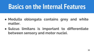
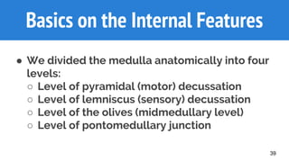
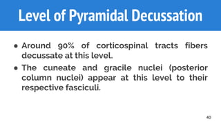


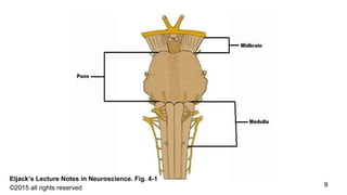
The document provides a comprehensive overview of the brainstem, particularly the medulla oblongata, including its anatomy, functions, cranial nerves, and clinical syndromes associated with it. It details the internal and external structures of the brainstem and highlights important clinical conditions such as Arnold-Chiari syndrome and lateral medullary syndrome. The document serves as an educational resource for understanding cranial nerve classification and the complexities of the brainstem's development and function.


![ONFH[AVN HIP] -TRIPLE REGIME -A NOVAL SURGICAL CONCEPT .pptx](https://cdn.slidesharecdn.com/ss_thumbnails/onfhavnhip2026koaconcalicutdrgokuldevdrmashraf-260210064517-213ec005-thumbnail.jpg?width=640&height=640&fit=bounds)


