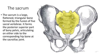
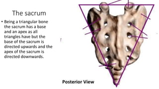
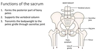
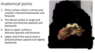
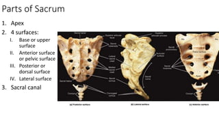
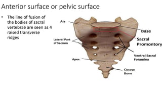
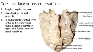
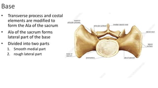
The Sacrum
Dr. Mushfiqul Hoque
Lecturer
Dhaka National Medical College
The sacrum
• The sacrum is a large,
flattened, triangular bone
formed by the fusion of five
sacral vertebrae. It forms
the posterior superior part
of bony pelvis, articulating
on either side to the
corresponding hip bone at
the sacroiliac joint.
The sacrum
• Being a triangular bone
the sacrum has a base
and an apex as all
triangles have but the
base of the sacrum is
directed upwards and the
apex of the sacrum is
directed downwards.
The sacrum
• The upper part is huge
because it supports body
weight and transmits it to
the hip bones. The lower
part is free from weight,
and therefore tapers
rapidly.
Functions of the sacrum
1. Forms the posterior part of bony
pelvis
2. Supports the vertebral column
3. Transmits the bodyweight to the
pelvic girdle through sacroiliac joint
Anatomical points
1. Pelvic surface which is convex and
smooth is directed downwards and
forwards
2. The Dorsal surface is rough and
convex and directed upwards and
backwards
3. Base or upper surface is
directed upwards and forwards
4. Upper end of the sacral canal is
directed almost upward and slightly
backwards
Parts of Sacrum
1. Apex
2. 4 surfaces:
I. Base or upper
surface
II. Anterior surface
or pelvic surface
III. Posterior or
dorsal surface
IV. Lateral surface
3. Sacral canal
Anterior surface or pelvic surface
1. Directed forwards and
downwards
2. Concave
3. Median area is marked
by four transverse
ridges which indicate
the lines of fusion of
the bodies of the
sacral vertebrae
4. These ridges end on
either side at the four
pelvic sacral foramina
Anterior surface or pelvic surface
1. It has 4 pairs of pelvic
sacral foramina which
communicate through
intervertebral
foramina with sacral
canal
2. Bars of bone which
separate the foramina
from each other
represent the costal
element
Anterior surface or pelvic surface
• Surface lateral to sacral
foramina on each side
are formed by the
fusion of costal
elements to one
another
Anterior surface or pelvic surface
• Sacral foramen
transmits ventral rami
of upper 4 sacral
nerves and branch
from lateral sacral
artery
Anterior surface or pelvic surface
• The line of fusion of
the bodies of sacral
vertebrae are seen as 4
raised transverse
ridges
Attachments of Anterior surface or pelvic surface
• The piriformis muscle arises from
the bodies of middle 3 sacral
vertebrae in an E shaped fashion
• Origin of iliacus is partly from this
surface close to the sacroiliac
joint on the rough part of the Ala of
the sacrum
• Insertion of cuccygeus muscle in
the lower lateral part
Ligaments on Anterior surface or pelvic surface
• Anterior Sacro iliac ligaments
• Sacro-spinous ligaments
• Sacro-coccygeal ligaments
Relations of Anterior surface or pelvic surface
1. Median sacral vessels in the
median plane
2. Sympathetic trunks along
medial margins of sacral
foramina
3. Upper two and half sacral
vertebrae are related to
parietal peritoneum
4. Lower two and half sacral
vertebrae are related to
rectum but separated from
S3 by bifurcation of superior
rectal artery
Dorsal surface or posterior surface
1. Rough, irregular, convex
2. Faces backwards and
upwards
3. Raised and interrupted crest
in the midline known as
median sacral crest which
represents fused spines of
sacral vertebrae
Dorsal surface or posterior surface
• Sacral hiatus, an inverted U
shaped gap below the 4th
spinous tubercle in the
posterior wall of sacral canal
which is due to failure of 5th
sacral vertebrae to meet in
the median plane
Dorsal surface or posterior surface
• Dorsal sacral foramina, 4 in
number lying on each side of
the median crest. They
communicate with the sacral
canal through intervertebral
foramen and transmit dorsal
rami of sacral nerves
Dorsal surface or posterior surface
• Intermediate sacral crest,
these are 4 small articular
tubercles lying just medial to
the dorsal sacral foramina.
These represent the articular
processes fused together
• Lateral sacral crest, lies
lateral to the dorsal sacral
foramina, formed by fusion
of transverse processes of
the sacral vertebrae
Dorsal surface or posterior surface
• Sacral corua, free and
projecting part of the lower
part of 5th sacral vertebrae
on the sides of the sacral
hiatus; it represents the
inferior articular processes of
the 5th sacral vertebrae
Attachments of Dorsal surface
• Erector spinae muscle arises
from an elongated U shaped
linear area involving the
continuous rows of spinous
and transverse tubercle
Attachments of Dorsal surface
• Multifidus arises from the
area enclosed by the U
shaped origin of Erector
spinae muscle
• Gluteus maximus arises from
lower and lateral part of this
surface
• Sacrotuberous ligament
attached to the lower and
lateral part below the
attachment of gluteus
maximus
Lateral surface
• Formed by fused transverse
process and costal elements
• Broader above and narrower
below
• Upper ear shaped part is
called the articular surface
which articulate with the
ilium to form the sacroiliac
joint. This articular surface is
formed entirely by costal
element of the sacral
vertebrae
Lateral surface
• The area behind the articular
surface is rough for
attachments of ligaments
Lateral surface
• At the lower part of the lateral surface it suddenly bends and forms an angle
which is called inferior lateral angle.
• Below the angle the lateral surface becomes very narrow and almost like a
border.
Attachments of Lateral surface
• The rough area behind the
articular surface gives
attachment to Interosseous
Sacro-iliac ligament
• Lower narrow part below the
articular surface gives
attachment to 4 structures
from behind to forwards:
1.Gluteus Maximus
2.Sacrotuberous and
sacrospinous ligaments
3.coccygeus
Attachments of Lateral surface
• Inferior lateral angle gives
attachment to lateral
sacrococcygeal ligament
Base
• Directed upwards and
forwards
• Formed by superior surface
of 1st sacral vertebrae
• It articulates with 5th lumbar
vertebrae to form sacro-
vertebral angle
• Divided into three parts
1.Median part
2.Right lateral part
3.Left lateral part
Base
• It has the parts of a typical
vertebrae in modified form
1. Body:
I. Transverse measurement
is more that anterior-
posterior measurement
II. Sacral promontory:
anterior projecting part of the
body
2. Vertebral foramen: lies behind
the body and leads to sacral
canal
3. Superior articular process: to
articulate with the Lumbar 5
vertebrae
Base
• Transverse process and costal
elements are modified to
form the Ala of the sacrum
• Ala of the sacrum forms
lateral part of the base
• Divided into two parts
1. Smooth medial part
2. rough lateral part
Attachments of Base
• Rough lateral part of the Ala
of the sacrum gives origin to
iliacus muscle anteriorly and
gives attachment to
lumbosacral ligaments
posteriorly
Relations of Base
• Smooth medial surface of the
Ala of sacrum is related to
following 4 structures from
medial to lateral side:
1. Sympathetic chain
2. Lumbosacral trunk
3. Iliolumbar artery
4. Obturator nerve
Joints formed with sacrum
• Between the upper surface
of sacrum and body of
lumbar 5th vertebrae,
secondary cartilaginous joint
• Between apex of sacrum and
coccyx, secondary
cartilaginous joint
• Between lateral articular
surface of sacrum and ilium
of hip bone on both sides,
plane variety of synovial
joint
Sacral canal
• Formed by vertebral
foramina of sacral vertebrae
• Triangular in shape
• Lateral wall of the canal has 4
intervertebral foramens
which communicates in front
with pelvic sacral foramina
and in the back with dorsal
sacral foramina
Sacral canal
Contents:
1. Cauda equina including filum
terminale
2. Spinal meninges
3. Sacral and coccygeal nerve
roots
Sacral hiatus transmits:
1. 5th pair of sacral nerves
2. Coccygeal nerves
3. Filum terminale
Sacral canal
Contents:
1. Cauda equina including filum
terminale
2. Spinal meninges
3. Sacral and coccygeal nerve
roots
Sacral hiatus transmits:
1. 5th pair of sacral nerves
2. Coccygeal nerves
3. Filum terminale
Sacral canal
Contents:
1. Cauda equina including filum
terminale
2. Spinal meninges
3. Sacral and coccygeal nerve
roots
Sacral hiatus transmits:
1. 5th pair of sacral nerves
2. Coccygeal nerves
3. Filum terminale
End of discussion
Any questions??

Sacrum

  • 1.
    The Sacrum Dr. MushfiqulHoque Lecturer Dhaka National Medical College
  • 3.
    The sacrum • Thesacrum is a large, flattened, triangular bone formed by the fusion of five sacral vertebrae. It forms the posterior superior part of bony pelvis, articulating on either side to the corresponding hip bone at the sacroiliac joint.
  • 4.
    The sacrum • Beinga triangular bone the sacrum has a base and an apex as all triangles have but the base of the sacrum is directed upwards and the apex of the sacrum is directed downwards.
  • 5.
    The sacrum • Theupper part is huge because it supports body weight and transmits it to the hip bones. The lower part is free from weight, and therefore tapers rapidly.
  • 6.
    Functions of thesacrum 1. Forms the posterior part of bony pelvis 2. Supports the vertebral column 3. Transmits the bodyweight to the pelvic girdle through sacroiliac joint
  • 7.
    Anatomical points 1. Pelvicsurface which is convex and smooth is directed downwards and forwards 2. The Dorsal surface is rough and convex and directed upwards and backwards 3. Base or upper surface is directed upwards and forwards 4. Upper end of the sacral canal is directed almost upward and slightly backwards
  • 8.
    Parts of Sacrum 1.Apex 2. 4 surfaces: I. Base or upper surface II. Anterior surface or pelvic surface III. Posterior or dorsal surface IV. Lateral surface 3. Sacral canal
  • 9.
    Anterior surface orpelvic surface 1. Directed forwards and downwards 2. Concave 3. Median area is marked by four transverse ridges which indicate the lines of fusion of the bodies of the sacral vertebrae 4. These ridges end on either side at the four pelvic sacral foramina
  • 10.
    Anterior surface orpelvic surface 1. It has 4 pairs of pelvic sacral foramina which communicate through intervertebral foramina with sacral canal 2. Bars of bone which separate the foramina from each other represent the costal element
  • 11.
    Anterior surface orpelvic surface • Surface lateral to sacral foramina on each side are formed by the fusion of costal elements to one another
  • 12.
    Anterior surface orpelvic surface • Sacral foramen transmits ventral rami of upper 4 sacral nerves and branch from lateral sacral artery
  • 13.
    Anterior surface orpelvic surface • The line of fusion of the bodies of sacral vertebrae are seen as 4 raised transverse ridges
  • 14.
    Attachments of Anteriorsurface or pelvic surface • The piriformis muscle arises from the bodies of middle 3 sacral vertebrae in an E shaped fashion • Origin of iliacus is partly from this surface close to the sacroiliac joint on the rough part of the Ala of the sacrum • Insertion of cuccygeus muscle in the lower lateral part
  • 15.
    Attachments of Anteriorsurface or pelvic surface • The piriformis muscle arises from the bodies of middle 3 sacral vertebrae in an E shaped fashion • Origin of iliacus is partly from this surface close to the sacroiliac joint on the rough part of the Ala of the sacrum • Insertion of cuccygeus muscle in the lower lateral part
  • 16.
    Ligaments on Anteriorsurface or pelvic surface • Anterior Sacro iliac ligaments • Sacro-spinous ligaments • Sacro-coccygeal ligaments
  • 17.
    Relations of Anteriorsurface or pelvic surface 1. Median sacral vessels in the median plane 2. Sympathetic trunks along medial margins of sacral foramina 3. Upper two and half sacral vertebrae are related to parietal peritoneum 4. Lower two and half sacral vertebrae are related to rectum but separated from S3 by bifurcation of superior rectal artery
  • 18.
    Relations of Anteriorsurface or pelvic surface 1. Median sacral vessels in the median plane 2. Sympathetic trunks along medial margins of sacral foramina 3. Upper two and half sacral vertebrae are related to parietal peritoneum 4. Lower two and half sacral vertebrae are related to rectum but separated from S3 by bifurcation of superior rectal artery
  • 19.
    Dorsal surface orposterior surface 1. Rough, irregular, convex 2. Faces backwards and upwards 3. Raised and interrupted crest in the midline known as median sacral crest which represents fused spines of sacral vertebrae
  • 20.
    Dorsal surface orposterior surface • Sacral hiatus, an inverted U shaped gap below the 4th spinous tubercle in the posterior wall of sacral canal which is due to failure of 5th sacral vertebrae to meet in the median plane
  • 21.
    Dorsal surface orposterior surface • Dorsal sacral foramina, 4 in number lying on each side of the median crest. They communicate with the sacral canal through intervertebral foramen and transmit dorsal rami of sacral nerves
  • 22.
    Dorsal surface orposterior surface • Intermediate sacral crest, these are 4 small articular tubercles lying just medial to the dorsal sacral foramina. These represent the articular processes fused together • Lateral sacral crest, lies lateral to the dorsal sacral foramina, formed by fusion of transverse processes of the sacral vertebrae
  • 23.
    Dorsal surface orposterior surface • Sacral corua, free and projecting part of the lower part of 5th sacral vertebrae on the sides of the sacral hiatus; it represents the inferior articular processes of the 5th sacral vertebrae
  • 24.
    Attachments of Dorsalsurface • Erector spinae muscle arises from an elongated U shaped linear area involving the continuous rows of spinous and transverse tubercle
  • 25.
    Attachments of Dorsalsurface • Multifidus arises from the area enclosed by the U shaped origin of Erector spinae muscle • Gluteus maximus arises from lower and lateral part of this surface • Sacrotuberous ligament attached to the lower and lateral part below the attachment of gluteus maximus
  • 26.
    Lateral surface • Formedby fused transverse process and costal elements • Broader above and narrower below • Upper ear shaped part is called the articular surface which articulate with the ilium to form the sacroiliac joint. This articular surface is formed entirely by costal element of the sacral vertebrae
  • 27.
    Lateral surface • Thearea behind the articular surface is rough for attachments of ligaments
  • 28.
    Lateral surface • Atthe lower part of the lateral surface it suddenly bends and forms an angle which is called inferior lateral angle. • Below the angle the lateral surface becomes very narrow and almost like a border.
  • 29.
    Attachments of Lateralsurface • The rough area behind the articular surface gives attachment to Interosseous Sacro-iliac ligament • Lower narrow part below the articular surface gives attachment to 4 structures from behind to forwards: 1.Gluteus Maximus 2.Sacrotuberous and sacrospinous ligaments 3.coccygeus
  • 30.
    Attachments of Lateralsurface • Inferior lateral angle gives attachment to lateral sacrococcygeal ligament
  • 31.
    Base • Directed upwardsand forwards • Formed by superior surface of 1st sacral vertebrae • It articulates with 5th lumbar vertebrae to form sacro- vertebral angle • Divided into three parts 1.Median part 2.Right lateral part 3.Left lateral part
  • 32.
    Base • It hasthe parts of a typical vertebrae in modified form 1. Body: I. Transverse measurement is more that anterior- posterior measurement II. Sacral promontory: anterior projecting part of the body 2. Vertebral foramen: lies behind the body and leads to sacral canal 3. Superior articular process: to articulate with the Lumbar 5 vertebrae
  • 33.
    Base • Transverse processand costal elements are modified to form the Ala of the sacrum • Ala of the sacrum forms lateral part of the base • Divided into two parts 1. Smooth medial part 2. rough lateral part
  • 34.
    Attachments of Base •Rough lateral part of the Ala of the sacrum gives origin to iliacus muscle anteriorly and gives attachment to lumbosacral ligaments posteriorly
  • 35.
    Relations of Base •Smooth medial surface of the Ala of sacrum is related to following 4 structures from medial to lateral side: 1. Sympathetic chain 2. Lumbosacral trunk 3. Iliolumbar artery 4. Obturator nerve
  • 37.
    Joints formed withsacrum • Between the upper surface of sacrum and body of lumbar 5th vertebrae, secondary cartilaginous joint • Between apex of sacrum and coccyx, secondary cartilaginous joint • Between lateral articular surface of sacrum and ilium of hip bone on both sides, plane variety of synovial joint
  • 38.
    Sacral canal • Formedby vertebral foramina of sacral vertebrae • Triangular in shape • Lateral wall of the canal has 4 intervertebral foramens which communicates in front with pelvic sacral foramina and in the back with dorsal sacral foramina
  • 39.
    Sacral canal Contents: 1. Caudaequina including filum terminale 2. Spinal meninges 3. Sacral and coccygeal nerve roots Sacral hiatus transmits: 1. 5th pair of sacral nerves 2. Coccygeal nerves 3. Filum terminale
  • 40.
    Sacral canal Contents: 1. Caudaequina including filum terminale 2. Spinal meninges 3. Sacral and coccygeal nerve roots Sacral hiatus transmits: 1. 5th pair of sacral nerves 2. Coccygeal nerves 3. Filum terminale
  • 41.
    Sacral canal Contents: 1. Caudaequina including filum terminale 2. Spinal meninges 3. Sacral and coccygeal nerve roots Sacral hiatus transmits: 1. 5th pair of sacral nerves 2. Coccygeal nerves 3. Filum terminale
  • 42.