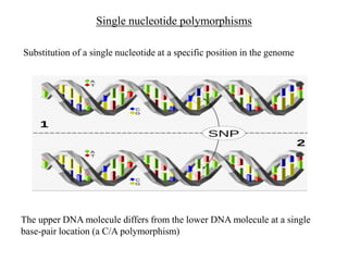
Blood Group Genotyping
M.G.R.R. Muhandiram (MBBS, DTM, MD)
Consultant in Transfusion Medicine (Acting)
National Blood Transfusion Service
Sri Lanka
Introduction
• Genomics is impacting in all areas of medicine
• In transfusion medicine,
DNA-based genotyping is being used as an alternative to
serological antibody-based methods
• Single nucleotide changes (SNP's) in the respective genes responsible for
most antigenic polymorphisms
• Validated by comparison with antibody-based typing
• Provide the patient’s comprehensive extended blood group profile as part
of their medical record by the growth of whole genome sequencing
(WGS)
• Since the discovery of the ABO system, blood typing by antibody-
based methods has been the standard for determining ABO, Rh, &
ā€œextendedā€ blood group antigens
• A blood group antigen (Ag)
polymorphism on RBCs (platelets and neutrophils) that differ between
individuals and stimulates production of an immune antibody following
exposure via pregnancy or blood transfusion
• The association of a specific polymorphism with antibody production
is central to defining an antigen
• Most of these blood group systems were discovered when Ag-specific
abs present in the human sera were identified by agglutination-based
methods
These serology-based techniques are simple and inexpensive
and when correctly performed, have specificity and sensitivity
appropriate for the clinical care of the majority of patients
Why need genotyping?
ā€œHaemagglutination methods have certain limitationsā€
1. A subjective test, labor-intensive and time-consuming
2. High cost of reagents and labor & lack of specificities – routine typing for
transfusion, has been limited to ABO and RhD as the most important blood
groups
3. Commercial antibody reagents for ABO and Rh (D, C,c, E,e) and
approximately 18 other common blood group antigen specificities are
available, but no typing reagents for many clinically significant antigens
4. For some blood group Ags, the antisera prepared from polyclonal human
serum are poorly standardized & available in limited stocks
5. Reagents’ sensitivity and specificity can vary
6. ā€œExtended or minorā€ Ag typing has been challenging for automation - the
carrier protein and/or carbohydrate are often part of trans-membrane
complexes – therefore the recognition of antigenic epitope often requires
establishing the native conformation
7. Does not reliably predict a fetus at risk of HDFN.
8. Haemagglutination technique has restricted the ability to
• Predict RhD zygosity,
• Define weaker variants of ABO and Rh,
• Red cell phenotyping in multi-transfused and DAT positive
patients
Molecular basis of blood group antigen expression
Different mechanisms responsible for the molecular diversity of the blood
group systems
However, most blood groups are encoded by single nucleotide polymorphisms
(SNPs)
Gene conversion or recombination events (MNs, Rh, Ch/Rg
blood group system)
Duplication of an exon (Gerbich)
Deletion of a gene, exon, or nucleotide(s) (ABO, Rh, MNS, Kel,
Duffy, Dombrock)
Insertion of a nucleotide(s) (Rh, Colton)
Single nucleotide substitutions (Most blood group systems)
Single nucleotide polymorphisms
Substitution of a single nucleotide at a specific position in the genome
The upper DNA molecule differs from the lower DNA molecule at a single
base-pair location (a C/A polymorphism)
Table 1: Different molecular events giving rise to blood group phenotypes (Adapted
from Hillyer CD 2007)
Event Mechanism Blood group phenotype
Promoter mutation Nucleotide change in GATA box Fy(b-)
Alternative spicing Nucleotide change in splice site S-s-, Gy(a-)
Premature stop codon 1.Deletion of nucleotides Fy(a-b-), Rh null, Ge2, Ge3, Ge4,
K0
2. Insertion of nucleotides D-, Co(a-b-)
Amino acid change Missense mutation D-, Rh null, K0
Hybrid genes 1.Crossover GP.Vw, GP.Hil
2.Gene conversion GP.Mur, GP.Hop, D --
Interacting protein 1. Absence of RHAG Rh null
2. Absence of Kx Weak expression of Kell antigen
Modifying gene IN(LU) LU(a-b-)
These variations that encode different blood group Ags are documented in the
Blood Group Antigen Gene Mutation Database (dbRBC)
It was set up in 1999 through an initiative of the Human Genome Variation
Society (HGVS)
A part of the resource of the National Center for Biotechnology Information
(NCBI) of USA. It consists in an online repository that includes about 1251
alleles of all 40 gene loci
1. ABO blood group system
The ABO gene, located on 9q34.1-q34.2
Contains 7 exons ranging from 28 to 688 bp
Exon 7 is the largest and exon 6 & 7 contains most of the coding
sequence ,encode for the catalytic domain of the ABO
glycosyltransferases
More than 200 ABO alleles have been identified by molecular Ix
3 main allelic forms: known as IA, IB, and i.
• All ABO Ags arise from mutations in the single ABO gene;
3 specific mutations, which show a high frequency in the population,
indirectly cause changes in the epitope structures resulting A, B specificities
• Frame-shift mutations -inactivates the enzyme altogether, leaving the H
glycan unmodified
• Recombination or gene conversion between common alleles may lead to
hybrids
• Numerous mutations associated with weak subgroups, non-deletional null (O)
alleles
• Deletions, mutations, or recombination within two exons of the coding DNA
cause less efficient transfer of the immunodominant sugar to H substance-
ABO subgroups
The B antigen allele is a mutation/polymorphic form of A antigen allele. Location 9q34.1-34.2
Differs from allele A in terms of four AAs
O allele also a modification of allele for A antigen. Due to a deletion in the
allele for A Ag creating frame-shift mutation
Frame-shift mutations due to deletions or additions of 1, 2, or 4 nucleotides that
change the ribosome reading frame and cause premature termination of translation
• The Bombay phenotype is caused by point mutation in the coding region
of FUT1(H) gene. The mutation introduces a stop codon rendering the
enzyme inactive.
(codon- a sequence of 3 DNA or RNA nucleotides that corresponds with a
specific AA or stop signal during protein synthesis)
• In Caucasians, the Bombay phenotype may be caused by a number of
mutations. Likewise, a number of mutations have been reported to
underlie the para-Bombay phenotype
2. Rh blood group system
• The Rh blood group system is encoded by two closely linked and highly
homologous RH genes (RHD & RHCE) present at chromosome location
1p36.1
• Both RHD and RHCE genes are inherited together and consists of 10 exons
each that encode a transmembrane protein with 6 extracellular loops
• RHD and RHCE genes strongly favors the formation of hybrid alleles of
RHD-CE-D or RHCE-D-CE type.
RHD negativity- 3 molecular mechanisms of RHD negativity:
1.Total deletion of RHD gene
2.RHD pseudogene - causes the creation of a stop codon signal
3.Hybrid gene
3. Other Blood Group Systems
• Most of the Ags of other Bg systems are encoded by SNPs
• These SNPs can be either
– Silent (do not alter the AA sequence)
– Missense (cause change of one AA to another)
– Nonsense (change the codon to stop codon)
– May alter splice sites -DNA sequence that occurs at the boundary of an
exon and an intron. This change can disrupt RNA splicing & altered
protein-coding sequence
DNA-arrays (specific DNA sequences either deposited or synthesized & attached to a solid
surface) for extended RBC typing do not routinely include ABO and RhD
• A single mutation anywhere in an A or B allele, or in RHD allele, can result
in an inactive transferase, i.e. a group O phenotype, or RhD-negative
phenotype.
• DNA-arrays cannot target every nucleotide in the gene, a novel or
uncommon mutation would be undetected and the possibility of not
getting the ABO or RhD correctly
• Serologic ABO/RhD typing is
fast
Accurate and
Relatively inexpensive
So , in contrast to other blood group antigens genotyping will not be relied
on as the sole means for determining ABO/RhD
Even with these limitations ABO genotyping is increasingly being used
for
• Transplant registries to aid in donor selection
• Useful to resolve patient and donor typing discrepancies
• Determine the original blood type of patients massively transfused to
conserve Group O donor inventories
• To determine the original blood type of transplant recipients by testing a
buccal sample
• To confirm A2 subgroup in kidney donors who may have been transfused
or whose RBCs give discordant reactivity in serologic testing with anti-A1
reagents-
(Transplantation of living donor A2 renal allografts into non-A recipients produces
excellent long-term allograft survival and expands the potential living donor pool for
non-group A recipients)
Uses of DNA-based genotyping for Transfusion Medicine
1) Type patients for multiple antigens in a single assay
Extended antigen profiling is best done by genotyping as testing involves
a single automated assay
More accurate and provides more information
Testing only needs to be performed once (when made part of the patient
transfusion record)
2) RBC genotyping in Sickle Cell Disease (SCD)
Alloimmunization is a serious complication
Performing an extended antigen profile on patients with SCD prior to the
first transfusion is recommended
Genotyping improves accuracy and provides more information
e.g
RBC genotyping identifies ~20% of patients with SCD whose RBCs type
C+ by serology, but who have altered C antigen.
They should receive C- donor units as it has been shown in several cohorts
that one third make anti-C if transfused with C+ donor units
Overall ~ 6% of African-American RhD+ patients with SCD are at risk
for clinically significant anti-D; ~ 21% of C+ patients are at risk for
anti-C,& 21% are at risk for anti-c or -e when prophylactic matching
is based on serology
• Genotyping also identifies patients who lack high-prevalence antigens,
avoids misdiagnosis as a warm autoantibody
• Genotyping also alerts the transfusion service to incompatibilities in the
Dombrock system (Doa/b, Joa,Hy) Kell system (Kpa/b, Jsa/b)
Colton (Coa/b)
Yt (a/b) Lutheran (Lua/b)
Diego (Dia/b) Scianna (Sc1/2)
• Incompatibility in these systems should be considered when patients are
hemolyzing transfused RBCs in the absence of detectable new antibodies
These antibodies can be clinically significant and life-threatening but
Difficult to identify
No methods were previously available to type donors to find
compatible units
• SCD patients sometimes make Rh antibodies despite D, C, and E antigen
matching by serology.( Frequently in Africans)
This occurs because Rh epitopes are complex and RH genes in individuals
of African Black background are diverse
• To avoid alloimmunization : High-resolution typing is required
3) Type patients receiving monoclonal antibody therapies that interfere
with pre-transfusion testing
Some monoclonal antibody drug therapies cause interference in pre-
transfusion testing
• Most often encountered is anti-CD38 (daratumumab) approved in 2015 for
treatment of MM. Anti-CD38 interferes in the indirect antiglobulin testing
(IAT)
• A monoclonal antibody targeting CD47 is also in phase I clinical trials-
anti-CD47 interferes with all phases of pre-transfusion testing including
ABO typing
4) Type patients who have been recently transfused or RBCs coated with
immunoglobulin e. g. autoimmune hemolytic anemia
When RBCs are coated with IgG, RBC antigens cannot be typed accurately
 particularly when directly agglutinating antibodies are not available or IgG
removal by chemical treatment of RBCs is insufficient
5) Genotyping for Blood Donors
DNA-based typing can also be used to type blood donor for
• Transfusion
• For reagent panels
Particularly useful when antisera are not available or are weakly reactive
e.g. Dombrock blood group polymorphism ,to type patients and donors for
Doa and Dob to overcome the dearth of typing reagents
Healthy donors sometime have depressed ABO antigens (subgroups) or low
titers of natural ABO antibodies.
The products cannot be labeled for transfusion because the RBC and plasma
ABO typing appears to be discordant and products are often discarded
ABO genotyping can determine the donor’s inherited ABO type
DNA methods offer the potential to be a ā€œconfirmatory testā€
This avoid discarding safe & effective donor products & retain donors with
depressed antibody titers (whose products are superior when out-of-ABO
group plasma and platelet transfusions needed)
With DNA-based genotyping, extended antigen typing of donors is becoming
practical, more economical, and readily available
6) Resolution of weak A, B, and D typing discrepancies
The bases of many of weak subgroups of A and B are associated with
altered transferase genes
PCR-based assays can be used to define the transferase gene and thus the
ABO group
Similarly with the D Ag of the Rh blood group system, a proportion of
blood donors that historically have been typed as D-negative are now
reclassified as D-positive, due to monoclonal reagents that detect small and
specific parts of the D antigen.
The molecular basis of numerous D variants can be used to identify the
genes encoding altered RhD protein in these individuals
7) RBC genotyping in Bone Marrow Transplantation
Most antibodies to RBCs including ABO are not a barrier to engraftment,
but alloantibodies can cause complications
For transplant patients who have existing RBC alloantibodies
1) Genotyping both the donor and recipient for extended blood group
antigens &
2) Evaluating donor/recipient compatibility prior to transplantation
proactively informs donor selection and transfusion support
3) RBC genotyping can guide donor selection if several HLA-identical
donors are eligible, i.e. siblings in a related transplant
This is particularly relevant for patients with SCD undergoing
transplantation who are often highly RBC alloimmunized
4) Post-transplantation, genotyping of the recipient peripheral blood sample
and buccal cells can be done to determine the origin of new antibodies
i.e. donor-derived or
recipient-derived,
to inform selection of units for transfusion to provide the best
transfusion support
8) Prenatal Testing (FNAIT, HDFN and RHD typing)
Paternal testing
Paternal genotype is key to assessing the risk for complications
If the father is antigen positive, the paternal gene copy number (zygosity) is
determined
Fetal testing
Circulating free fetal DNA is released from fetal &/or placental cells which
undergoing apoptosis
The majority of fetal DNA is circulate in membrane-bound vesicles (apoptotic
bodies)
Present by 5 weeks of gestation. Gestational age correlates positively with amount
of fetal DNA in plasma; higher detection rates reported with increased
gestation and rapid clearance following delivery
During the last 8 weeks of pregnancy ,sharp increase of fetal DNA in maternal
plasma- might be related to gradual breakdown of the maternal–fetal interface/
placental barrier
Increased fetal DNA concentrations
Pre-eclampsia
Intrauterine growth restriction (IUGR)
Preterm labour
Invasive placentation
Hyperemesis gravidarum
Feto-maternal haemorrhage
Polyhydramnious
Methods
Amniocentesis
Chorionic villous sampling
cff-DNA
In Europe, to type the fetus for most clinical blood group antigens
(D, c, C, E, e, K, HPA-1a) use genotyping
If the father is heterozygous or paternity is uncertain or unknown -fetal DNA
determine fetal antigen status
If fetal antigen negative- mother need not undergo invasive and costly
monitoring or receive immune-modulating agents
If the fetus is antigen positive- Appropriate maternal and fetal monitoring
To determine fetal RhD type
Antenatal RhIg is routinely administered to all RhDāˆ’ women as the fetal
blood type is not known
Postnatal immunoprophylaxis given based on the RhD type of the
Newborn
Now routinely in several European countries use cffDNA to determine fetal
RhD type and avoid unnecessary administration of antenatal RhIG ,no cord
blood serology is performed
( ~ 38-40% of women who carry a RhDāˆ’ fetus)
This approach has been implemented in
Netherlands
Denmark
Sweden
UK
France
to avoid unnecessary administration of RhIg, a human blood product with
limited supply
Challenges in identifying women who are candidates for RhIg by Ag-Ab
based method
• 1-6% of individuals (depending on their ethnicity )have inherited alleles
encoding altered RhD
• RBCs type by antibody-based methods as RhD+ or RhDāˆ’, or much weaker
than expected, - depending on the method and/or the reagent used for
typing
• Some of them lack major epitopes of RhD & at risk for anti-D directed to
missing RhD epitopes on their RBCs
• which women are at risk of clinically significant anti-D -cannot be
identified by antibody-based typing.
Due to limitations of serology
• Some women who would benefit from RhIg are missed (RBCs type
strongly RhD+)
• Some women still receive unnecessary RhIg
Transfusion services often on the side of caution and treat women with RBCs
that
• type weaker than expected (<2+ reactivity) or D type positive with some
reagents and negative with others RhDāˆ’
• This results in the unnecessary use of RhIg and of RhDāˆ’ blood
RHD genotyping can discriminate partial D phenotypes at risk for clinically
significant anti-D
In 2015 a workgroup representing a number of clinical standard setting
organizations recommended that RHD genotyping be incorporated in the
management of pregnant women
It is anticipated that in future it will be routine to determine RhD type of all
pregnant women by RHD genotyping to guide transfusion and RhIg
prophylaxis.
Comparison of methods for blood group antigen typing
Antibody-based typing DNA-based typing
1-hour or less turn-around 24-hour turn-around
Manual Automated
Fresh RBCs required Any cell source* (including cffDNA)
Existing equipment Specialized equipment and
environment
Direct detection of antigen
expression
Indirect ā€œpredictedā€
antigen expression
Interference from
transfused RBCs
or bound IgG
No interference from
transfused RBCs
or bound IgG
No reagents for some
clinically significant
antigens
Type for any antigen
whose genetic basis is
known
Weak/variable antigen
expression may be missed
Detection of weak antigen
expression
Low resolution High resolution possible
Molecular methods
The molecular genetics was revolutionized in 1983 with the advent of
polymerase chain reaction
Various molecular methods used for red cell typing
Low throughput Medium
throughput
High throughput
Technique PCR-SSP, PCR -RFLP, Nested
PCR, Multiplex PCR etc.
PCR-Sequencing,
Quantitative PCR
etc.
Microarrays, Mini
sequencing etc.
Applications Genotyping common
antigens
of Rh, Duffy, Kell, Kidd etc.,
weaker variants etc.
Identification of
weaker variants of
ABO & Rh,
noninvasive
fetal RhD
typing, Rh zygosity
testing, SNP
genotyping etc.
Genotyping of
multiple blood
group and platelet
antigens
Low throughput Medium
throughput
High throughput
Advantages Simple and easy,
Cost
effective
More sensitive,
no post
PCR processing
require
Large scale donor
typing for many
antigens in a
single
test, automated
1. Low throughput methods
SSP-PCR
PCR-RFLP (restriction fragment length polymorphism)
Multiplex PCR
SSP-PCR
• This technique is used do differentiate antithetical blood group antigens
differing by a SNP
• Requires prior knowledge of the target DNA sequence including
differences between alleles
• Primers were designed according to the Blood Group Antigen Database
• Ready-to-use PCR-SSP typing kits allow the determination of common,
rare, or weak alleles of the ABO blood group, Rh, and Kell/Kidd/Duffy
systems as well as alleles of the human platelet antigens
e.g. RBC genotyping for MNS ,Kidd, Duffy and Diego blood group systems
using PCR with sequence-specific primers (PCR-SSP), has successfully
been performed in Thai blood donors
• Allele sequence-specific primer pairs are designed to selectively amplify
target sequences which are specific to a single allele or group of alleles
• Only primers with completely matched sequences to the target sequences
result in amplified products under controlled PCR conditions
• The presence of amplified DNA fragment is a positive indication of the
existence of allele specific sequence in the genomic DNA
• Gel electrophoresis
PCR-RFLP (restriction fragment length polymorphism)
• Involves fragmenting a sample of DNA with the application of a restriction
enzyme, which can selectively cleave a DNA molecule - A short, specific
sequence.
• DNA fragments then separated by length through agarose gel
electrophoresis and transferred to a membrane via the southern
blot procedure
• Hybridization of the membrane to a labeled DNA probe & determines the
length of the fragments which are complementary to the probe
• Each fragment length is considered an allele, whether it actually contains
a coding region or not, and can be used in subsequent genetic analysis
2. Medium throughput methods
• PCR-Sequencing
• Quantitative PCR etc.
In contrast to conventional PCR-based methods, Real-Time PCR, DNA
sequencing are more robust methods for genotyping
Real-Time PCR method is based on detection of fluorescence signals in the
PCR cycler. It measures the amplified DNA quantitatively in real time i.e.
as the reaction progresses
This method eliminates the need to perform post PCR steps
Sanger sequencing is being widely used in blood group genotyping. This is
based on the selective incorporation of chain-terminating
dideoxynucleotides (ddNTPs) during in vitro DNA replication
3. High-throughput methods
• Microarrays
• BeadChip array
• Mini sequencing etc.
Microarray based methods that integrate thousands of reactions in a single test
Can be used to test multiple blood group antigens at different loci
simultaneously
Next Generation Sequencing (NGS) /massively parallel
sequencing
• DNA-arrays interrogate only a limited number of SNPs (Microarray- specific DNA
sequence either deposited or synthesized on a surface. use to assess the gene expression level)
• Comprehensive analysis of all blood groups of interest in a single assay is not
currently possible- to date there are 360 defined RBC antigens and 33 platelet
antigens encoded by genetic changes in 45 RBC and 6 platelet genes
More than 2000 alleles have been identified associated with differences in RBC
phenotype
It is possible to determine all RBC and platelet antigens of interest from next
generation sequencing (NGS) of whole genomes (WGS) or exomes (WES) or
by targeting the specific blood group loci
A number of publications over the past 6 years have shown that NGS provides
adequate coverage of the genes for blood group extended antigen typing and
for RH genotyping in SCD
Resolve complex cases
Capability of identifying rare and novel blood group variants that would
otherwise be unrecognized
NGS provides extensive genotyping details with high resolution at the
single base level
Three basic steps:
Library preparation
Clonal amlification
Sequencing
NGS-fast turnaround time and takes only about 4 hours to complete a run
In contrast to patient typing, NGS is ideal for donor typing:
• NGS –
optimal number of samples can processed calmly, and large number of blood
group alleles can be tested for each donor simultaneously- Interrogating >100
genes at a time cost effectively
• Additional primers to screen for very rare phenotypes, (e.g. Vel negative, Lan
negative, or In(b–) phenotypes,) can be easily included in the panel
• Small amounts of starting material (theoretically only a single molecule may be
required for sequencing) needed.
• Finding novel variants by expanding the number of targets sequenced in a
single run
• Higher throughput, faster turnaround time , higher consensus accuracy
• Blood group genotyping with NGS has been applied to
– Patients, donors and even pregnant women
– Blood group allele frequency establishment
– Proper blood group antigen prediction
– Discrimination between auto- or allo-antibodies
– Guidance for clinical intervention for fetuses
• Also sequencing microbial genomes for pathogen subtyping to enable
research of critical outbreak situations
Summary
Uses of DNA-based genotyping for Transfusion Medicine
1) Type patients for multiple antigens in a single assay
2) Type patients who have been recently transfused or RBCs coated with
immunoglobulin
3) Type patients with autoimmune hemolytic anemia
4) Type patients receiving monoclonal antibody therapies that interfere with
pre-transfusion testing
5) Type RBCs when commercial antisera are not available
6) To identify weak D and partial D phenotypes to determine candidates for
Rh immune globulin and to avoid use of limited RhD negative blood
7) Resolve blood group typing discrepancies
8) Determine paternal zygosity for RHD and HPA
9) Type fetus to determine risk for HDFN or FNAIT
10) Accurate Rh antigen matching in SCD
References:
1. Westhoff CM. Blood group genotyping. blood. 2019 Apr 25;133(17):1814-
20.
2. Applications of blood group genotyping
Mariza A. Mota1, Araci Sakashita2, JosƩ Mauro Kutner3, Lilian Castilho4
3. Molecular genotyping of blood group antigens
3. Wu PC, Pai SC, Chen PL. Blood group genotyping goes next generation:
featuring ABO, RH and MNS. ISBT Science Series. 2018 Aug;13(3):290-7
5. Blood group genotyping Genotipagem de grupos sangüíneos
5. Fürst D, Tsamadou C, Neuchel C, Schrezenmeier H, Mytilineos J,
Weinstock C. Next-Generation Sequencing Technologies in Blood Group
Typing. Transfusion Medicine and Hemotherapy. 2020;47(1):4-13.
Thank You

Blood Group Genotyping

  • 1.
    Blood Group Genotyping M.G.R.R.Muhandiram (MBBS, DTM, MD) Consultant in Transfusion Medicine (Acting) National Blood Transfusion Service Sri Lanka
  • 3.
    Introduction • Genomics isimpacting in all areas of medicine • In transfusion medicine, DNA-based genotyping is being used as an alternative to serological antibody-based methods • Single nucleotide changes (SNP's) in the respective genes responsible for most antigenic polymorphisms • Validated by comparison with antibody-based typing • Provide the patient’s comprehensive extended blood group profile as part of their medical record by the growth of whole genome sequencing (WGS)
  • 4.
    • Since thediscovery of the ABO system, blood typing by antibody- based methods has been the standard for determining ABO, Rh, & ā€œextendedā€ blood group antigens • A blood group antigen (Ag) polymorphism on RBCs (platelets and neutrophils) that differ between individuals and stimulates production of an immune antibody following exposure via pregnancy or blood transfusion • The association of a specific polymorphism with antibody production is central to defining an antigen • Most of these blood group systems were discovered when Ag-specific abs present in the human sera were identified by agglutination-based methods
  • 5.
    These serology-based techniquesare simple and inexpensive and when correctly performed, have specificity and sensitivity appropriate for the clinical care of the majority of patients
  • 6.
    Why need genotyping? ā€œHaemagglutinationmethods have certain limitationsā€ 1. A subjective test, labor-intensive and time-consuming 2. High cost of reagents and labor & lack of specificities – routine typing for transfusion, has been limited to ABO and RhD as the most important blood groups 3. Commercial antibody reagents for ABO and Rh (D, C,c, E,e) and approximately 18 other common blood group antigen specificities are available, but no typing reagents for many clinically significant antigens 4. For some blood group Ags, the antisera prepared from polyclonal human serum are poorly standardized & available in limited stocks
  • 7.
    5. Reagents’ sensitivityand specificity can vary 6. ā€œExtended or minorā€ Ag typing has been challenging for automation - the carrier protein and/or carbohydrate are often part of trans-membrane complexes – therefore the recognition of antigenic epitope often requires establishing the native conformation 7. Does not reliably predict a fetus at risk of HDFN. 8. Haemagglutination technique has restricted the ability to • Predict RhD zygosity, • Define weaker variants of ABO and Rh, • Red cell phenotyping in multi-transfused and DAT positive patients
  • 8.
    Molecular basis ofblood group antigen expression Different mechanisms responsible for the molecular diversity of the blood group systems However, most blood groups are encoded by single nucleotide polymorphisms (SNPs) Gene conversion or recombination events (MNs, Rh, Ch/Rg blood group system) Duplication of an exon (Gerbich) Deletion of a gene, exon, or nucleotide(s) (ABO, Rh, MNS, Kel, Duffy, Dombrock) Insertion of a nucleotide(s) (Rh, Colton) Single nucleotide substitutions (Most blood group systems)
  • 9.
    Single nucleotide polymorphisms Substitutionof a single nucleotide at a specific position in the genome The upper DNA molecule differs from the lower DNA molecule at a single base-pair location (a C/A polymorphism)
  • 10.
    Table 1: Differentmolecular events giving rise to blood group phenotypes (Adapted from Hillyer CD 2007) Event Mechanism Blood group phenotype Promoter mutation Nucleotide change in GATA box Fy(b-) Alternative spicing Nucleotide change in splice site S-s-, Gy(a-) Premature stop codon 1.Deletion of nucleotides Fy(a-b-), Rh null, Ge2, Ge3, Ge4, K0 2. Insertion of nucleotides D-, Co(a-b-) Amino acid change Missense mutation D-, Rh null, K0 Hybrid genes 1.Crossover GP.Vw, GP.Hil 2.Gene conversion GP.Mur, GP.Hop, D -- Interacting protein 1. Absence of RHAG Rh null 2. Absence of Kx Weak expression of Kell antigen Modifying gene IN(LU) LU(a-b-)
  • 11.
    These variations thatencode different blood group Ags are documented in the Blood Group Antigen Gene Mutation Database (dbRBC) It was set up in 1999 through an initiative of the Human Genome Variation Society (HGVS) A part of the resource of the National Center for Biotechnology Information (NCBI) of USA. It consists in an online repository that includes about 1251 alleles of all 40 gene loci
  • 12.
    1. ABO bloodgroup system The ABO gene, located on 9q34.1-q34.2 Contains 7 exons ranging from 28 to 688 bp Exon 7 is the largest and exon 6 & 7 contains most of the coding sequence ,encode for the catalytic domain of the ABO glycosyltransferases
  • 13.
    More than 200ABO alleles have been identified by molecular Ix 3 main allelic forms: known as IA, IB, and i. • All ABO Ags arise from mutations in the single ABO gene; 3 specific mutations, which show a high frequency in the population, indirectly cause changes in the epitope structures resulting A, B specificities • Frame-shift mutations -inactivates the enzyme altogether, leaving the H glycan unmodified • Recombination or gene conversion between common alleles may lead to hybrids • Numerous mutations associated with weak subgroups, non-deletional null (O) alleles • Deletions, mutations, or recombination within two exons of the coding DNA cause less efficient transfer of the immunodominant sugar to H substance- ABO subgroups
  • 16.
    The B antigenallele is a mutation/polymorphic form of A antigen allele. Location 9q34.1-34.2 Differs from allele A in terms of four AAs
  • 17.
    O allele alsoa modification of allele for A antigen. Due to a deletion in the allele for A Ag creating frame-shift mutation
  • 18.
    Frame-shift mutations dueto deletions or additions of 1, 2, or 4 nucleotides that change the ribosome reading frame and cause premature termination of translation
  • 19.
    • The Bombayphenotype is caused by point mutation in the coding region of FUT1(H) gene. The mutation introduces a stop codon rendering the enzyme inactive. (codon- a sequence of 3 DNA or RNA nucleotides that corresponds with a specific AA or stop signal during protein synthesis) • In Caucasians, the Bombay phenotype may be caused by a number of mutations. Likewise, a number of mutations have been reported to underlie the para-Bombay phenotype
  • 20.
    2. Rh bloodgroup system • The Rh blood group system is encoded by two closely linked and highly homologous RH genes (RHD & RHCE) present at chromosome location 1p36.1 • Both RHD and RHCE genes are inherited together and consists of 10 exons each that encode a transmembrane protein with 6 extracellular loops • RHD and RHCE genes strongly favors the formation of hybrid alleles of RHD-CE-D or RHCE-D-CE type. RHD negativity- 3 molecular mechanisms of RHD negativity: 1.Total deletion of RHD gene 2.RHD pseudogene - causes the creation of a stop codon signal 3.Hybrid gene
  • 21.
    3. Other BloodGroup Systems • Most of the Ags of other Bg systems are encoded by SNPs • These SNPs can be either – Silent (do not alter the AA sequence) – Missense (cause change of one AA to another) – Nonsense (change the codon to stop codon) – May alter splice sites -DNA sequence that occurs at the boundary of an exon and an intron. This change can disrupt RNA splicing & altered protein-coding sequence
  • 22.
    DNA-arrays (specific DNAsequences either deposited or synthesized & attached to a solid surface) for extended RBC typing do not routinely include ABO and RhD • A single mutation anywhere in an A or B allele, or in RHD allele, can result in an inactive transferase, i.e. a group O phenotype, or RhD-negative phenotype. • DNA-arrays cannot target every nucleotide in the gene, a novel or uncommon mutation would be undetected and the possibility of not getting the ABO or RhD correctly • Serologic ABO/RhD typing is fast Accurate and Relatively inexpensive So , in contrast to other blood group antigens genotyping will not be relied on as the sole means for determining ABO/RhD
  • 23.
    Even with theselimitations ABO genotyping is increasingly being used for • Transplant registries to aid in donor selection • Useful to resolve patient and donor typing discrepancies • Determine the original blood type of patients massively transfused to conserve Group O donor inventories • To determine the original blood type of transplant recipients by testing a buccal sample • To confirm A2 subgroup in kidney donors who may have been transfused or whose RBCs give discordant reactivity in serologic testing with anti-A1 reagents- (Transplantation of living donor A2 renal allografts into non-A recipients produces excellent long-term allograft survival and expands the potential living donor pool for non-group A recipients)
  • 24.
    Uses of DNA-basedgenotyping for Transfusion Medicine 1) Type patients for multiple antigens in a single assay Extended antigen profiling is best done by genotyping as testing involves a single automated assay More accurate and provides more information Testing only needs to be performed once (when made part of the patient transfusion record) 2) RBC genotyping in Sickle Cell Disease (SCD) Alloimmunization is a serious complication Performing an extended antigen profile on patients with SCD prior to the first transfusion is recommended
  • 25.
    Genotyping improves accuracyand provides more information e.g RBC genotyping identifies ~20% of patients with SCD whose RBCs type C+ by serology, but who have altered C antigen. They should receive C- donor units as it has been shown in several cohorts that one third make anti-C if transfused with C+ donor units Overall ~ 6% of African-American RhD+ patients with SCD are at risk for clinically significant anti-D; ~ 21% of C+ patients are at risk for anti-C,& 21% are at risk for anti-c or -e when prophylactic matching is based on serology • Genotyping also identifies patients who lack high-prevalence antigens, avoids misdiagnosis as a warm autoantibody
  • 26.
    • Genotyping alsoalerts the transfusion service to incompatibilities in the Dombrock system (Doa/b, Joa,Hy) Kell system (Kpa/b, Jsa/b) Colton (Coa/b) Yt (a/b) Lutheran (Lua/b) Diego (Dia/b) Scianna (Sc1/2) • Incompatibility in these systems should be considered when patients are hemolyzing transfused RBCs in the absence of detectable new antibodies These antibodies can be clinically significant and life-threatening but Difficult to identify No methods were previously available to type donors to find compatible units
  • 27.
    • SCD patientssometimes make Rh antibodies despite D, C, and E antigen matching by serology.( Frequently in Africans) This occurs because Rh epitopes are complex and RH genes in individuals of African Black background are diverse • To avoid alloimmunization : High-resolution typing is required
  • 28.
    3) Type patientsreceiving monoclonal antibody therapies that interfere with pre-transfusion testing Some monoclonal antibody drug therapies cause interference in pre- transfusion testing • Most often encountered is anti-CD38 (daratumumab) approved in 2015 for treatment of MM. Anti-CD38 interferes in the indirect antiglobulin testing (IAT) • A monoclonal antibody targeting CD47 is also in phase I clinical trials- anti-CD47 interferes with all phases of pre-transfusion testing including ABO typing
  • 29.
    4) Type patientswho have been recently transfused or RBCs coated with immunoglobulin e. g. autoimmune hemolytic anemia When RBCs are coated with IgG, RBC antigens cannot be typed accurately  particularly when directly agglutinating antibodies are not available or IgG removal by chemical treatment of RBCs is insufficient 5) Genotyping for Blood Donors DNA-based typing can also be used to type blood donor for • Transfusion • For reagent panels Particularly useful when antisera are not available or are weakly reactive e.g. Dombrock blood group polymorphism ,to type patients and donors for Doa and Dob to overcome the dearth of typing reagents
  • 30.
    Healthy donors sometimehave depressed ABO antigens (subgroups) or low titers of natural ABO antibodies. The products cannot be labeled for transfusion because the RBC and plasma ABO typing appears to be discordant and products are often discarded ABO genotyping can determine the donor’s inherited ABO type DNA methods offer the potential to be a ā€œconfirmatory testā€ This avoid discarding safe & effective donor products & retain donors with depressed antibody titers (whose products are superior when out-of-ABO group plasma and platelet transfusions needed) With DNA-based genotyping, extended antigen typing of donors is becoming practical, more economical, and readily available
  • 31.
    6) Resolution ofweak A, B, and D typing discrepancies The bases of many of weak subgroups of A and B are associated with altered transferase genes PCR-based assays can be used to define the transferase gene and thus the ABO group Similarly with the D Ag of the Rh blood group system, a proportion of blood donors that historically have been typed as D-negative are now reclassified as D-positive, due to monoclonal reagents that detect small and specific parts of the D antigen. The molecular basis of numerous D variants can be used to identify the genes encoding altered RhD protein in these individuals
  • 32.
    7) RBC genotypingin Bone Marrow Transplantation Most antibodies to RBCs including ABO are not a barrier to engraftment, but alloantibodies can cause complications For transplant patients who have existing RBC alloantibodies 1) Genotyping both the donor and recipient for extended blood group antigens & 2) Evaluating donor/recipient compatibility prior to transplantation proactively informs donor selection and transfusion support 3) RBC genotyping can guide donor selection if several HLA-identical donors are eligible, i.e. siblings in a related transplant This is particularly relevant for patients with SCD undergoing transplantation who are often highly RBC alloimmunized
  • 33.
    4) Post-transplantation, genotypingof the recipient peripheral blood sample and buccal cells can be done to determine the origin of new antibodies i.e. donor-derived or recipient-derived, to inform selection of units for transfusion to provide the best transfusion support
  • 34.
    8) Prenatal Testing(FNAIT, HDFN and RHD typing) Paternal testing Paternal genotype is key to assessing the risk for complications If the father is antigen positive, the paternal gene copy number (zygosity) is determined Fetal testing Circulating free fetal DNA is released from fetal &/or placental cells which undergoing apoptosis The majority of fetal DNA is circulate in membrane-bound vesicles (apoptotic bodies) Present by 5 weeks of gestation. Gestational age correlates positively with amount of fetal DNA in plasma; higher detection rates reported with increased gestation and rapid clearance following delivery During the last 8 weeks of pregnancy ,sharp increase of fetal DNA in maternal plasma- might be related to gradual breakdown of the maternal–fetal interface/ placental barrier
  • 35.
    Increased fetal DNAconcentrations Pre-eclampsia Intrauterine growth restriction (IUGR) Preterm labour Invasive placentation Hyperemesis gravidarum Feto-maternal haemorrhage Polyhydramnious Methods Amniocentesis Chorionic villous sampling cff-DNA
  • 36.
    In Europe, totype the fetus for most clinical blood group antigens (D, c, C, E, e, K, HPA-1a) use genotyping If the father is heterozygous or paternity is uncertain or unknown -fetal DNA determine fetal antigen status If fetal antigen negative- mother need not undergo invasive and costly monitoring or receive immune-modulating agents If the fetus is antigen positive- Appropriate maternal and fetal monitoring To determine fetal RhD type Antenatal RhIg is routinely administered to all RhDāˆ’ women as the fetal blood type is not known Postnatal immunoprophylaxis given based on the RhD type of the Newborn
  • 37.
    Now routinely inseveral European countries use cffDNA to determine fetal RhD type and avoid unnecessary administration of antenatal RhIG ,no cord blood serology is performed ( ~ 38-40% of women who carry a RhDāˆ’ fetus) This approach has been implemented in Netherlands Denmark Sweden UK France to avoid unnecessary administration of RhIg, a human blood product with limited supply
  • 38.
    Challenges in identifyingwomen who are candidates for RhIg by Ag-Ab based method • 1-6% of individuals (depending on their ethnicity )have inherited alleles encoding altered RhD • RBCs type by antibody-based methods as RhD+ or RhDāˆ’, or much weaker than expected, - depending on the method and/or the reagent used for typing • Some of them lack major epitopes of RhD & at risk for anti-D directed to missing RhD epitopes on their RBCs • which women are at risk of clinically significant anti-D -cannot be identified by antibody-based typing. Due to limitations of serology • Some women who would benefit from RhIg are missed (RBCs type strongly RhD+) • Some women still receive unnecessary RhIg
  • 39.
    Transfusion services oftenon the side of caution and treat women with RBCs that • type weaker than expected (<2+ reactivity) or D type positive with some reagents and negative with others RhDāˆ’ • This results in the unnecessary use of RhIg and of RhDāˆ’ blood RHD genotyping can discriminate partial D phenotypes at risk for clinically significant anti-D In 2015 a workgroup representing a number of clinical standard setting organizations recommended that RHD genotyping be incorporated in the management of pregnant women It is anticipated that in future it will be routine to determine RhD type of all pregnant women by RHD genotyping to guide transfusion and RhIg prophylaxis.
  • 40.
    Comparison of methodsfor blood group antigen typing Antibody-based typing DNA-based typing 1-hour or less turn-around 24-hour turn-around Manual Automated Fresh RBCs required Any cell source* (including cffDNA) Existing equipment Specialized equipment and environment Direct detection of antigen expression Indirect ā€œpredictedā€ antigen expression Interference from transfused RBCs or bound IgG No interference from transfused RBCs or bound IgG No reagents for some clinically significant antigens Type for any antigen whose genetic basis is known Weak/variable antigen expression may be missed Detection of weak antigen expression Low resolution High resolution possible
  • 41.
    Molecular methods The moleculargenetics was revolutionized in 1983 with the advent of polymerase chain reaction Various molecular methods used for red cell typing Low throughput Medium throughput High throughput Technique PCR-SSP, PCR -RFLP, Nested PCR, Multiplex PCR etc. PCR-Sequencing, Quantitative PCR etc. Microarrays, Mini sequencing etc. Applications Genotyping common antigens of Rh, Duffy, Kell, Kidd etc., weaker variants etc. Identification of weaker variants of ABO & Rh, noninvasive fetal RhD typing, Rh zygosity testing, SNP genotyping etc. Genotyping of multiple blood group and platelet antigens
  • 42.
    Low throughput Medium throughput Highthroughput Advantages Simple and easy, Cost effective More sensitive, no post PCR processing require Large scale donor typing for many antigens in a single test, automated
  • 43.
    1. Low throughputmethods SSP-PCR PCR-RFLP (restriction fragment length polymorphism) Multiplex PCR SSP-PCR • This technique is used do differentiate antithetical blood group antigens differing by a SNP • Requires prior knowledge of the target DNA sequence including differences between alleles • Primers were designed according to the Blood Group Antigen Database • Ready-to-use PCR-SSP typing kits allow the determination of common, rare, or weak alleles of the ABO blood group, Rh, and Kell/Kidd/Duffy systems as well as alleles of the human platelet antigens e.g. RBC genotyping for MNS ,Kidd, Duffy and Diego blood group systems using PCR with sequence-specific primers (PCR-SSP), has successfully been performed in Thai blood donors
  • 44.
    • Allele sequence-specificprimer pairs are designed to selectively amplify target sequences which are specific to a single allele or group of alleles • Only primers with completely matched sequences to the target sequences result in amplified products under controlled PCR conditions • The presence of amplified DNA fragment is a positive indication of the existence of allele specific sequence in the genomic DNA • Gel electrophoresis
  • 46.
    PCR-RFLP (restriction fragmentlength polymorphism) • Involves fragmenting a sample of DNA with the application of a restriction enzyme, which can selectively cleave a DNA molecule - A short, specific sequence. • DNA fragments then separated by length through agarose gel electrophoresis and transferred to a membrane via the southern blot procedure • Hybridization of the membrane to a labeled DNA probe & determines the length of the fragments which are complementary to the probe • Each fragment length is considered an allele, whether it actually contains a coding region or not, and can be used in subsequent genetic analysis
  • 47.
    2. Medium throughputmethods • PCR-Sequencing • Quantitative PCR etc. In contrast to conventional PCR-based methods, Real-Time PCR, DNA sequencing are more robust methods for genotyping Real-Time PCR method is based on detection of fluorescence signals in the PCR cycler. It measures the amplified DNA quantitatively in real time i.e. as the reaction progresses This method eliminates the need to perform post PCR steps Sanger sequencing is being widely used in blood group genotyping. This is based on the selective incorporation of chain-terminating dideoxynucleotides (ddNTPs) during in vitro DNA replication
  • 48.
    3. High-throughput methods •Microarrays • BeadChip array • Mini sequencing etc. Microarray based methods that integrate thousands of reactions in a single test Can be used to test multiple blood group antigens at different loci simultaneously
  • 49.
    Next Generation Sequencing(NGS) /massively parallel sequencing • DNA-arrays interrogate only a limited number of SNPs (Microarray- specific DNA sequence either deposited or synthesized on a surface. use to assess the gene expression level) • Comprehensive analysis of all blood groups of interest in a single assay is not currently possible- to date there are 360 defined RBC antigens and 33 platelet antigens encoded by genetic changes in 45 RBC and 6 platelet genes More than 2000 alleles have been identified associated with differences in RBC phenotype It is possible to determine all RBC and platelet antigens of interest from next generation sequencing (NGS) of whole genomes (WGS) or exomes (WES) or by targeting the specific blood group loci A number of publications over the past 6 years have shown that NGS provides adequate coverage of the genes for blood group extended antigen typing and for RH genotyping in SCD
  • 50.
    Resolve complex cases Capabilityof identifying rare and novel blood group variants that would otherwise be unrecognized NGS provides extensive genotyping details with high resolution at the single base level Three basic steps: Library preparation Clonal amlification Sequencing NGS-fast turnaround time and takes only about 4 hours to complete a run
  • 52.
    In contrast topatient typing, NGS is ideal for donor typing: • NGS – optimal number of samples can processed calmly, and large number of blood group alleles can be tested for each donor simultaneously- Interrogating >100 genes at a time cost effectively • Additional primers to screen for very rare phenotypes, (e.g. Vel negative, Lan negative, or In(b–) phenotypes,) can be easily included in the panel • Small amounts of starting material (theoretically only a single molecule may be required for sequencing) needed. • Finding novel variants by expanding the number of targets sequenced in a single run • Higher throughput, faster turnaround time , higher consensus accuracy
  • 53.
    • Blood groupgenotyping with NGS has been applied to – Patients, donors and even pregnant women – Blood group allele frequency establishment – Proper blood group antigen prediction – Discrimination between auto- or allo-antibodies – Guidance for clinical intervention for fetuses • Also sequencing microbial genomes for pathogen subtyping to enable research of critical outbreak situations
  • 54.
    Summary Uses of DNA-basedgenotyping for Transfusion Medicine 1) Type patients for multiple antigens in a single assay 2) Type patients who have been recently transfused or RBCs coated with immunoglobulin 3) Type patients with autoimmune hemolytic anemia 4) Type patients receiving monoclonal antibody therapies that interfere with pre-transfusion testing 5) Type RBCs when commercial antisera are not available 6) To identify weak D and partial D phenotypes to determine candidates for Rh immune globulin and to avoid use of limited RhD negative blood 7) Resolve blood group typing discrepancies 8) Determine paternal zygosity for RHD and HPA 9) Type fetus to determine risk for HDFN or FNAIT 10) Accurate Rh antigen matching in SCD
  • 55.
    References: 1. Westhoff CM.Blood group genotyping. blood. 2019 Apr 25;133(17):1814- 20. 2. Applications of blood group genotyping Mariza A. Mota1, Araci Sakashita2, José Mauro Kutner3, Lilian Castilho4 3. Molecular genotyping of blood group antigens 3. Wu PC, Pai SC, Chen PL. Blood group genotyping goes next generation: featuring ABO, RH and MNS. ISBT Science Series. 2018 Aug;13(3):290-7 5. Blood group genotyping Genotipagem de grupos sangüíneos 5. Fürst D, Tsamadou C, Neuchel C, Schrezenmeier H, Mytilineos J, Weinstock C. Next-Generation Sequencing Technologies in Blood Group Typing. Transfusion Medicine and Hemotherapy. 2020;47(1):4-13.
  • 57.