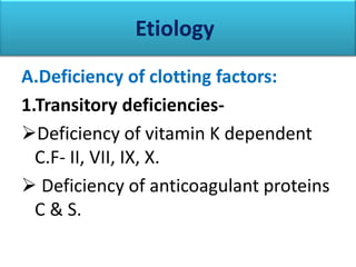
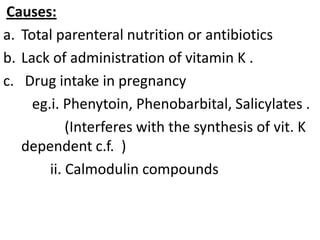
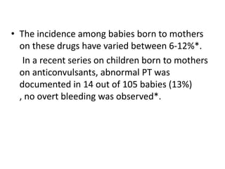
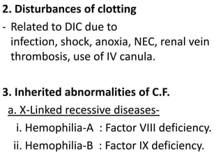
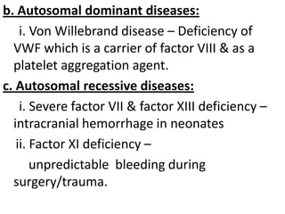
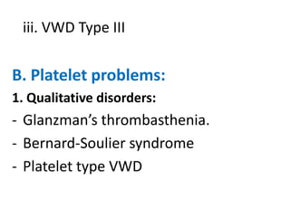
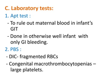
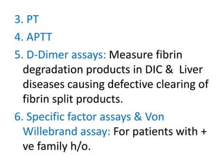
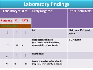
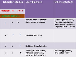
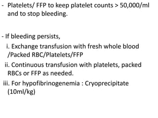
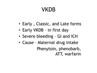
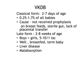
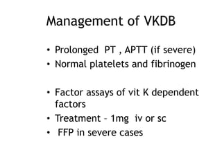
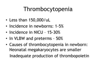
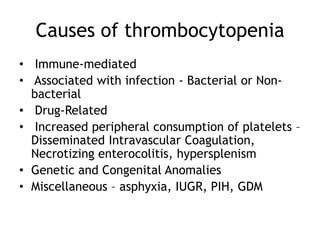
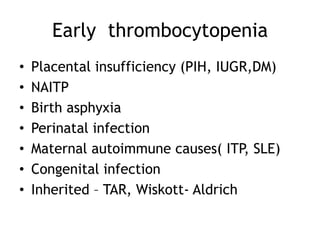
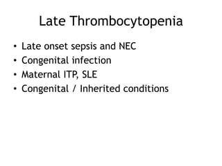
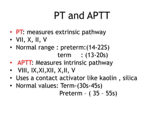
Bleeding neonate
Dr.Sandip Gupta
PGT,PEDIATRICS
B.S.M.C.H.
Introduction
•

Neonates are susceptible to bleeding for
various reasons
Immaturity of the haemostatic system
because of quantitative and qualitative
deficiency of coagulation factors
Maternal disease and drugs
Birth trauma

Other conditions - sepsis and asphyxia
Clinical presentation
•

Bleeding in neonates may present with
Oozing from the umbilical stump
Cephalhaematoma
Bruising , Petechiae
Bleeding from peripheral venipuncture or
procedure sites
Bleeding following circumcision
Intracranial haemorrhage
Bleeding from mucous membranes
Unexplained anemia and hypotension
Etiology
A.Deficiency of clotting factors:
1.Transitory deficienciesDeficiency of vitamin K dependent
C.F- II, VII, IX, X.
 Deficiency of anticoagulant proteins
C & S.
Causes:
a. Total parenteral nutrition or antibiotics
b. Lack of administration of vitamin K .
c. Drug intake in pregnancy
eg.i. Phenytoin, Phenobarbital, Salicylates .
(Interferes with the synthesis of vit. K
dependent c.f. )
ii. Calmodulin compounds
• The incidence among babies born to mothers
on these drugs have varied between 6-12%*.
In a recent series on children born to mothers
on anticonvulsants, abnormal PT was
documented in 14 out of 105 babies (13%)
, no overt bleeding was observed*.
2. Disturbances of clotting
- Related to DIC due to
infection, shock, anoxia, NEC, renal vein
thrombosis, use of IV canula.
3. Inherited abnormalities of C.F.
a. X-Linked recessive diseasesi. Hemophilia-A : Factor VIII deficiency.
ii. Hemophilia-B : Factor IX deficiency.
b. Autosomal dominant diseases:
i. Von Willebrand disease – Deficiency of
VWF which is a carrier of factor VIII & as a
platelet aggregation agent.
c. Autosomal recessive diseases:
i. Severe factor VII & factor XIII deficiency –
intracranial hemorrhage in neonates
ii. Factor XI deficiency –
unpredictable bleeding during
surgery/trauma.
iii. VWD Type III

B. Platelet problems:
1. Qualitative disorders:

- Glanzman’s thrombasthenia.
- Bernard-Soulier syndrome
- Platelet type VWD
2. Quantitive disorders:
- Immune thrombocytopenia
- Matrnal Preeclampsia, HELLP syndrome
or severe uteroplacental insuffuciency.
- DIC due to infection or asphyxia.
- Inherited marrow failure syndromes :
Fanconi anemia & congenital
amegakaryocytic thrombocytopenia
- Congenital leukemia
- Inherited thrombocytopenia syndromes
: gray platelet syndrome
- Macrothrombocytopenias : MayHegglin syndr.
- Platelet consumption in clots/ vascular
disorders eg. Vascular
malformations, NEC.
C. Vascular origin:
- Pulmonary haemorrhage
- A-V malformations
- CNS haemorrhage
- Hemangiomas.
Diagnostic workup
• HISTORY: A detailed history and examination
essential in the assessment of bleeding neonate
History includes

• Maternal diseases as ITP, preeclampsia .
• Maternal exposure to drugs as aspirin,
anticonvulsants, rifampicin and isoniazid

• Family history of bleeding disorders
• Previous affected sibling
B. Examination:
First diagnose whether the infant is Sick or Well
1. Sick infant:
- DIC
- Bacterial/ viral infections.
2. Well infant:
- Vit K deficiency
- Isolated C.F. deficiencies
- Immune thrombocytopenia
- Maternal blood in infant’s GIT.
3. Patchiae, ecchymosis, mucosal
bleeding: Platelet problem
4. Large bruises: DIC, C.F deficiencies,
liver diseases
5. Enlarged spleen : Possible congenital
infections or erythroblastosis.
6. Jaundice : Sepsis, liver diseases,
resorption of large hematoma.
C. Laboratory tests:
1. Apt test :
- To rule out maternal blood in infant’s
GIT
- Done in otherwise well infant with
only GI bleeding.
2. PBS :
- DIC- fragmented RBCs
- Congenital macrothrombocytopenias –
large platelets.
3. PT
4. APTT
5. D-Dimer assays: Measure fibrin
degradation products in DIC & Liver
diseases causing defective clearing of
fibrin split products.
6. Specific factor assays & Von
Willebrand assay: For patients with +
ve family h/o.
Laboratory findings
Laboratory Studies

Other useful tests

DIC

Platelets PT

Likely Diagnosis

Fibrinogen, FDP, Sepsis
screen

Platelet consumption
(NEC, Renal vein thrombosis,
marrow infiltration, Sepsis)

LFT, Albumin

APTT

SICK INFANTS

N

N

Liver disease
N

N

N

N

Compromised vascular integrity
(hypoxia, prematurity, acidosis)
Laboratory Studies
Platelets PT

Likely Diagnosis

Other useful tests

Immune thrombocytopenia
Bone marrow hypoplasia

Maternal platelet count,
Platelet antigen typing,
Bone marrow, Fibrinogen,
FDP, Factor VII & IX assays

APTT

HEALTHY INFANTS
N

N

N

N

N

Vitamin K Deficiency

N

Heriditory C.F. deficiencies

N

Bleeding d/t local factors,
Plt function anomalies,
Factor XIII deficiency(rare)

N

Platelet aggregometry
Urea clot solubility
Treatment Of Bleeding
A. Inj Vitamin K1 (Aquaminophyton)
- 1 mg IV or IM if not given at birth.
- Infants on TPN
- Infants on Antibiotics > 2 weeks: at
least 0.5mg Vit K weekly.
- Preferred rather than FFP for prolonged
PT & PTT, FFP should be reserved for
emergencies.
B. FFP:
- 10ml/kg IV for active bleeding
- Repeated 8-12 hrly as needed.
- Replaces C.F. immediately.
C. Platelets:
- 1 Unit of platelet raises count by
50,000-100,000/mm3 in a 3kg
newborn.
- Platelet count slowly decreases if stores
3-5 days.
D. Fresh whole blood:
- 10ml/kg
- Can be repeated after 6-8 hrs as needed.
E. Clotting factor concetrates
- Severe VWD :
- VWF containing plasma derived factor VIII
concetrate.
- Known deficiency of factor VIII or IX :
Recombinent DNA derived factor VIII and
IX concetrate
F. Disorders due to problems other than hemostatic
proteins :
- Rule out the underlying possibilities
- eg. Infection, Liver rupture, catheter, NEC.
G. T/t of specific disorders :
1. DIC :
- Treat the underlying cause i.e. sepsis, NEC
- Make sure that Vit K1 has been given.
- Platelets/ FFP to keep platelet counts > 50,000/ml
and to stop bleeding.
- If bleeding persists,
i. Exchange transfusion with fresh whole blood
/Packed RBC/Platelets/FFP
ii. Continuous transfusion with platelets, packed
RBCs or FFP as needed.
iii. For hypofibrinogenemia : Cryoprecipitate
(10ml/kg)
VKDB
•
•
•
•

Early , Classic, and Late forms
Early VKDB – in first day
Severe bleeding – GI and ICH
Cause – Maternal drug intake
Phenytoin, phenobarb,
ATT, warfarin
VKDB
Classical form: 2-7 days of age
• 0.25-1.7% of all babies
• Cause – not received prophylaxis
on breast feeds, sterile gut, lack of
placental transfer
Late form : 2-8 weeks of age
• Boys > girls, 5-10/1 lac
• Well , breastfed, term baby
• Liver disease
• Malabsorption
Management of VKDB
• Prolonged PT , APTT (if severe)
• Normal platelets and fibrinogen

• Factor assays of vit K dependent
factors
• Treatment – 1mg iv or sc
• FFP in severe cases
Prophylaxis of VKDB
• Early VKDB- single IM inj of vit K at
birth and oral Vit K to mother for
last 4 weeks
• Classical and Late forms –
IM Vit K at birth
oral Vit K at 0 , 4 days and 4 weeks
In preterms – Weekly iv Vit K
Hemophilia in the Newborn
• Factor VIII or XI deficiency
– A good family history goes a long way
Hemophilia A
Most common inherited clotting factor def
X linked recessive, 1 in 4000 males
1/3rd of cases present in newborn period
ICH(25%), cephalhematoma(10-15%)
Post circumcision bleed is characteristic
Family history – absent in 30%
Inv – prolonged APTT, normal PT, normal
platelets.
• Factor VIIIc assay level <2% severe, 2-10%
moderate, >10% mild
•
•
•
•
•
•
•
Hemophilia B
•
•
•
•
•
•

XLR
Deficiency of Factor IX
Less common than the classical form
Prolonged APTT and low Factor IX
Rx- 100u/k iv OD , to raise levels to 100%
Avoid lumbar punctures, IM injections
Thrombocytopenia
•
•
•
•
•

Less than 150,000/uL
Incidence in newborns: 1-5%
Incidence in NICU – 15-30%
In VLBW and preterms – 50%
Causes of thrombocytopenia in newborn:
Neonatal megakaryocytes are smaller
Inadequate production of thrombopoietin
Causes of thrombocytopenia
• Immune-mediated
• Associated with infection - Bacterial or Nonbacterial
• Drug-Related
• Increased peripheral consumption of platelets –
Disseminated Intravascular Coagulation,
Necrotizing enterocolitis, hypersplenism
• Genetic and Congenital Anomalies
• Miscellaneous – asphyxia, IUGR, PIH, GDM
Early thrombocytopenia
•
•
•
•
•
•
•

Placental insufficiency (PIH, IUGR,DM)
NAITP
Birth asphyxia
Perinatal infection
Maternal autoimmune causes( ITP, SLE)
Congenital infection
Inherited – TAR, Wiskott- Aldrich
Late Thrombocytopenia
•
•
•
•

Late onset sepsis and NEC
Congenital infection
Maternal ITP, SLE
Congenital / Inherited conditions
Immune Thrombocytopenia
• Neonatal allo-immune thrombocytopenia
(NAIT)
• Incidental thrombocytopenia of
pregnancy or Gestational
thrombocytopenia
• Autoimmune thrombocytopenic purpura
Neonatal allo-immune
thrombocytopenia (NAIT )
•
•
•
•
•
•

Incompatibility between mother and baby
Similar to Rh disease
Antibodies against HPA – 1 (most common)
In utero bleed can occur
Manifests with first pregnancy in 50%
Postnatal : petechiae, purpura
ICH in 10% with sequelae
NAIT
• Management – fetal blood sampling and
platelet transfusion or maternal IVIG
• If previous sibling had a significant bleed
• Caesarian section
• In newborn – maternal platelets or HPA
compatible platelets
• IVIG 1gm/k for 2 days or 0.5g/k for 4 days
Congenital causes
•
•
•
•
•

TAR , Fanconis anemia,
Congenital amegakaryocytic anemia
Trisomy 21, 18,13
Wiskott Aldrich syndrome
Noonan’s and Apert’s Syndromes
TAR (Thrombocytopenia & Absent
Radii)
• Congenital
• Findings
–
–
–
–
–

Thrombocytopenia
Absent radii bilaterally
Small shoulders
Abnormal knees
Malabsorption

• History
– Platelets stabilize
– ? Leukemia
PT and APTT
• PT: measures extrinsic pathway
• VII, X, II, V
• Normal range : preterm:(14-22S)
term
: (13-20s)
• APTT: Measures intrinsic pathway
• VIII, IX,XI,XII, X,II, V
• Uses a contact activator like kaolin , silica
• Normal values: Term-(30s-45s)
Preterm – ( 35 – 55s)
Thank You…
Bleedingneonate sandip1

Bleedingneonate sandip1

  • 1.
  • 2.
    Introduction • Neonates are susceptibleto bleeding for various reasons Immaturity of the haemostatic system because of quantitative and qualitative deficiency of coagulation factors Maternal disease and drugs Birth trauma Other conditions - sepsis and asphyxia
  • 3.
    Clinical presentation • Bleeding inneonates may present with Oozing from the umbilical stump Cephalhaematoma Bruising , Petechiae Bleeding from peripheral venipuncture or procedure sites Bleeding following circumcision Intracranial haemorrhage Bleeding from mucous membranes Unexplained anemia and hypotension
  • 4.
    Etiology A.Deficiency of clottingfactors: 1.Transitory deficienciesDeficiency of vitamin K dependent C.F- II, VII, IX, X.  Deficiency of anticoagulant proteins C & S.
  • 5.
    Causes: a. Total parenteralnutrition or antibiotics b. Lack of administration of vitamin K . c. Drug intake in pregnancy eg.i. Phenytoin, Phenobarbital, Salicylates . (Interferes with the synthesis of vit. K dependent c.f. ) ii. Calmodulin compounds
  • 6.
    • The incidenceamong babies born to mothers on these drugs have varied between 6-12%*. In a recent series on children born to mothers on anticonvulsants, abnormal PT was documented in 14 out of 105 babies (13%) , no overt bleeding was observed*.
  • 7.
    2. Disturbances ofclotting - Related to DIC due to infection, shock, anoxia, NEC, renal vein thrombosis, use of IV canula. 3. Inherited abnormalities of C.F. a. X-Linked recessive diseasesi. Hemophilia-A : Factor VIII deficiency. ii. Hemophilia-B : Factor IX deficiency.
  • 8.
    b. Autosomal dominantdiseases: i. Von Willebrand disease – Deficiency of VWF which is a carrier of factor VIII & as a platelet aggregation agent. c. Autosomal recessive diseases: i. Severe factor VII & factor XIII deficiency – intracranial hemorrhage in neonates ii. Factor XI deficiency – unpredictable bleeding during surgery/trauma.
  • 9.
    iii. VWD TypeIII B. Platelet problems: 1. Qualitative disorders: - Glanzman’s thrombasthenia. - Bernard-Soulier syndrome - Platelet type VWD
  • 10.
    2. Quantitive disorders: -Immune thrombocytopenia - Matrnal Preeclampsia, HELLP syndrome or severe uteroplacental insuffuciency. - DIC due to infection or asphyxia. - Inherited marrow failure syndromes : Fanconi anemia & congenital amegakaryocytic thrombocytopenia
  • 11.
    - Congenital leukemia -Inherited thrombocytopenia syndromes : gray platelet syndrome - Macrothrombocytopenias : MayHegglin syndr. - Platelet consumption in clots/ vascular disorders eg. Vascular malformations, NEC.
  • 12.
    C. Vascular origin: -Pulmonary haemorrhage - A-V malformations - CNS haemorrhage - Hemangiomas.
  • 13.
    Diagnostic workup • HISTORY:A detailed history and examination essential in the assessment of bleeding neonate History includes • Maternal diseases as ITP, preeclampsia . • Maternal exposure to drugs as aspirin, anticonvulsants, rifampicin and isoniazid • Family history of bleeding disorders • Previous affected sibling
  • 14.
    B. Examination: First diagnosewhether the infant is Sick or Well 1. Sick infant: - DIC - Bacterial/ viral infections. 2. Well infant: - Vit K deficiency - Isolated C.F. deficiencies - Immune thrombocytopenia - Maternal blood in infant’s GIT.
  • 15.
    3. Patchiae, ecchymosis,mucosal bleeding: Platelet problem 4. Large bruises: DIC, C.F deficiencies, liver diseases 5. Enlarged spleen : Possible congenital infections or erythroblastosis. 6. Jaundice : Sepsis, liver diseases, resorption of large hematoma.
  • 16.
    C. Laboratory tests: 1.Apt test : - To rule out maternal blood in infant’s GIT - Done in otherwise well infant with only GI bleeding. 2. PBS : - DIC- fragmented RBCs - Congenital macrothrombocytopenias – large platelets.
  • 17.
    3. PT 4. APTT 5.D-Dimer assays: Measure fibrin degradation products in DIC & Liver diseases causing defective clearing of fibrin split products. 6. Specific factor assays & Von Willebrand assay: For patients with + ve family h/o.
  • 18.
    Laboratory findings Laboratory Studies Otheruseful tests DIC Platelets PT Likely Diagnosis Fibrinogen, FDP, Sepsis screen Platelet consumption (NEC, Renal vein thrombosis, marrow infiltration, Sepsis) LFT, Albumin APTT SICK INFANTS N N Liver disease N N N N Compromised vascular integrity (hypoxia, prematurity, acidosis)
  • 19.
    Laboratory Studies Platelets PT LikelyDiagnosis Other useful tests Immune thrombocytopenia Bone marrow hypoplasia Maternal platelet count, Platelet antigen typing, Bone marrow, Fibrinogen, FDP, Factor VII & IX assays APTT HEALTHY INFANTS N N N N N Vitamin K Deficiency N Heriditory C.F. deficiencies N Bleeding d/t local factors, Plt function anomalies, Factor XIII deficiency(rare) N Platelet aggregometry Urea clot solubility
  • 20.
    Treatment Of Bleeding A.Inj Vitamin K1 (Aquaminophyton) - 1 mg IV or IM if not given at birth. - Infants on TPN - Infants on Antibiotics > 2 weeks: at least 0.5mg Vit K weekly. - Preferred rather than FFP for prolonged PT & PTT, FFP should be reserved for emergencies.
  • 21.
    B. FFP: - 10ml/kgIV for active bleeding - Repeated 8-12 hrly as needed. - Replaces C.F. immediately. C. Platelets: - 1 Unit of platelet raises count by 50,000-100,000/mm3 in a 3kg newborn. - Platelet count slowly decreases if stores 3-5 days.
  • 22.
    D. Fresh wholeblood: - 10ml/kg - Can be repeated after 6-8 hrs as needed. E. Clotting factor concetrates - Severe VWD : - VWF containing plasma derived factor VIII concetrate. - Known deficiency of factor VIII or IX : Recombinent DNA derived factor VIII and IX concetrate
  • 23.
    F. Disorders dueto problems other than hemostatic proteins : - Rule out the underlying possibilities - eg. Infection, Liver rupture, catheter, NEC. G. T/t of specific disorders : 1. DIC : - Treat the underlying cause i.e. sepsis, NEC - Make sure that Vit K1 has been given.
  • 24.
    - Platelets/ FFPto keep platelet counts > 50,000/ml and to stop bleeding. - If bleeding persists, i. Exchange transfusion with fresh whole blood /Packed RBC/Platelets/FFP ii. Continuous transfusion with platelets, packed RBCs or FFP as needed. iii. For hypofibrinogenemia : Cryoprecipitate (10ml/kg)
  • 25.
    VKDB • • • • Early , Classic,and Late forms Early VKDB – in first day Severe bleeding – GI and ICH Cause – Maternal drug intake Phenytoin, phenobarb, ATT, warfarin
  • 26.
    VKDB Classical form: 2-7days of age • 0.25-1.7% of all babies • Cause – not received prophylaxis on breast feeds, sterile gut, lack of placental transfer Late form : 2-8 weeks of age • Boys > girls, 5-10/1 lac • Well , breastfed, term baby • Liver disease • Malabsorption
  • 27.
    Management of VKDB •Prolonged PT , APTT (if severe) • Normal platelets and fibrinogen • Factor assays of vit K dependent factors • Treatment – 1mg iv or sc • FFP in severe cases
  • 28.
    Prophylaxis of VKDB •Early VKDB- single IM inj of vit K at birth and oral Vit K to mother for last 4 weeks • Classical and Late forms – IM Vit K at birth oral Vit K at 0 , 4 days and 4 weeks In preterms – Weekly iv Vit K
  • 29.
    Hemophilia in theNewborn • Factor VIII or XI deficiency – A good family history goes a long way
  • 30.
    Hemophilia A Most commoninherited clotting factor def X linked recessive, 1 in 4000 males 1/3rd of cases present in newborn period ICH(25%), cephalhematoma(10-15%) Post circumcision bleed is characteristic Family history – absent in 30% Inv – prolonged APTT, normal PT, normal platelets. • Factor VIIIc assay level <2% severe, 2-10% moderate, >10% mild • • • • • • •
  • 31.
    Hemophilia B • • • • • • XLR Deficiency ofFactor IX Less common than the classical form Prolonged APTT and low Factor IX Rx- 100u/k iv OD , to raise levels to 100% Avoid lumbar punctures, IM injections
  • 32.
    Thrombocytopenia • • • • • Less than 150,000/uL Incidencein newborns: 1-5% Incidence in NICU – 15-30% In VLBW and preterms – 50% Causes of thrombocytopenia in newborn: Neonatal megakaryocytes are smaller Inadequate production of thrombopoietin
  • 33.
    Causes of thrombocytopenia •Immune-mediated • Associated with infection - Bacterial or Nonbacterial • Drug-Related • Increased peripheral consumption of platelets – Disseminated Intravascular Coagulation, Necrotizing enterocolitis, hypersplenism • Genetic and Congenital Anomalies • Miscellaneous – asphyxia, IUGR, PIH, GDM
  • 34.
    Early thrombocytopenia • • • • • • • Placental insufficiency(PIH, IUGR,DM) NAITP Birth asphyxia Perinatal infection Maternal autoimmune causes( ITP, SLE) Congenital infection Inherited – TAR, Wiskott- Aldrich
  • 35.
    Late Thrombocytopenia • • • • Late onsetsepsis and NEC Congenital infection Maternal ITP, SLE Congenital / Inherited conditions
  • 36.
    Immune Thrombocytopenia • Neonatalallo-immune thrombocytopenia (NAIT) • Incidental thrombocytopenia of pregnancy or Gestational thrombocytopenia • Autoimmune thrombocytopenic purpura
  • 37.
    Neonatal allo-immune thrombocytopenia (NAIT) • • • • • • Incompatibility between mother and baby Similar to Rh disease Antibodies against HPA – 1 (most common) In utero bleed can occur Manifests with first pregnancy in 50% Postnatal : petechiae, purpura ICH in 10% with sequelae
  • 38.
    NAIT • Management –fetal blood sampling and platelet transfusion or maternal IVIG • If previous sibling had a significant bleed • Caesarian section • In newborn – maternal platelets or HPA compatible platelets • IVIG 1gm/k for 2 days or 0.5g/k for 4 days
  • 39.
    Congenital causes • • • • • TAR ,Fanconis anemia, Congenital amegakaryocytic anemia Trisomy 21, 18,13 Wiskott Aldrich syndrome Noonan’s and Apert’s Syndromes
  • 40.
    TAR (Thrombocytopenia &Absent Radii) • Congenital • Findings – – – – – Thrombocytopenia Absent radii bilaterally Small shoulders Abnormal knees Malabsorption • History – Platelets stabilize – ? Leukemia
  • 41.
    PT and APTT •PT: measures extrinsic pathway • VII, X, II, V • Normal range : preterm:(14-22S) term : (13-20s) • APTT: Measures intrinsic pathway • VIII, IX,XI,XII, X,II, V • Uses a contact activator like kaolin , silica • Normal values: Term-(30s-45s) Preterm – ( 35 – 55s)
  • 42.