This Presentation is on Bleeding and Coagulation Disorders like Haemophilia A, Haemophilia B, Von Wilboard Disease. This also contains Lab investigations and management of diseases.
Coagulation Disorders
Dr. AijazAli Tunio
MBBS, MCPS, MD Paediatrics
Consultant Child Specialist & Neonatologist
Unit III CMC Children Hospital Larkana
2.
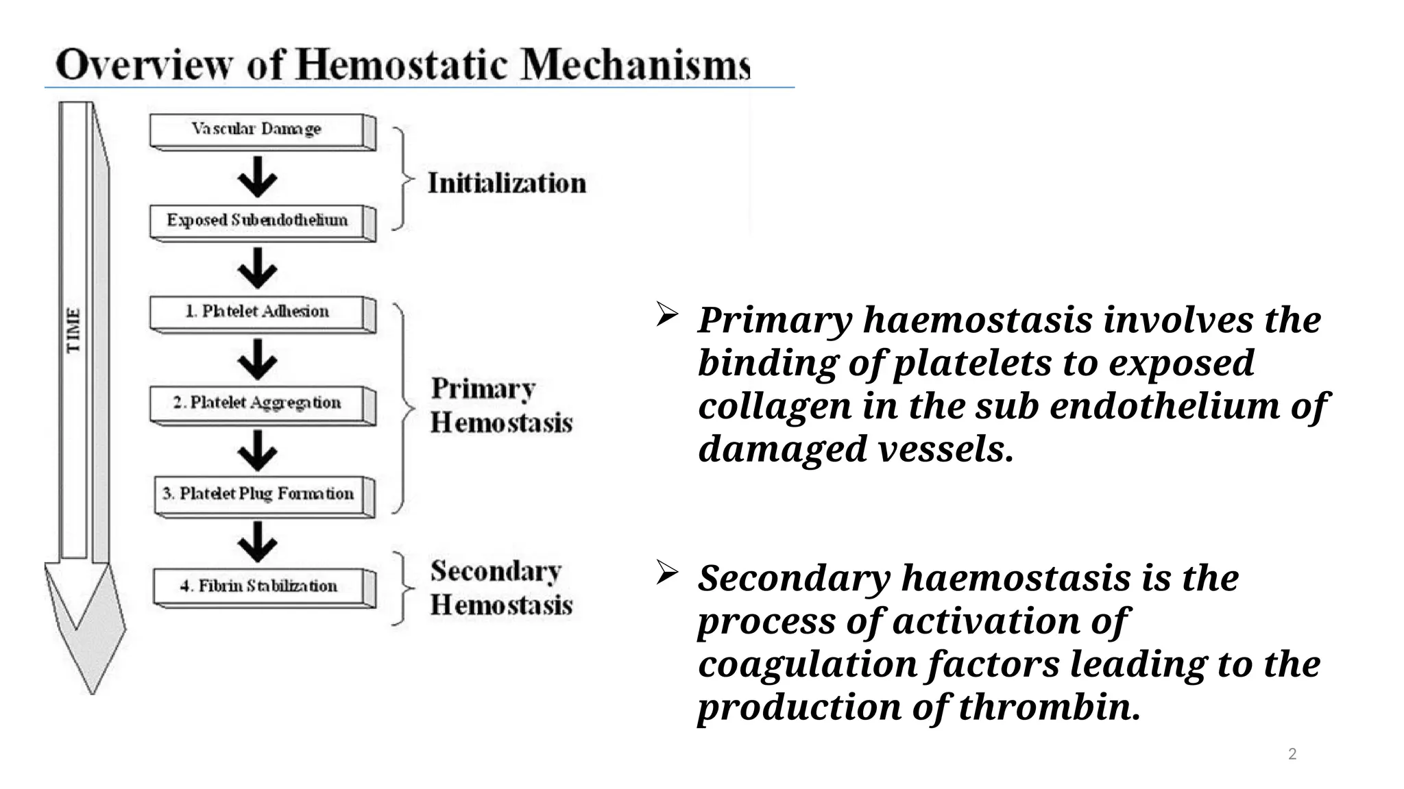
2
Primary haemostasisinvolves the
binding of platelets to exposed
collagen in the sub endothelium of
damaged vessels.
Secondary haemostasis is the
process of activation of
coagulation factors leading to the
production of thrombin.
3.
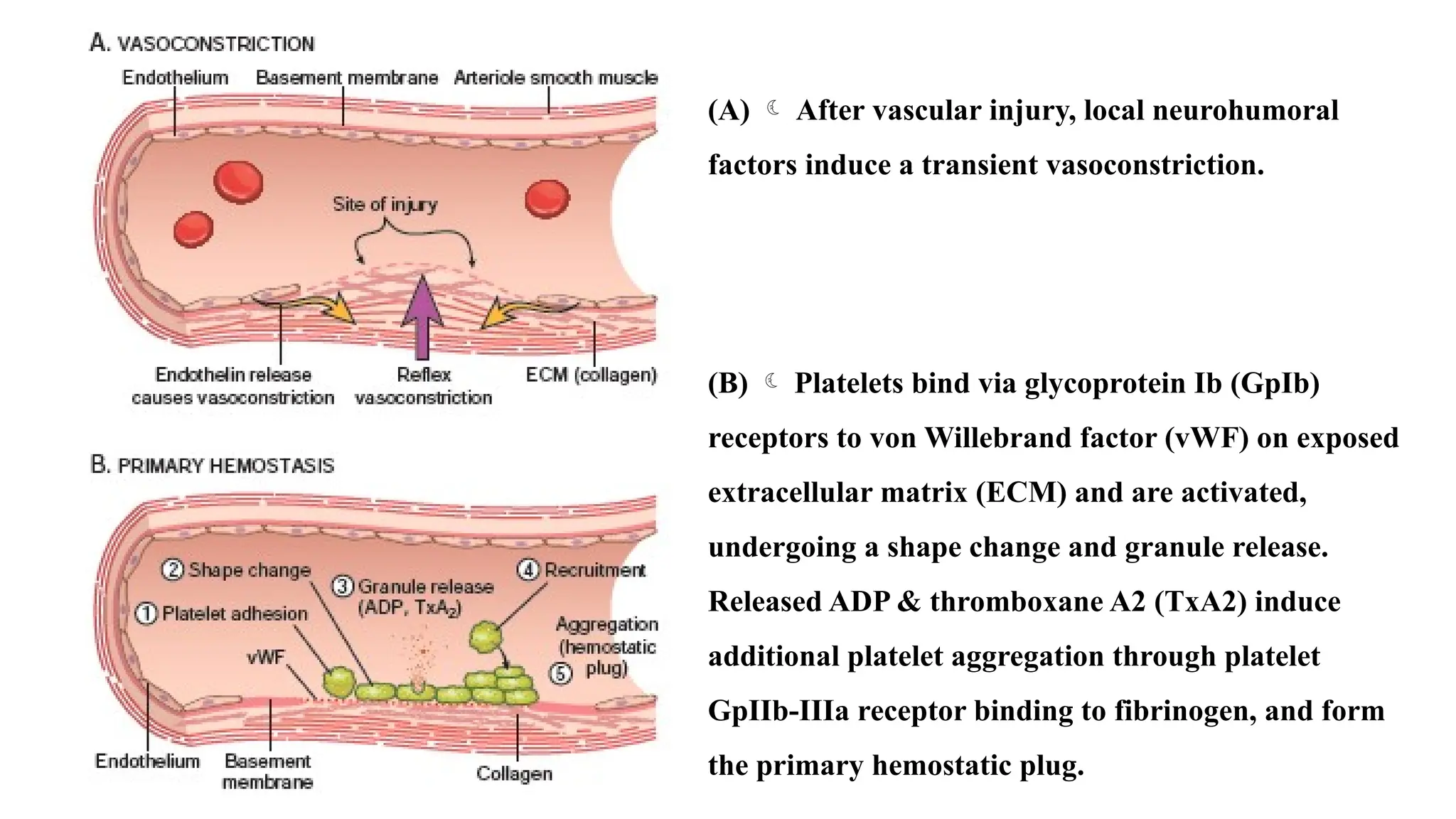
(A) Aftervascular injury, local neurohumoral
factors induce a transient vasoconstriction.
(B) Platelets bind via glycoprotein Ib (GpIb)
receptors to von Willebrand factor (vWF) on exposed
extracellular matrix (ECM) and are activated,
undergoing a shape change and granule release.
Released ADP & thromboxane A2 (TxA2) induce
additional platelet aggregation through platelet
GpIIb-IIIa receptor binding to fibrinogen, and form
the primary hemostatic plug.
4.
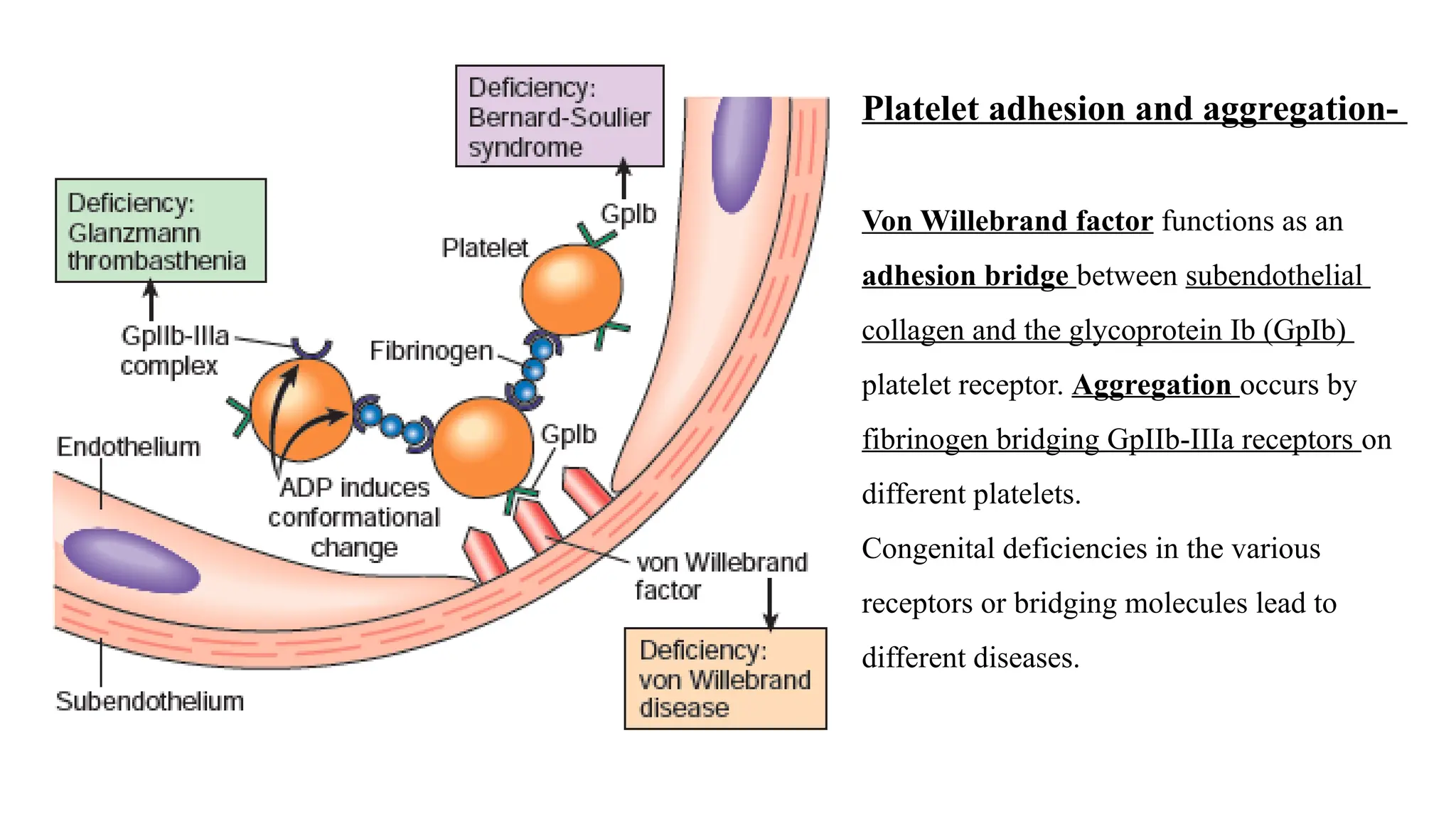
Platelet adhesion andaggregation-
Von Willebrand factor functions as an
adhesion bridge between subendothelial
collagen and the glycoprotein Ib (GpIb)
platelet receptor. Aggregation occurs by
fibrinogen bridging GpIIb-IIIa receptors on
different platelets.
Congenital deficiencies in the various
receptors or bridging molecules lead to
different diseases.
5.
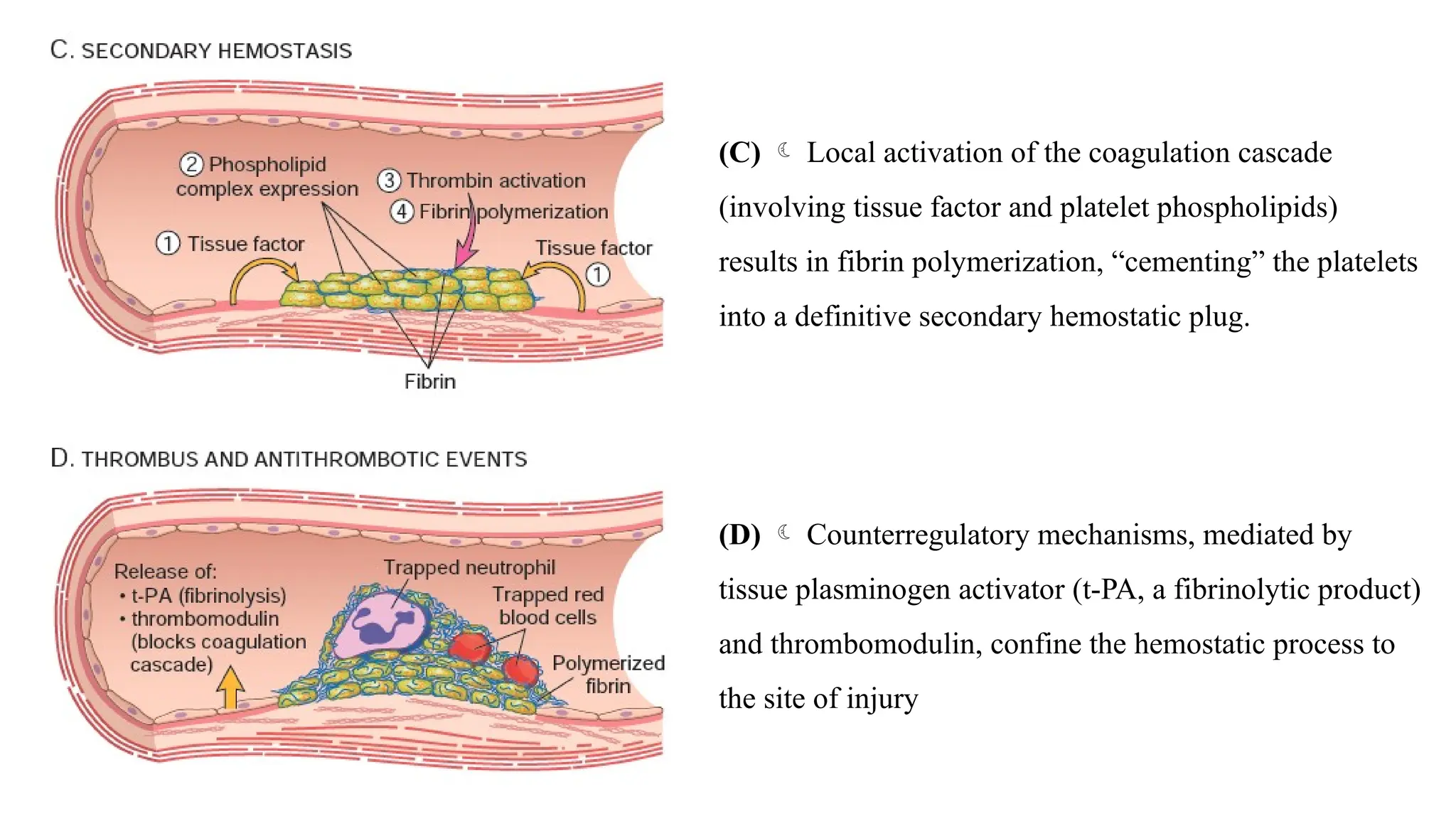
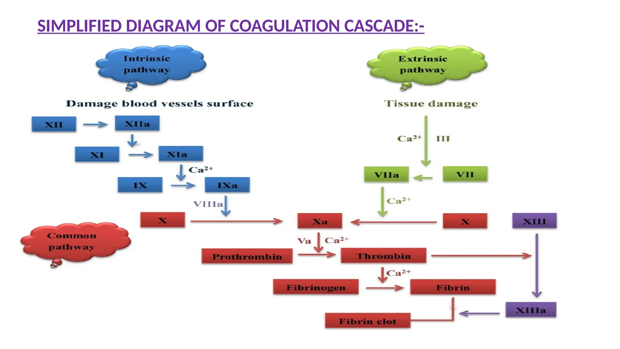
(C) Localactivation of the coagulation cascade
(involving tissue factor and platelet phospholipids)
results in fibrin polymerization, “cementing” the platelets
into a definitive secondary hemostatic plug.
(D) Counterregulatory mechanisms, mediated by
tissue plasminogen activator (t-PA, a fibrinolytic product)
and thrombomodulin, confine the hemostatic process to
the site of injury
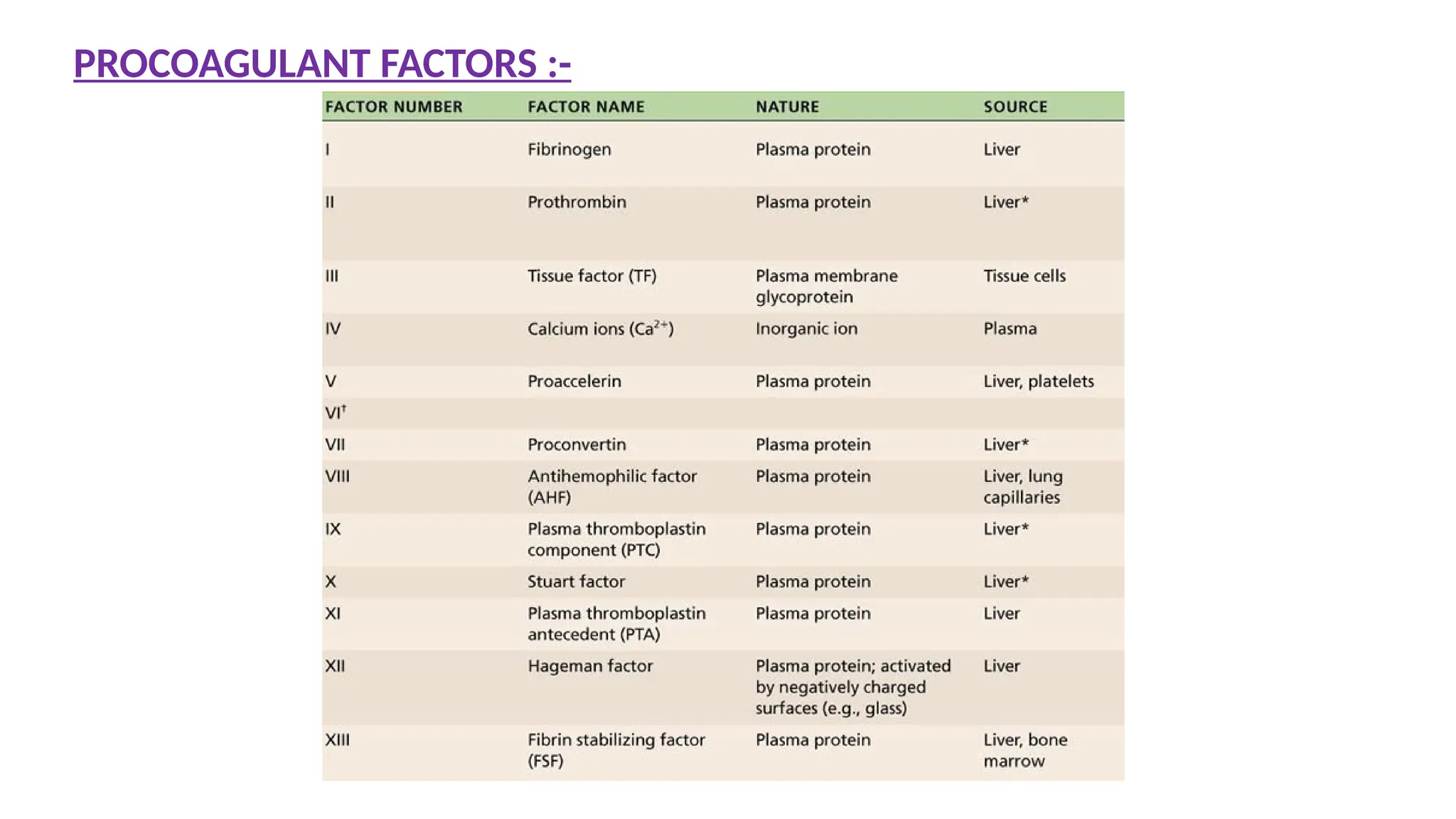
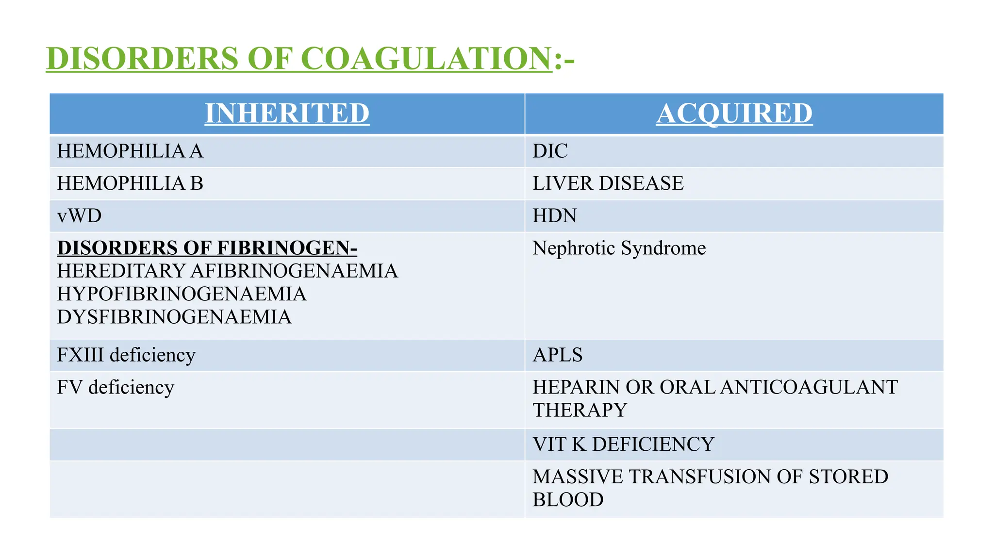
Most Common hereditarydisease
Reduced activity of F – VIII
X – linked recessive trait
30% of patients have no positive family history
<1% of normal F-VIII activity – Severe disease
2 – 5% of normal F-VIII activity – Moderate disease
6 – 50% of normal F-VIII activity – Mild disease
HEMOPHILIA – A
(F – VIII deficiency)
20.
Clinical Features:
• normalhemostasis require 25% factor VIII activity
• Symptomatic patients mostly have < 5% factor VIII activity
• Easy bruising
• Massive Hemorrhage after mild trauma / operation
• Joint bleeding – Haemarthrosis – Deformities
21.
Lab Features
• BleedingTime - Normal
• Prothrombin Time - Normal
• Platelet Count - Normal
• APTT - Increased
• Diagnosis can be confirmed by F-VIII assay.
Therapy
F-VIII Infusion
15% of severely affected patients –developed Antibodies against F - VIII
22.
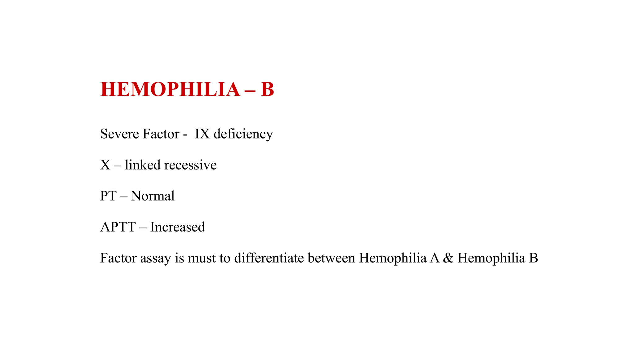
HEMOPHILIA – B
SevereFactor - IX deficiency
X – linked recessive
PT – Normal
APTT – Increased
Factor assay is must to differentiate between Hemophilia A & Hemophilia B
23.
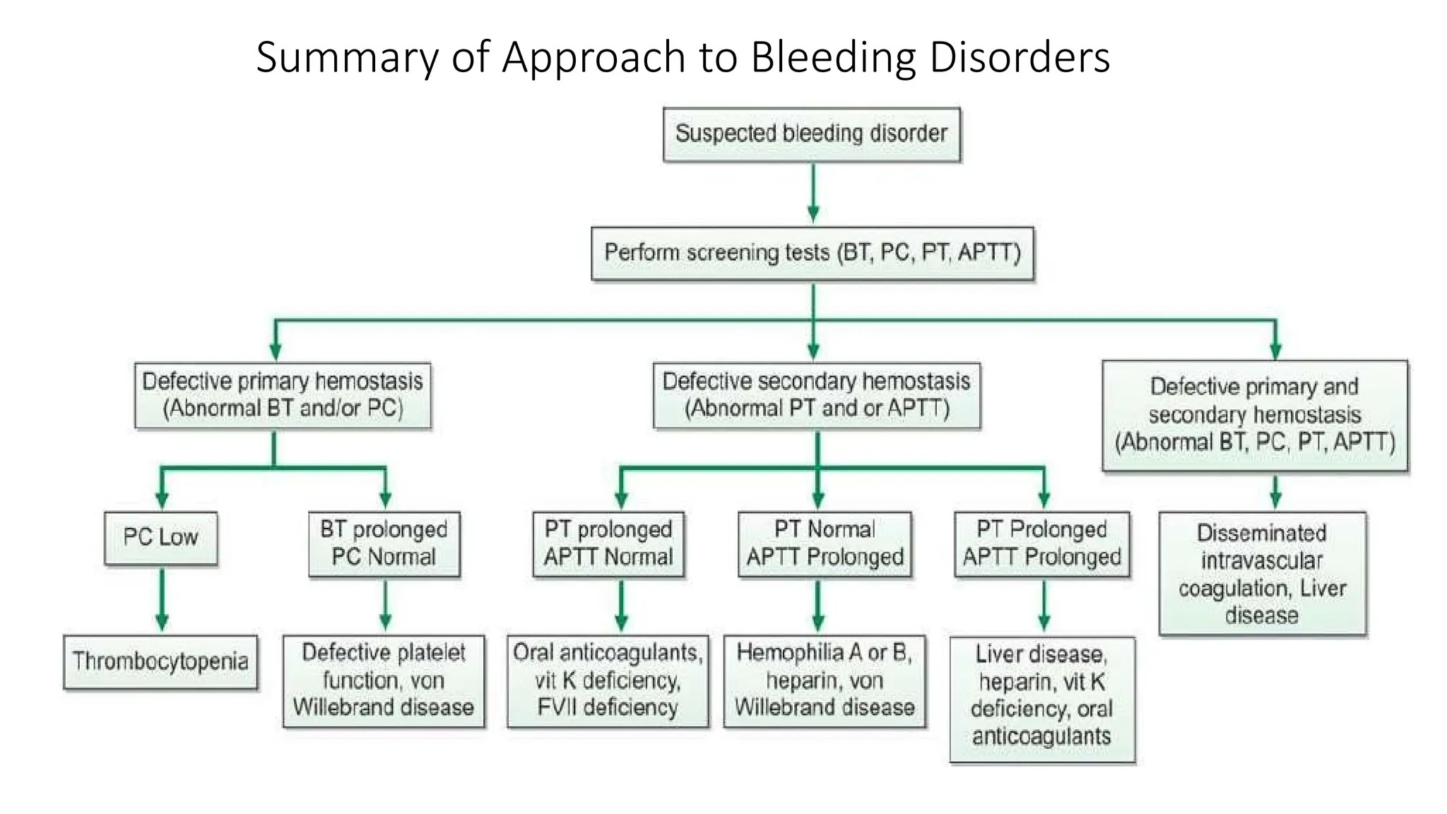
Screening tests forprimary hemostasis are -
I. Bleeding time- Assesses adequate functioning of platelets and blood vessels
II. Peripheral blood smear examination
III. Platelet count
IV. Mean Platelet volume
V. Reticulated platelets
VI. Platelet function analysis
VII. Tests for Vessel wall disorder
24.
Tests for Vesselwall disorder
HESS` CAPILLARY FRAGILITY TEST:
Cuff is wrapped in upper arm and pressure is maintained midway b/w systolic and
diastolic BP for 15 minutes, 4 cm below the elbow joint, a circle of 2.5 cm diameter
is drawn on the anterior aspect of forearm.
Upto 10 new hemorrhagic spots are normal.
But >20 new spots are always
pathological.
This is positive in increased capillary
fragility, ITP.
25.
Screening tests forsecondary hemostasis are -
I. Clotting time
II. Prothrombin time (PT) and Activated partial thromboplastin time (aPTT)
III. Thrombin Time (TT)
26.
Collection of bloodfor coagulation studies
The anticoagulant used for coagulation studies is trisodium
citrate (3.2%), with anticoagulant to blood proportion being
1:9.
27.
Clotting Time
Thisis a crude test and is now replaced by activated partial thromboplastin time.
Prolongation of clotting time only occurs in severe deficiency of a clotting factor and
is normal in mild or moderate deficiency.
28.
PROTHROMBIN TIME(PT)
PT assessescoagulation factors in extrinsic pathway (F VII) and
common pathway.
Principle:- Tissue thromboplastin and calcium are added to
platelet poor plasma and clotting time is determined.
29.
CONCEPT OF INR
1.Theinternational normalized ratio (INR) was introduced in an attempt to standardize the
PT.
2.Calculation ~ INR = [ PT (patient) / PT (Control) ]ISI
The INR has no units (it is a ratio)
**ISI, or international sensitivity index is a function of the thromboplastin reagent.
** NORMAL RANGE PT 11-16 seconds INR 0.9 – 1.1.
30.
Uses of PT
1.To monitor patients who are on oral anticoagulant therapy
2. To assess liver function
3. Detection of vitamin K deficiency
4. To screen for hereditary deficiency of coagulation factors
Causes of prolongation of PT
5.Treatment with oral anticoagulants
6.Liver disease
7.Vitamin K deficiency
8.Disseminated intravascular coagulation
9.Inherited deficiency of factors in extrinsic and common pathways.
31.
Significance
Reflects efficiency ofIntrinsic and Common pathway.
Principle
The test measures the clotting time of plasma after the activation of contact
factors (Kaolin/Silica/Ellagic acid) and the addition of phospholipid and
CaCl2, but without added tissue thromboplastin.
So it indicates the overall efficiency of the Intrinsic pathway.
Normal range
26 to 40 seconds.
ACTIVATED PARTIAL THROMBOPLASTIN TIME (APTT)
32.
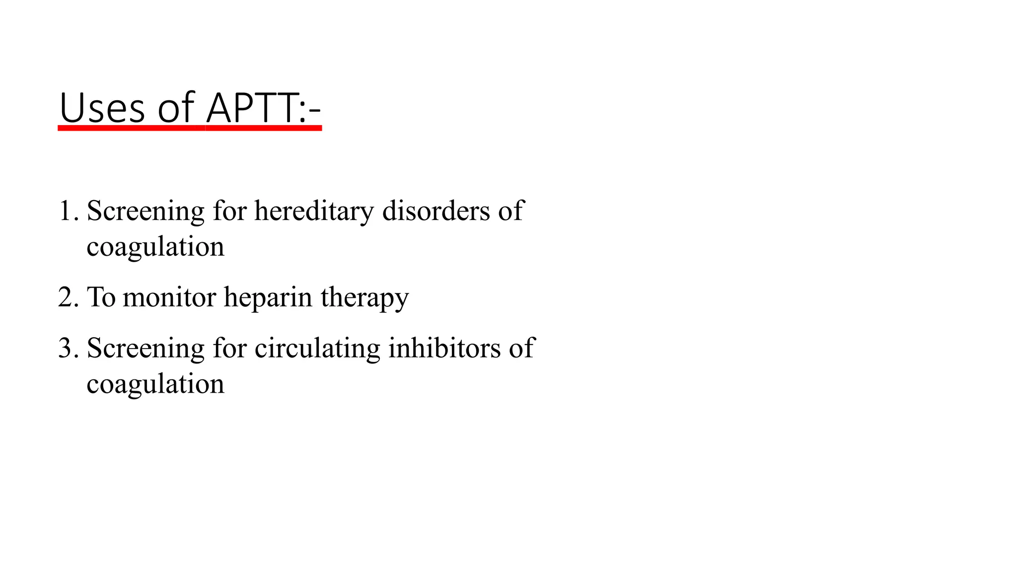
Uses of APTT:-
1.Screening for hereditary disorders of
coagulation
2. To monitor heparin therapy
3. Screening for circulating inhibitors of
coagulation
33.
APTT is prolongedin:-
1.Inherited deficiencies of factor VIII (Hemophilia A) and Factor IX (Hemophilia B)
2.Non specific inhibitor antibodies against F VIII e.g. Lupus inhibitor
(Don’t act directly but block interaction of FVIII with other clotting factors)
3.DIC
4.Heparin
( Inhibits factor XII, XI and X through antithrombin III & Heparin therapy is monitored
through aPTT)
5. Vit K deficiency
6.Massive transfusion of plasma depleted stored blood.
34.
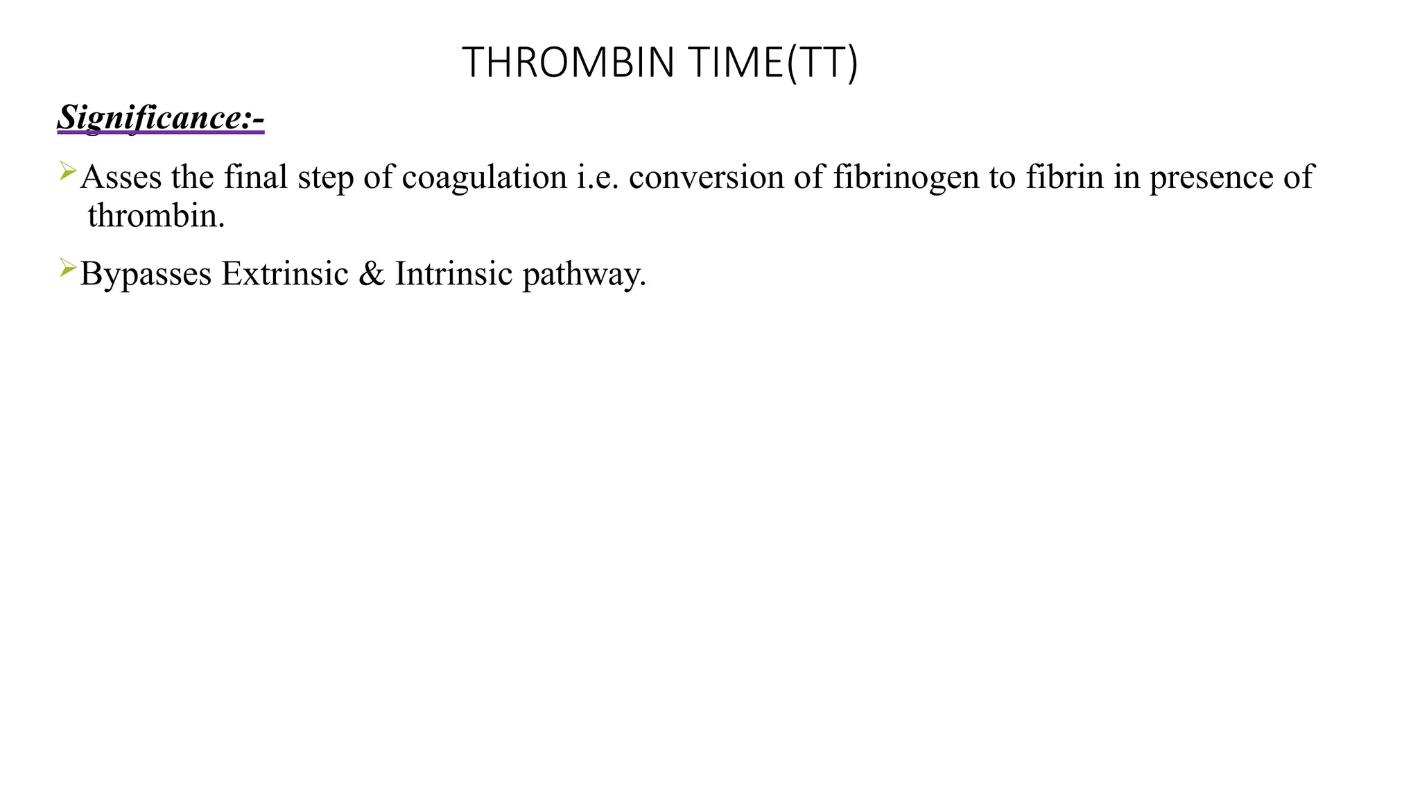
Significance:-
Asses the finalstep of coagulation i.e. conversion of fibrinogen to fibrin in presence of
thrombin.
Bypasses Extrinsic & Intrinsic pathway.
THROMBIN TIME(TT)
35.
Causes of prolongedTT
1. Disorders of fibrinogen-
i) Afibrinogenaemia
ii) Hypofibrinogenaemia
3. Chronic liver disease
36.
Abnormal platelet aggregationtests ~
Scenario 1. ~ Glanzman Thrombasthenia
1. In Glanzmann’s thrombasthenia, Platelet GPIIb/IIIa (important for platelet aggregation) is
defective.
3. All agents induce aggregation through this receptor except ristocetin.
4. Hence in Glanzmann’s thrombasthenia, aggregation is defective for all agents except ristocetin.
37.
Abnormal platelet
aggregation tests~
Scenario 2. ~ BERNARD SOULIER SYNDROME AND VON WILLEBRAND DISEASE
1.In Bernard soulier syndrome there is deficiency of GpIb/IX, whereas in von willebrand disease, there
is deficiency of von willebrand factor.
2. Aggregation is there in response to all agents except ristocetin
Then how can we
differentiate between these 2
conditions?
BS and vWD can be
differentiated by addition
of normal plasma.
If the aggregation is seen now,
it means the disease is vWD
because normal plasma is a
38.
Abnormal platelet aggregationtests ~
Scenario 3. ~ STORAGE POOL DEFECTS
1.In this disorder defective granule release from platelets
2.Due to defective release reaction, secondary aggregation is defective
3.Hence there is no secondary wave in response to ADP/epinephrine/collagen and only partial
aggregation in response to ristocetin.
FXIII Qualitative assay(Urea clot lysis test)
Done when all other tests for hemostasis are normal.
FXIII provides stability to clot formed.
Method:-


![CONCEPT OF INR
1.The international normalized ratio (INR) was introduced in an attempt to standardize the
PT.
2.Calculation ~ INR = [ PT (patient) / PT (Control) ]ISI
The INR has no units (it is a ratio)
**ISI, or international sensitivity index is a function of the thromboplastin reagent.
** NORMAL RANGE PT 11-16 seconds INR 0.9 – 1.1.](https://image.slidesharecdn.com/leccoagulationdisordersppt-250407182127-fad3b592/75/Bleeding-and-Coagulation-Disorders-Slides-29-2048.jpg)