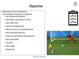
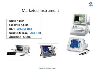
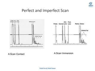
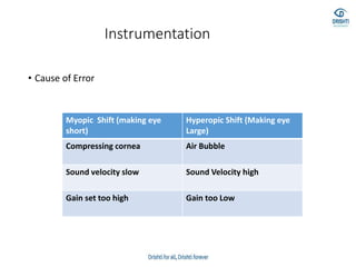
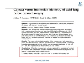
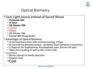
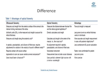
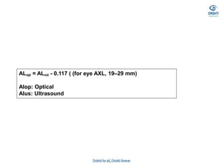
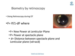
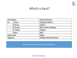
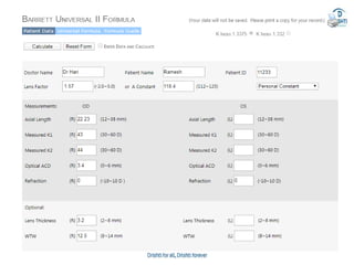
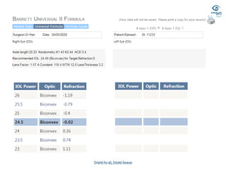
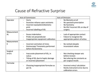
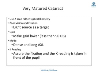
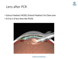
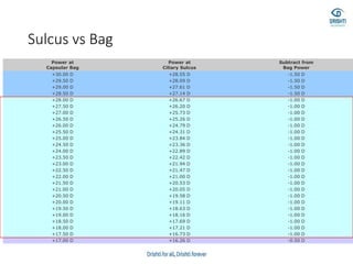
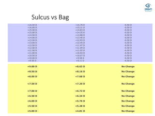
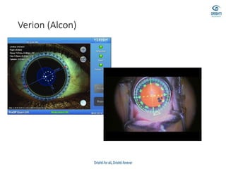
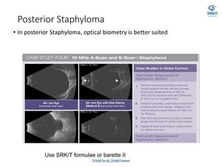
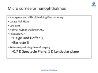
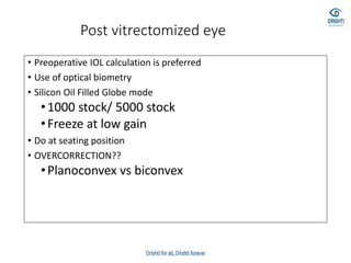
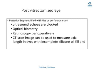
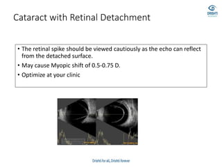
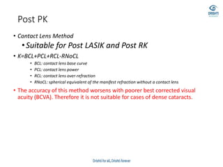
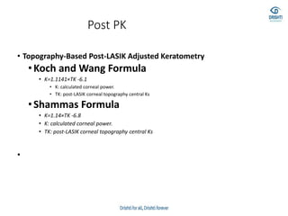
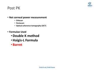
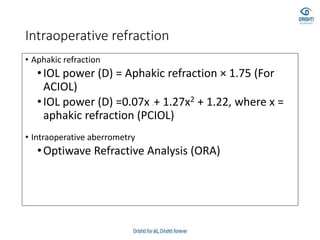
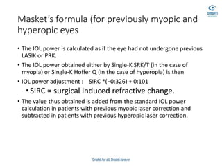
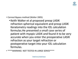
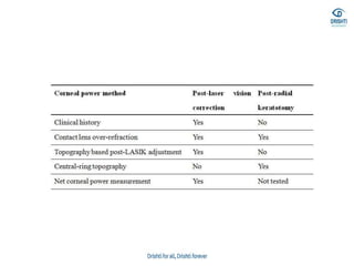
This document discusses various biometry techniques and considerations for accurate intraocular lens (IOL) power calculation in complex cases. It describes tools and methods for measuring ocular parameters in conditions like mature cataracts, posterior staphylomas, and post-refractive surgery eyes. Formulas for calculating IOL power for different IOL types and surgical procedures are presented. Intraoperative techniques like retinoscopy and aberrometry are also covered to optimize refractive outcomes.
This document discusses various techniques for measuring eye dimensions needed for accurate intraocular lens (IOL) power calculations, including:
- Applanation A-scan, immersion A-scan, and immersion vector A/B-scan techniques
- Using the IOL Master, which utilizes partial coherence interferometry, to obtain highly precise measurements
- Factors that can affect IOL power calculations like axial length, corneal power, anterior chamber depth, prior refractive surgery, and placement of IOL in bag vs sulcus
- Recommendations for validating biometry measurements and identifying potential errors in IOL power calculations based on certain parameters outside of expected ranges.
This document provides an overview of biometry and intraocular lens (IOL) power calculation. It discusses the history of IOL implantation and the evolution of crude IOL power calculations to modern biometry methods and formulas. Key aspects covered include the components of IOL power calculation like axial length, corneal power, and effective lens position. Various biometry techniques like ultrasonic measurement, optical biometry, and keratometry are described. Common formulas used in IOL power calculation and the variables that affect calculations are also summarized.
1. Accurate IOL power calculations require precise measurements of axial length and corneal power using modern devices like optical biometers. Errors in these measurements can lead to incorrect IOL powers.
2. Newer theoretical formulas like Holladay II and Haigis are generally more accurate than older formulas or regression formulas. They take into account additional parameters like anterior chamber depth.
3. Special considerations are needed for calculating IOL power in children, eyes with previous refractive surgery or conditions like high myopia which can affect biometry measurements. Repeat measurements may be needed if initial values are outside normal ranges.
Biometry is used to measure the eye's axial length and corneal power to determine the correct intraocular lens (IOL) power for cataract surgery. There are two main types of biometry - ultrasound and optical coherence interferometry. Ultrasound biometry uses sound waves to measure axial length and was introduced in the 1970s, allowing more accurate IOL power calculation than previous empirical methods. Accuracy is important as a 1mm error can lead to 2.88-3 diopters of IOL power difference. Special cases like high myopia, posterior staphyloma, or dense cataracts may require different biometry techniques.
This document discusses various methods for calculating intraocular lens (IOL) power in patients who have previously undergone laser eye surgery such as LASIK. It notes that accurately measuring corneal power and predicting refractive outcome is challenging in these patients due to changes induced by the previous surgery. Several methods are described that use pre-operative data, post-operative measurements, or a combination to calculate IOL power, including the clinical history method, Feiz-Mannis method, corneal bypass method, Aramberri "double K" method, and others. Accurately accounting for factors like the effective lens position is important to achieve the desired refractive outcome.
SRK/T power of +4D is more accurate for this patient since their axial length is within the normal range where third generation formulas like SRK/T are more accurate than second generation formulas like SRK II.
Biometry uses ultrasound A-scans to measure the eye's dimensions, which are used to calculate the ideal intraocular lens power for cataract surgery. The A-scan procedure involves taking a patient history, preparing the patient, and using ultrasound to measure the axial length of the eye. Accurate measurements require using the proper gain and sound velocity settings for each patient's eye condition, and ensuring the probe is correctly positioned on the cornea pointing towards the macula. The immersion technique, where the probe is submerged in fluid coupled to the eye, provides more precise measurements than the contact technique.
This document discusses biometry techniques used for precise intraocular lens (IOL) power calculations. Keratometry and axial length measurements are essential but prone to errors. Manual keratometry uses fixed or variable object sizes while automated keratometry uses reflected images. A-scan ultrasound can overestimate axial length due to corneal compression. Immersion and optical biometry are more accurate. IOL power formulas continue improving but require adjustments for high myopia, silicone oil, pediatric or post-surgical eyes. Accurate biometry is critical for optimal IOL calculations and patient outcomes.
This document discusses tips for calculating intraocular lens (IOL) power in difficult situations. It begins by outlining situations that can make IOL power calculation challenging, such as previous refractive surgery, high astigmatism, previous keratoplasty, pediatric cases, eyes with silicone oil or posterior staphyloma.
It then provides details on methods to calculate IOL power in each situation, including using previous refractive data, topography readings, and specialized formulas. Optical biometry is generally preferred over ultrasound biometry in difficult cases. The document emphasizes using third and fourth generation formulas and online calculators.
Special considerations are discussed for cases like piggyback IOLs, aphakia,
This document discusses the history of intraocular lens (IOL) implantation and development of technologies for calculating IOL power. It begins with Sir Harold Ridley implanting the first IOL in 1949 using the human lens as a model. Over subsequent decades, improvements were made such as developing foldable lenses to allow for smaller incisions. Advances in biometry technologies like ultrasound A-scan and optical biometry using partial coherence interferometry allowed for more accurate measurements of eye dimensions needed for precise IOL power calculations.
1) The Holladay 2 formula is considered one of the most accurate IOL calculation formulas today. It was developed in 1993 by Dr. Holladay based on a study of over 34,000 eyes to determine the key variables that predict effective lens position.
2) The Holladay 2 formula utilizes 7 parameters - axial length, keratometry, white-to-white, lens thickness, and anterior chamber depth - to calculate the effective lens position and appropriate IOL power.
3) The Holladay 2 formula is recommended for its high accuracy and predictability. The IOLMaster 500 is currently the only device that has the Holladay 2 formula integrated to automatically calculate the IOL
This document summarizes key details about optical coherence biometry, a new optical method for measuring eye dimensions as an alternative to ultrasound biometry. It discusses the measurement principles, comparisons to ultrasound measurements, and evaluation of the Zeiss IOLMaster device which combines optical coherence biometry with keratometry and anterior chamber depth measurements. Studies found excellent correlation between optical and ultrasound axial length measurements, with optical lengths on average 0.3mm longer than ultrasound lengths. Reproducibility of measurements was high for both optical coherence biometry and ultrasound.
IOL POWER CALCULATION IN CHILDREN-Dr.Preetiilal.pptxdrPreetiilal
This document discusses intraocular lens (IOL) power calculation in children. It begins by outlining normal eye development in children and the myopic shift that occurs. It then discusses important considerations for IOL power calculation including accurate measurement of axial length and keratometry, choosing an appropriate IOL formula, determining the target postoperative refraction based on age, and making intraoperative adjustments if needed. Tables provide estimated axial length and keratometry values by age and examples of IOL powers to achieve emmetropia or a desired refraction to minimize myopic shift. The document emphasizes that IOL power calculation in growing children requires a multifaceted approach to determine the optimal lens power.
The Implantable Collamer Lens (ICL) is a soft, flexible, posterior chamber phakic intraocular lens made of collagen-copolymer material called Collamer. Studies have shown ICL implantation is safe and effective for correcting myopia between -3 to -25 diopters and astigmatism up to -6 diopters. It provides stable refractive results with few complications over 4 years. Toric ICL models were found to be superior to LASIK in safety, efficacy, predictability and stability for high myopic astigmatism. The procedure is reversible and preserves corneal tissue, reducing risks compared to LASIK.
The document discusses various formulas used for calculating intraocular lens (IOL) power, including SRK, SRK2, Holladay, Haigis, and Holladay 2. It explains the factors these formulas account for such as axial length, corneal power, anterior chamber depth, and how they have evolved over generations to improve accuracy. Special considerations for calculating IOL power in cases involving prior refractive surgery, silicone oil filling, posterior staphyloma, and using optical biometry devices are also summarized.
This document discusses different types of multifocal intraocular lenses (IOLs) used in cataract surgery. There are three main types: refractive, diffractive, and a combination. Refractive IOLs use concentric rings of different optical powers while diffractive IOLs use diffraction optics to create two focal points. Combination IOLs can provide the advantages of both refractive and diffractive technologies. The document also covers specific multifocal IOL models and considerations for patient selection.
Clinical discussion on Biometry for IOL power calculationRezwanul Hasan
Biometry is the process of measuring the cornea's power and axial length of the eye to determine the ideal intraocular lens (IOL) power for cataract surgery. It involves keratometry to measure corneal power, axial length measurement, and determining the effective IOL position. Different IOL power calculation formulas are used, ranging from theoretical to regression-based generations, with newer generations incorporating additional eye parameters for improved accuracy. Factors like corneal conditions, poor fixation, prior refractive surgery, or pediatric cases can make accurate biometry more challenging.
This document discusses ways to avoid errors in biometry measurements and intraocular lens (IOL) power calculations that can lead to postoperative refractive surprises. It provides tips for accurately measuring the eye's axial length and corneal power, using the appropriate IOL calculation formulas for different eye characteristics, and identifying sources of human error. Minimizing errors requires proper training, auditing outcomes, learning from mistakes, and following checklist procedures to confirm the correct IOL power and patient before surgery.
IOL power calculation is challenging in eyes with prior refractive surgery or other special situations. In eyes with prior radial keratotomy, standard keratometry overestimates corneal power due to flattening outside the central optical zone. Multiple methods of IOL power calculation should be used, including topography to measure the flattest central corneal power. A study comparing methods in eyes with prior RK found IOL power calculation using topographic keratometry was least accurate compared to formulas from the ESCRS calculator. No single method provided reliable results, highlighting the difficulty in IOL power calculation for eyes with prior refractive surgery.
The document discusses various methods for calculating intraocular lens (IOL) power for cataract surgery, including the importance of accurately measuring corneal power, axial length, and lens position. It describes older applanation ultrasound techniques and newer non-contact technologies like the IOLMaster, noting the IOLMaster provides the most accurate axial length measurements. The document also reviews generations of IOL power calculation formulas and factors to consider like capsule status when selecting the appropriate formula.
This presentation covers the Optics & application of Jackson Cross Cylinder | Jackson Cross Cylinder works on an optical principle that constricts & expands the sturm's conoid.
This document discusses various methods and considerations for measuring ocular biometry and intraocular lens (IOL) power calculation. It describes average eye measurements, different devices for keratometry and axial length measurement, sources of error, and formulas for calculating IOL power for different eye types and surgical conditions. Proper technique and accounting for patient factors are important for obtaining accurate measurements and refractive outcomes.
This document discusses the measurement and management of aniseikonia. It describes several instruments used to measure aniseikonia, including the standard eikonometer and space eikonometer. It also discusses predicting aniseikonia through ocular component analysis using spectacle prescriptions, keratometry, A-scan ultrasound, and IOL status. Simple tests like size lenses, Maddox rod, and penlights as well as special tests like the Leaf Room effects and Awaya Aniseikonia test are outlined. Management of aniseikonia includes iseikonic lenses, toric lenses, doublet lenses, and fused bifocal lenses.
The document discusses implantable contact lenses (ICL) for correcting high refractive errors. Key points include:
- ICL is preferred over LASIK for high myopia or thin corneas, as it has fewer risks of complications.
- ICL is made of a biocompatible collamer material and is implanted in the posterior chamber behind the iris.
- A clinical trial found nearly 60% of eyes achieved 20/20 vision and 95% achieved 20/40 vision or better after 3 years with ICL implantation.
- Complications are generally low but can include cataracts, increased eye pressure, damage to the natural lens, and other rare issues like infections. Most risks are
This document provides guidelines for prescribing glasses in children. It discusses that the pediatric eye is different from the adult eye in terms of axial length, corneal curvature, and lens power. The goals of prescribing glasses in children are to provide a focused retinal image and achieve optimal balance between accommodation and convergence. It is more difficult to prescribe glasses for children due to lack of subjective response and poor attention. American guidelines provide recommendations on refractive errors that warrant correction at different ages. Factors like emmetropization, amblyopia risk, and presence of strabismus are considered. Frame selection depends on the child's condition and age, aiming for correct fit, comfort, safety, and not hindering nasal development.
This document discusses A-scan biometry, which uses ultrasound to measure eye structures. It describes the A-scan and B-scan display formats, how ultrasound works, and how biometry is used to measure distances in the eye. Key information obtained includes axial length, anterior chamber depth, and lens thickness, which are used in intraocular lens power calculation formulas to predict a patient's post-operative refraction. The document outlines factors that can impact biometry accuracy and discusses managing surprises after cataract surgery.
This document discusses comanagement of cataract surgery and premium intraocular lenses (IOLs) such as toric and multifocal IOLs. It provides guidance on criteria for ethical and legal comanagement including profitability. Reasons for comanaging with Visionary Ophthalmology include their reputation for excellent outcomes and being a leader in technology. The document reviews options for correcting astigmatism during cataract surgery such as toric IOLs. It discusses patient selection criteria and pearls for optimal results with toric IOLs including importance of accurate keratometry and marking the correct axis. The document also reviews multifocal IOL options and important tips for patient counseling, management of side effects
This document discusses accurate biometry and its role in determining the correct intraocular lens power for cataract surgery. It covers techniques for measuring axial length and keratometry, different formulas used in biometric calculations, tips for obtaining accurate measurements, and common sources of error. The goal is to provide surgeons with information to optimize biometry and achieve the best possible postoperative vision outcomes for patients.
This document discusses biometry IOL calculation, which involves measuring the axial length and corneal power of the eye to determine the correct intraocular lens power for cataract surgery. It describes various methods of measuring axial length, including ultrasonic A-scan biometry and optical methods like IOL Master. Corneal power can be measured via keratometry, corneal topography, or Pentacam. Different formulas are used to calculate IOL power based on these measurements and other factors. Special considerations for biometry in conditions like aphakia, pseudophakia, and after refractive surgery are also outlined.
The document discusses intraocular lens (IOL) power calculation methods. It begins by introducing IOL implantation and the key measurements involved in IOL power calculation: keratometry, axial length, and anterior chamber depth. It then describes early theoretical formulas and regression formulas like SRK/T. The document outlines improvements made in newer generation formulas like Holladay's formula which accounts for additional ocular variables to improve IOL power prediction accuracy, especially in extreme eyes. Overall, the document provides an overview of the evolution of IOL power calculation formulas from early theoretical to modern regression-based approaches.
Utilizing topolyzer vario & oculyzer ii for accurate refractiveMichael Mrochen
This document discusses the use of TopolyzerVario and Oculyzer II devices to provide accurate measurements for refractive laser eye surgery. It provides details on the essential data required by the ex500 laser including pre-operative and post-operative corneal measurements. The accuracy of the TopolyzerVario and Oculyzer II is impacted by factors like corneal irregularities, eyelashes, and light scattering. Ensuring accurate measurements from these devices is important for achieving good refractive predictability and visual outcomes with laser eye surgery.
Utilizing topolyzer vario & oculyzer ii for accurate refractive outcomesMichael Mrochen
This document discusses the use of TopolyzerVario and Oculyzer II devices to provide accurate measurements for refractive laser eye surgery. It provides details on the essential data required by the ex500 laser including pre-operative and post-operative corneal measurements. The accuracy of the TopolyzerVario and Oculyzer II is impacted by factors like corneal irregularities, eyelashes, and light scattering. Ensuring accurate measurements from these devices is important for achieving good refractive predictability and visual outcomes with laser eye surgery.
This document discusses biometry, which involves measuring the eye to determine the ideal intraocular lens power for cataract surgery. It notes that biometry errors are the second most common cause of claims in cataract malpractice cases. It describes various techniques for measuring the corneal curvature and axial length of the eye, including manual and automated keratometry, ultrasound A-scan, and optical biometers. It also discusses considerations for biometry in special cases and different intraocular lens calculation formulas.
The document discusses the AcrySof IQ ReSTOR Multifocal Toric IOL. It provides details on the product timeline and design features, including its apodized diffractive optic and toric posterior surface. Clinical data shows the IOL provides good distance and near vision with reduced spectacle dependence. It can correct presbyopia and astigmatism in a single procedure. Ideal patients are those without ocular issues and realistic expectations about potential visual disturbances.
Biometria casi complessi ome 2016 dott nicola canali Nicola Canali
1. The document discusses biometry techniques such as laser interferometry and axial length measurement. It compares biometry data from various devices and analyzes axial length distributions of cataract patients.
2. Formulas for calculating intraocular lens power are discussed, including theoretical formulas and regression formulas derived from empirical analysis. Accuracy of modern formulas is evaluated for different eye parameters like axial length and corneal curvature.
3. Techniques for improving biometry in eyes with conditions like high myopia, keratoconus, and vitrectomized eyes are covered. Accuracy of measurements from different devices is also assessed.
-IOL formula
1st generation formula : SRK, Binkhost
2nd generation formula : SRK II
3rd generation formula: Hoffer Q, Holladay 1, SRK/T
4th generation formula: Haigis, Holladay 2, Olsen
-The Hoffer Q, Holladay I, and SRK/T formula are all commonly used.
Biometry- Iol power and calculation final ppt.pptxKervi Mehta
Biometry- IOL power formulae and calculations
This presentation describes about different generations of IOL formulae and newer formulae. It also gives information how to calculate IOL power in special situations
This document summarizes research conducted on developing an aperture partitioning optic using four articulating mirrors positioned using piezo-ceramic actuators. Testing characterized the open-loop behavior of the actuators and explored methods to detect errors in mirror configuration. It was found that the actuators behaved as expected with hysteresis and creep within tolerable levels. Detecting piston and tilt errors was most effective using an annular intensity mask in the focal plane data, which could then be used to correct the mirror positions. With the piezo behavior characterized and an error detection method identified, the optic device can be used to improve satellite imagery quality.
Biometry is used to measure the eye to determine the correct intraocular lens power for cataract surgery. It involves measuring the corneal power with keratometry and the eye length with axial length measurement. The optimal method is optical biometry which measures both simultaneously while allowing the patient to fixate, improving accuracy. Special cases like high myopia, prior refractive surgery, or pathology require adjusted measurement techniques or formulas to calculate the lens power accurately.
Contact lenses are optical devices placed directly on the cornea to correct refractive errors, provide protection, or improve appearance. Leonardo da Vinci first sketched the concept in 1508, but the first successful fitting was by Adolf Fick in 1887 using glass lenses. Materials have advanced from glass and cellulose to polymers like PMMA, silicone acrylate, and hydrogels. Classification is based on position, material, wear schedule, refractive correction, and FDA group. Indications include optical correction, therapy for corneal diseases, prevention of complications, cosmesis, and occupations. Advantages over glasses include wider visual field and less optical aberration. Proper fitting considers parameters like base curve, power, diameter and type. Comp
Cataract surgery and refractive surgery are now seen as a surgical spectrum
Significant advances in safety, technology, techniques and results
2006 200,000 Cataract operations
2006 50,000 Refractive operations
>10% of >60yo have IOLs
Cataract surgery is very cost effective surgery
Optical Biometry Measurements For Future Iol’Smeikocat
This document discusses optical biometry measurements and their use in determining appropriate intraocular lens (IOL) calculations and selections. It provides an overview of biometry techniques such as ultrasound and optical methods, compares their accuracy advantages, and outlines considerations for different eye anatomies and conditions. Formulas for calculating IOL power are examined, including preferences for different eye lengths. Challenges in post-refractive surgery patients are also addressed.
Similar to Biometry: Not only a mere Measurement (20)
Rasamanikya is a excellent preparation in the field of Rasashastra, it is used in various Kushtha Roga, Shwasa, Vicharchika, Bhagandara, Vatarakta, and Phiranga Roga. In this article Preparation& Comparative analytical profile for both Formulationon i.e Rasamanikya prepared by Kushmanda swarasa & Churnodhaka Shodita Haratala. The study aims to provide insights into the comparative efficacy and analytical aspects of these formulations for enhanced therapeutic outcomes.
These lecture slides, by Dr Sidra Arshad, offer a quick overview of the physiological basis of a normal electrocardiogram.
Learning objectives:
1. Define an electrocardiogram (ECG) and electrocardiography
2. Describe how dipoles generated by the heart produce the waveforms of the ECG
3. Describe the components of a normal electrocardiogram of a typical bipolar lead (limb II)
4. Differentiate between intervals and segments
5. Enlist some common indications for obtaining an ECG
6. Describe the flow of current around the heart during the cardiac cycle
7. Discuss the placement and polarity of the leads of electrocardiograph
8. Describe the normal electrocardiograms recorded from the limb leads and explain the physiological basis of the different records that are obtained
9. Define mean electrical vector (axis) of the heart and give the normal range
10. Define the mean QRS vector
11. Describe the axes of leads (hexagonal reference system)
12. Comprehend the vectorial analysis of the normal ECG
13. Determine the mean electrical axis of the ventricular QRS and appreciate the mean axis deviation
14. Explain the concepts of current of injury, J point, and their significance
Study Resources:
1. Chapter 11, Guyton and Hall Textbook of Medical Physiology, 14th edition
2. Chapter 9, Human Physiology - From Cells to Systems, Lauralee Sherwood, 9th edition
3. Chapter 29, Ganong’s Review of Medical Physiology, 26th edition
4. Electrocardiogram, StatPearls - https://www.ncbi.nlm.nih.gov/books/NBK549803/
5. ECG in Medical Practice by ABM Abdullah, 4th edition
6. Chapter 3, Cardiology Explained, https://www.ncbi.nlm.nih.gov/books/NBK2214/
7. ECG Basics, http://www.nataliescasebook.com/tag/e-c-g-basics
Cell Therapy Expansion and Challenges in Autoimmune DiseaseHealth Advances
There is increasing confidence that cell therapies will soon play a role in the treatment of autoimmune disorders, but the extent of this impact remains to be seen. Early readouts on autologous CAR-Ts in lupus are encouraging, but manufacturing and cost limitations are likely to restrict access to highly refractory patients. Allogeneic CAR-Ts have the potential to broaden access to earlier lines of treatment due to their inherent cost benefits, however they will need to demonstrate comparable or improved efficacy to established modalities.
In addition to infrastructure and capacity constraints, CAR-Ts face a very different risk-benefit dynamic in autoimmune compared to oncology, highlighting the need for tolerable therapies with low adverse event risk. CAR-NK and Treg-based therapies are also being developed in certain autoimmune disorders and may demonstrate favorable safety profiles. Several novel non-cell therapies such as bispecific antibodies, nanobodies, and RNAi drugs, may also offer future alternative competitive solutions with variable value propositions.
Widespread adoption of cell therapies will not only require strong efficacy and safety data, but also adapted pricing and access strategies. At oncology-based price points, CAR-Ts are unlikely to achieve broad market access in autoimmune disorders, with eligible patient populations that are potentially orders of magnitude greater than the number of currently addressable cancer patients. Developers have made strides towards reducing cell therapy COGS while improving manufacturing efficiency, but payors will inevitably restrict access until more sustainable pricing is achieved.
Despite these headwinds, industry leaders and investors remain confident that cell therapies are poised to address significant unmet need in patients suffering from autoimmune disorders. However, the extent of this impact on the treatment landscape remains to be seen, as the industry rapidly approaches an inflection point.
TEST BANK For Basic and Clinical Pharmacology, 14th Edition by Bertram G. Kat...rightmanforbloodline
TEST BANK For Basic and Clinical Pharmacology, 14th Edition by Bertram G. Katzung, Verified Chapters 1 - 66, Complete Newest Version.
Here is the updated list of Top Best Ayurvedic medicine for Gas and Indigestion and those are Gas-O-Go Syp for Dyspepsia | Lavizyme Syrup for Acidity | Yumzyme Hepatoprotective Capsules etc
Integrating Ayurveda into Parkinson’s Management: A Holistic ApproachAyurveda ForAll
Explore the benefits of combining Ayurveda with conventional Parkinson's treatments. Learn how a holistic approach can manage symptoms, enhance well-being, and balance body energies. Discover the steps to safely integrate Ayurvedic practices into your Parkinson’s care plan, including expert guidance on diet, herbal remedies, and lifestyle modifications.
Local Advanced Lung Cancer: Artificial Intelligence, Synergetics, Complex Sys...Oleg Kshivets
Overall life span (LS) was 1671.7±1721.6 days and cumulative 5YS reached 62.4%, 10 years – 50.4%, 20 years – 44.6%. 94 LCP lived more than 5 years without cancer (LS=2958.6±1723.6 days), 22 – more than 10 years (LS=5571±1841.8 days). 67 LCP died because of LC (LS=471.9±344 days). AT significantly improved 5YS (68% vs. 53.7%) (P=0.028 by log-rank test). Cox modeling displayed that 5YS of LCP significantly depended on: N0-N12, T3-4, blood cell circuit, cell ratio factors (ratio between cancer cells-CC and blood cells subpopulations), LC cell dynamics, recalcification time, heparin tolerance, prothrombin index, protein, AT, procedure type (P=0.000-0.031). Neural networks, genetic algorithm selection and bootstrap simulation revealed relationships between 5YS and N0-12 (rank=1), thrombocytes/CC (rank=2), segmented neutrophils/CC (3), eosinophils/CC (4), erythrocytes/CC (5), healthy cells/CC (6), lymphocytes/CC (7), stick neutrophils/CC (8), leucocytes/CC (9), monocytes/CC (10). Correct prediction of 5YS was 100% by neural networks computing (error=0.000; area under ROC curve=1.0).
Adhd Medication Shortage Uk - trinexpharmacy.comreignlana06
The UK is currently facing a Adhd Medication Shortage Uk, which has left many patients and their families grappling with uncertainty and frustration. ADHD, or Attention Deficit Hyperactivity Disorder, is a chronic condition that requires consistent medication to manage effectively. This shortage has highlighted the critical role these medications play in the daily lives of those affected by ADHD. Contact : +1 (747) 209 – 3649 E-mail : sales@trinexpharmacy.com
2. Objective
• Biometry and its importance
• Some difficult biometry conditions
– Very Matured Cataract
– IOL power calculation in Toric
– Lens after PCR
– Posterior Staphyloma
– Micro cornea or nanophthalmos
– Post vitrectomized eye
– Cataract with Retinal Detachment
– Toric Calculator
– Post PK
– Post LASIK
– Phakic IOL
3. Cataract Surgery
•Refractive Surgery
•Quality Vision
•Spectacle free world
•Intracapsular cataract
surgery to Femto-
second
•Multifocal IOL
•Toric IOL
•Implantable Contact
Lens (ICL)
4. Biometry
• The statistical analysis of biological
observations and phenomena
• Ocular Parameters Measurement
• Play Maker of successful Cataract
Surgery
5. Source of Refractive Surprise
• Measurement Error
•Axial Length: 54%
•K reading: 8%
• Formula Error (Olsen JCRS 1992)
• ELP (estimated lens position) error-38%
• Lack of Personalization
• IOL insertion Error
• IOL mislabeling
•improper IOL power insertion at time of surgery
6. Tool for Biometry
• Keratometer/Topographer and A scan
• Optical Biometry
• Retinoscope and trial lenses
7. Instrumentation
• A Scan
•Principle
•Benefits
•Way to improvise the
Precision
• Repeatability Vs Reliability
• SD????
•Source of Error
• Compression
• Misalignment
• Air trap
•Contact Vs Immersion
• Immersion mandatory
• High myopia
• Ectatic Corneal cases
• Paediatric Cases
• Refractive Upset with your contact technique
8. Marketed Instrument
• Nidek A Scan
• Sonomed A Scan
• DGH - 5000e A-scan
• Quantel Medical - Axis II PR
• Accutome - A-scan
11. Instrumentation
• Cause of Error
Myopic Shift (making eye
short)
Hyperopic Shift (Making eye
Large)
Compressing cornea Air Bubble
Sound velocity slow Sound Velocity high
Gain set too high Gain too Low
12.
13. Optical Biometry
• Uses Light source instead of Sound Waves
• Pentacam AXL
• Al Scan
• IOL Master 500
• Lenstar
• Alladin
• IOL Master 700
• Eyestar 900 (Haag-Streit)
• Advantages of Optical Biometry
• a) Increased precision with minimal training- ±25μm
• b) Consistency between testers- variability 21μm between 5 examiners
• c) Superior for Staphylomas, Pseudophakic eyes, Silicone Oil eyes
• Takes the K reading as well as AXL in One shot
• Disadvantages
• Dense cataract or media opacities
• Fixation error
•Cost
18. Biometry by retinoscopy
• Doing Retinoscopy during OT
•F= P/1-dF where
•F= New Power at Lenticular Plane
•P= Power at spectacle plane
• d= Distance between spectacle plane and
Lenticular plane (vd+acd)
19.
20.
21. • Chief Author : Dr. Hardik A. Shroff, Co-Authors : Dr. Ashok P. Shroff,
Dr. Dishita H. Shroff
• IOL power was selected by addition of 11D (surgeon’s factor) in the
refraction value.
• From our experience, we calculated IOL power from intraoperative
retinoscopy and we found that it was matching better with
postoperative BCVA rather than with IOL power calculated from
biometry.
22. Recommendation for Measurement
• Data screening criteria (Knox Cartwright Eye 2010)
•Calibrate your machine preferably at the start of
the day and with change in the observer
•Repeat measurements with second observer if:
• (1) Axial Length < 21.30 or >26.60mm
• (2) Avg Corneal Power < 41.00 or >47.00D and cylinder >2.50D
• (3) Between eyes: asymmetry of AL >0.70 mm
• (4) Between eyes: mean K >0.90D
25. IOL Calculating Formulae
• 1st Generation Formulae
•Theoretical Formula:Hoffer, Binkhorst
•Regression Formulae:SRK
• Second generation Formulae
•SRK II formula, Hoffer, Binkhorst II
• 3rd Generation Formulae
•SRK/T, Hoffer Q, Holladay
• 4th Generation Formula
•Holladay II Formula, Haigis Formula, Barrette
Formula, Olsen formula, Hill RBF formula
26. IOL Formula
• SRK formula
• Calculated IOL: A-2.5L-0.9 K
• A: A constant (ELP)
• L: Measured Axial Length
• K: Average K reading
• Mathematical Formulae (Fyodorov and its
modifications)
• P = (1336/[AL- ELP]) (1336/[1336/{1000/([1000/DPostRx] - V) + K} - ELP])
• K: Net corneal power
• AL: Axial length
• P: IOL power
• ELP: Effective lens position
• DPostRx : Desired refraction
• V: Vertex distance
27. A constant??
•Not A constant……
•Effective lens position
•A constant optimization
•How to do Optimization??
•Optimization on the basis of AXL and
specific Doctor, Lens type
36. Very Matured Cataract
• Use A scan rather Optical Biometry
• Poor Vision and Fixation
•Light source as a target
• Gain
•Make gain lower (less then 90 DB)
• Mode
•Dense and long AXL
• K Reading
•Assure the fixation and the K reading is taken in
front of the pupil
37. Lens after PCR
• Sulcus Fixation/ ACIOL/ Scleral Fixation/ Iris Claw Lens
• 0.5 to 1 D less then the PCIOL
38. Sulcus vs Bag
Power at
Capsular Bag
Power at
Ciliary Sulcus
Subtract from
Bag Power
+30.00 D +28.55 D -1.50 D
+29.50 D +28.09 D -1.50 D
+29.00 D +27.61 D -1.50 D
+28.50 D +27.14 D -1.50 D
+28.00 D +26.67 D -1.00 D
+27.50 D +26.20 D -1.00 D
+27.00 D +25.73 D -1.00 D
+26.50 D +25.26 D -1.00 D
+26.00 D +24.79 D -1.00 D
+25.50 D +24.31 D -1.00 D
+25.00 D +23.84 D -1.00 D
+24.50 D +23.36 D -1.00 D
+24.00 D +22.89 D -1.00 D
+23.50 D +22.42 D -1.00 D
+23.00 D +21.94 D -1.00 D
+22.50 D +21.47 D -1.00 D
+22.00 D +21.00 D -1.00 D
+21.50 D +20.53 D -1.00 D
+21.00 D +20.05 D -1.00 D
+20.50 D +19.58 D -1.00 D
+20.00 D +19.11 D -1.00 D
+19.50 D +18.63 D -1.00 D
+19.00 D +18.16 D -1.00 D
+18.50 D +17.69 D -1.00 D
+18.00 D +17.21 D -1.00 D
+17.50 D +16.73 D -1.00 D
+17.00 D +16.26 D -0.50 D
39. Sulcus vs Bag
+9.00 D +8.63 D No Change
+8.50 D +8.16 D No Change
+8.00 D +7.68 D No Change
+7.50 D +7.20 D No Change
+7.00 D +6.72 D No Change
+6.50 D +6.24 D No Change
+6.00 D +5.76 D No Change
+5.50 D +5.28 D No Change
+5.00 D +4.81 D No Change
+16.50 D +15.78 D -0.50 D
+16.00 D +15.31 D -0.50 D
+15.50 D +14.83 D -0.50 D
+15.00 D +14.35 D -0.50 D
+14.50 D +13.88 D -0.50 D
+14.00 D +13.40 D -0.50 D
+13.50 D +12.93 D -0.50 D
+13.00 D +12.45 D -0.50 D
+12.50 D +11.97 D -0.50 D
+12.00 D +11.49 D -0.50 D
+11.50 D +11.02 D -0.50 D
+11.00 D +10.54 D -0.50 D
+10.50 D +10.07 D -0.50 D
+10.00 D +9.58 D -0.50 D
+9.50 D +9.11 D -0.50 D
47. Posterior Staphyloma
• In posterior Staphyloma, optical biometry is better suited
Use SRK/T formulae or barette II
48. Micro cornea or nanophthalmos
• Nystagmus and difficult in doing Keratometery
• Locate Null Gaze
• Low gain
• Normal ACD or shallower ACD
• Formulae???
•Haigis and Hoffer Q
•Barrette II
• Retinoscopy during time of surgery
•0.7 D Spectacle Plane: 1 D Lenticular plane
49. Post vitrectomized eye
• Preoperative IOL calculation is preferred
• Use of optical biometry
• Silicon Oil Filled Globe mode
•1000 stock/ 5000 stock
•Freeze at low gain
• Do at seating position
• OVERCORRECTION??
•Planoconvex vs biconvex
50. Post vitrectomized eye
• Posterior Segment filled with Gas or perfluorocarbon
•ultrasound echoes are blocked
•Optical biometry
•Retinoscopy per operatively
•CT-scan imagecan be used to measure axial
length in eyes with incomplete silicone oil fill and
51. Cataract with Retinal Detachment
• The retinal spike should be viewed cautiously as the echo can reflect
from the detached surface.
• May cause Myopic shift of 0.5-0.75 D.
• Optimize at your clinic
52. Post LASIK/Post RK
• At least 30 methods
• Confusing for the clinicians
• The best ones
54. Post LASIK
• When preoperative Keratometery and/or refractive change are
available
55. Post PK
• Clinical History Method
•K=KPRE-RCC
• K: calculated corneal power
• KPRE: corneal power before refractive surgery
• RCC: change in manifest refraction at the corneal plane
•
56. Post PK
• Contact Lens Method
•Suitable for Post LASIK and Post RK
• K=BCL+PCL+RCL-RNoCL
• BCL: contact lens base curve
• PCL: contact lens power
• RCL: contact lens over-refraction
• RNoCL: spherical equivalent of the manifest refraction without a contact lens
• The accuracy of this method worsens with poorer best corrected visual
acuity (BCVA). Therefore it is not suitable for cases of dense cataracts.
57. Post PK
• Topography-Based Post-LASIK Adjusted Keratometry
•Koch and Wang Formula
• K=1.1141×TK -6.1
• K: calculated corneal power.
• TK: post-LASIK corneal topography central Ks
•Shammas Formula
• K=1.14×TK -6.8
• K: calculated corneal power.
• TK: post-LASIK corneal topography central Ks
•
58. Post PK
• Net corneal power measurement
• Orbscan
• Pentacam
• Optical coherence tomography (OCT)
• Formulae Used
•Double K method
•Haigis-L Formula
•Barret
59.
60. Intraoperative refraction
• Aphakic refraction
•IOL power (D) = Aphakic refraction × 1.75 (For
ACIOL)
•IOL power (D) =0.07x 2 + 1.27x2 + 1.22, where x =
aphakic refraction (PCIOL)
• Intraoperative aberrometry
•Optiwave Refractive Analysis (ORA)
61. Masket’s formula (for previously myopic and
hyperopic eyes
• The IOL power is calculated as if the eye had not undergone previous
LASIK or PRK.
• The IOL power obtained either by Single-K SRK/T (in the case of
myopia) or Single-K Hoffer Q (in the case of hyperopia) is then
• IOL power adjustment : SIRC *(−0:326) + 0:101
•SIRC = surgical induced refractive change.
• The value thus obtained is added from the standard IOL power
calculation in patients with previous myopic laser correction and
subtracted in patients with previous hyperopic laser correction.
62. • Corneal Bypass method (Walter 2005)
•Keith Walter et al proposed preop LASIK
refraction spherical equivalent and preop LASIK
Keratometry readings into the IOL calculation
formulas.He presented a small case series of
patient with myopic LASIK and found it to be very
accurate when you enter the preoperative LASIK
refraction as your target refraction or
postoperative target into your IOL calculation
formulas.
• ****WARNING- NOT TESTED IN LARGE SERIES****
63.
64. • Barrett true-K no history formula
• This version of Barrett’s formula has been developed to
• work without historical data and can be accessed via the
• same websites reported for the “historical” version (see
• 2.1.4). The formula has not been published, but the results
• are good [33].
68. IOL calculation for Phakic IOL
• The first pIOLs were placed in the anterior chamber angle as early as
1953 by Dr. Strampelli
• Phakic IOL
•ACIOL
•PCIOL
• Parameters
•Size of the lens: WTW distance and ACD
•Power