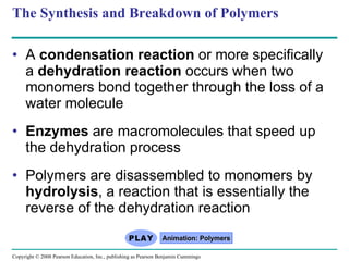
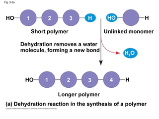
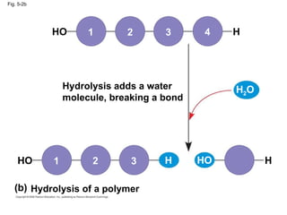
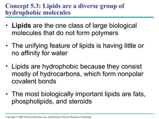
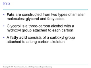
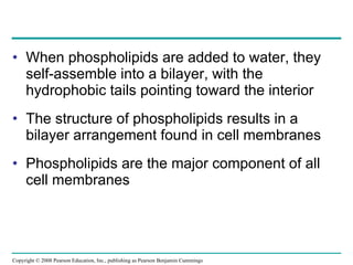
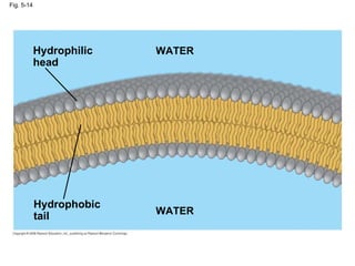
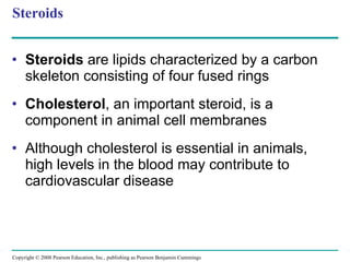
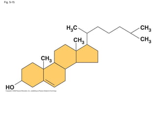
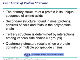
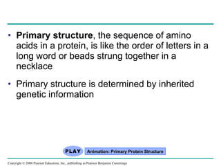
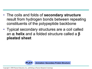
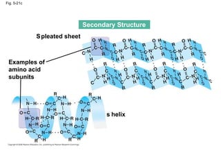
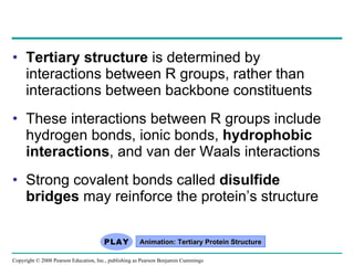
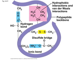
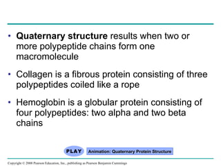
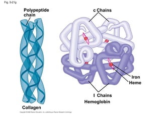
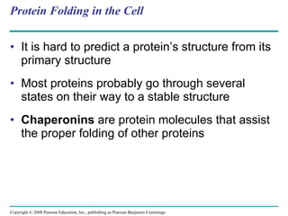
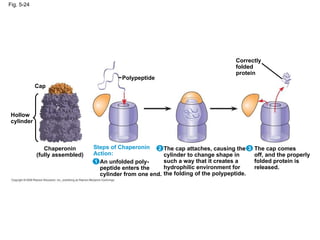
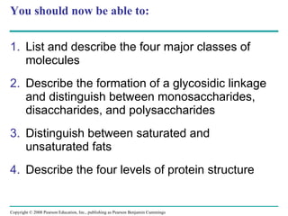
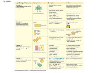
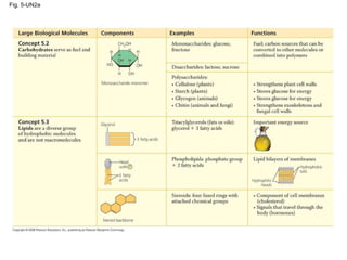
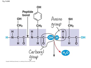
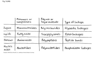
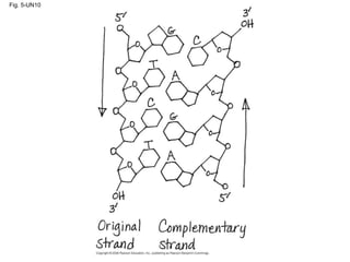
Chapter 5 The Structure and Function of Large Biological Molecules
Overview: The Molecules of Life All living things are made up of four classes of large biological molecules: carbohydrates, lipids, proteins, and nucleic acids Within cells, small organic molecules are joined together to form larger molecules Macromolecules  are large molecules composed of thousands of covalently connected atoms Molecular structure and function are inseparable Copyright © 2008 Pearson Education, Inc., publishing as Pearson Benjamin Cummings
Concept 5.1: Macromolecules are polymers, built from monomers A  polymer  is a long molecule consisting of many similar building blocks  These small building-block molecules are called  monomers Three of the four classes of life’s organic molecules are polymers: Carbohydrates Proteins Nucleic acids Copyright © 2008 Pearson Education, Inc., publishing as Pearson Benjamin Cummings
A  condensation reaction  or more specifically a  dehydration reaction  occurs when two monomers bond together through the loss of a water molecule Enzymes  are macromolecules that speed up the dehydration process Polymers are disassembled to monomers by  hydrolysis , a reaction that is essentially the reverse of the dehydration reaction The Synthesis and Breakdown of Polymers Animation: Polymers Copyright © 2008 Pearson Education, Inc., publishing as Pearson Benjamin Cummings
Fig. 5-2a Dehydration removes a water molecule, forming a new bond Short polymer Unlinked monomer Longer polymer Dehydration reaction in the synthesis of a polymer HO HO HO H 2 O H H H 4 3 2 1 1 2 3 (a)
Fig. 5-2b Hydrolysis adds a water molecule, breaking a bond Hydrolysis of a polymer HO HO HO H 2 O H H H 3 2 1 1 2 3 4 (b)
The Diversity of Polymers Each cell has thousands of different kinds of macromolecules  Macromolecules vary among cells of an organism, vary more within a species, and vary even more between species An immense variety of polymers can be built from a small set of monomers 2 3 HO H Copyright © 2008 Pearson Education, Inc., publishing as Pearson Benjamin Cummings
Concept 5.2: Carbohydrates serve as fuel and building material Carbohydrates  include sugars and the polymers of sugars The simplest carbohydrates are monosaccharides, or single sugars Carbohydrate macromolecules are polysaccharides, polymers composed of many sugar building blocks  Copyright © 2008 Pearson Education, Inc., publishing as Pearson Benjamin Cummings
Sugars Monosaccharides  have molecular formulas that are usually multiples of CH 2 O Glucose (C 6 H 12 O 6 ) is the most common monosaccharide Monosaccharides are classified by  The location of the carbonyl group (as aldose or ketose) The number of carbons in the carbon skeleton Copyright © 2008 Pearson Education, Inc., publishing as Pearson Benjamin Cummings
Fig. 5-3 Dihydroxyacetone Ribulose Ketoses Aldoses Fructose Glyceraldehyde Ribose Glucose Galactose Hexoses (C 6 H 12 O 6 ) Pentoses (C 5 H 10 O 5 ) Trioses (C 3 H 6 O 3 )
Though often drawn as linear skeletons, in aqueous solutions many sugars form rings Monosaccharides serve as a major fuel for cells and as raw material for building molecules  Copyright © 2008 Pearson Education, Inc., publishing as Pearson Benjamin Cummings
Fig. 5-4 (a) Linear and ring forms (b) Abbreviated ring structure
A  disaccharide  is formed when a dehydration reaction joins two monosaccharides  This covalent bond is called a  glycosidic linkage Animation: Disaccharides Copyright © 2008 Pearson Education, Inc., publishing as Pearson Benjamin Cummings
Fig. 5-5 (b) Dehydration reaction in the synthesis of sucrose Glucose Fructose Sucrose Maltose Glucose Glucose (a) Dehydration reaction in the synthesis of maltose 1–4 glycosidic linkage 1–2 glycosidic linkage
Polysaccharides Polysaccharides , the polymers of sugars, have storage and structural roles The structure and function of a polysaccharide are determined by its sugar monomers and the positions of glycosidic linkages Copyright © 2008 Pearson Education, Inc., publishing as Pearson Benjamin Cummings
Storage Polysaccharides Starch , a  storage polysaccharide of plants, consists entirely of glucose monomers Plants store surplus starch as granules within chloroplasts and other plastids  Copyright © 2008 Pearson Education, Inc., publishing as Pearson Benjamin Cummings
Fig. 5-6 (b) Glycogen: an animal polysaccharide Starch Glycogen Amylose Chloroplast (a) Starch: a plant polysaccharide Amylopectin Mitochondria Glycogen granules 0.5 µm 1 µm
Glycogen  is a storage polysaccharide in animals Humans and other vertebrates store glycogen mainly in liver and muscle cells Copyright © 2008 Pearson Education, Inc., publishing as Pearson Benjamin Cummings
Structural Polysaccharides The polysaccharide  cellulose  is a major component of the tough wall of plant cells Like starch, cellulose is a polymer of glucose, but the glycosidic linkages differ The difference is based on two ring forms for glucose: alpha (  ) and beta (  )  Animation: Polysaccharides Copyright © 2008 Pearson Education, Inc., publishing as Pearson Benjamin Cummings
Fig. 5-7 (a)    and    glucose ring structures    Glucose    Glucose (b) Starch: 1–4 linkage of    glucose monomers (b) Cellulose: 1–4 linkage of    glucose monomers
Polymers with    glucose are helical Polymers with    glucose are straight In straight structures, H atoms on one strand can bond with OH groups on other strands Parallel cellulose molecules held together this way are grouped into microfibrils, which form strong building materials for plants Copyright © 2008 Pearson Education, Inc., publishing as Pearson Benjamin Cummings
Fig. 5-8 Glucose monomer Cellulose molecules Microfibril Cellulose microfibrils in a plant cell wall 0.5 µm 10 µm Cell walls
Enzymes that digest starch by hydrolyzing    linkages can’t hydrolyze    linkages in cellulose Cellulose in human food passes through the digestive tract as insoluble fiber Some microbes use enzymes to digest cellulose Many herbivores, from cows to termites, have symbiotic relationships with these microbes Copyright © 2008 Pearson Education, Inc., publishing as Pearson Benjamin Cummings
Chitin , another structural polysaccharide, is found in the exoskeleton of arthropods Chitin also provides structural support for the cell walls of many fungi Copyright © 2008 Pearson Education, Inc., publishing as Pearson Benjamin Cummings
Fig. 5-10 The  structure of the chitin monomer. (a) (b) (c) Chitin forms the exoskeleton of arthropods. Chitin is used to make a strong and flexible surgical thread.
Concept 5.3: Lipids are a diverse group of hydrophobic molecules Lipids  are the one class of large biological molecules that do not form polymers The unifying feature of lipids is having little or no affinity for water Lipids are hydrophobic because  they consist mostly of hydrocarbons, which form nonpolar covalent bonds The most biologically important lipids are fats, phospholipids, and steroids Copyright © 2008 Pearson Education, Inc., publishing as Pearson Benjamin Cummings
Fats Fats  are constructed from two types of smaller molecules: glycerol and fatty acids Glycerol is a three-carbon alcohol with a hydroxyl group attached to each carbon A  fatty acid  consists of a carboxyl group attached to a long carbon skeleton Copyright © 2008 Pearson Education, Inc., publishing as Pearson Benjamin Cummings
Fig. 5-11 Fatty acid (palmitic acid) Glycerol (a) Dehydration reaction in the synthesis of a fat Ester linkage (b) Fat molecule (triacylglycerol)
Fats separate from water because water molecules form hydrogen bonds with each other and exclude the fats In a fat, three fatty acids are joined to glycerol by an ester linkage, creating a  triacylglycerol , or triglyceride Copyright © 2008 Pearson Education, Inc., publishing as Pearson Benjamin Cummings
Fatty acids vary in length (number of carbons) and in the number and locations of double bonds Saturated fatty acids  have the maximum number of hydrogen atoms possible and no double bonds Unsaturated fatty acids  have one or more double bonds Animation: Fats Copyright © 2008 Pearson Education, Inc., publishing as Pearson Benjamin Cummings
Fig. 5-12 Structural formula of a saturated fat molecule Stearic acid, a saturated fatty acid (a) Saturated fat Structural formula of an unsaturated fat molecule Oleic acid, an unsaturated fatty acid (b) Unsaturated fat cis  double bond causes bending
Fats made from saturated fatty acids are called saturated fats, and are solid at room temperature Most animal fats are saturated Fats made from unsaturated fatty acids are called unsaturated fats or oils, and are liquid at room temperature Plant fats and fish fats are usually unsaturated Copyright © 2008 Pearson Education, Inc., publishing as Pearson Benjamin Cummings
A diet rich in saturated fats may contribute to cardiovascular disease through plaque deposits  Hydrogenation is the process of converting unsaturated fats to saturated fats by adding hydrogen Hydrogenating vegetable oils also creates unsaturated fats with  trans  double bonds These  trans  fats may contribute more than saturated fats to cardiovascular disease Copyright © 2008 Pearson Education, Inc., publishing as Pearson Benjamin Cummings
The major function of fats is energy storage Humans and other mammals store their fat in adipose cells Adipose tissue also cushions vital organs and insulates the body Copyright © 2008 Pearson Education, Inc., publishing as Pearson Benjamin Cummings
Phospholipids In a  phospholipid , two fatty acids and a phosphate group are attached to glycerol  The two fatty acid tails are hydrophobic, but the phosphate group and its attachments form a hydrophilic head Copyright © 2008 Pearson Education, Inc., publishing as Pearson Benjamin Cummings
Fig. 5-13 (b) Space-filling model (a) (c) Structural formula Phospholipid symbol Fatty acids Hydrophilic head Hydrophobic tails Choline Phosphate Glycerol Hydrophobic tails Hydrophilic head
When phospholipids are added to water, they self-assemble into a bilayer, with the hydrophobic tails pointing toward the interior The structure of phospholipids results in a bilayer arrangement found in cell membranes Phospholipids are the major component of all cell membranes Copyright © 2008 Pearson Education, Inc., publishing as Pearson Benjamin Cummings
Fig. 5-14 Hydrophilic head Hydrophobic tail WATER WATER
Steroids Steroids  are lipids characterized by a carbon skeleton consisting of four fused rings Cholesterol , an important steroid, is a component in animal cell membranes Although cholesterol is essential in animals, high levels in the blood may contribute to cardiovascular disease Copyright © 2008 Pearson Education, Inc., publishing as Pearson Benjamin Cummings
Fig. 5-15
Concept 5.4: Proteins have many structures, resulting in a wide range of functions Proteins account for more than 50% of the dry mass of most cells Protein functions include structural support, storage, transport, cellular communications, movement, and defense against foreign substances Copyright © 2008 Pearson Education, Inc., publishing as Pearson Benjamin Cummings
Table 5-1
Enzymes  are a type of protein that acts as a  catalyst  to speed up chemical reactions Enzymes can perform their functions repeatedly, functioning as workhorses that carry out the processes of life Animation: Enzymes Copyright © 2008 Pearson Education, Inc., publishing as Pearson Benjamin Cummings
Fig. 5-16 Enzyme (sucrase) Substrate (sucrose) Fructose Glucose OH H O H 2 O
Polypeptides Polypeptides  are polymers built from the same set of 20 amino acids A  protein  consists of one or more polypeptides Copyright © 2008 Pearson Education, Inc., publishing as Pearson Benjamin Cummings
Amino Acid Monomers Amino acids  are organic molecules with carboxyl and amino groups Amino acids differ in their properties due to differing side chains, called R groups These R groups determine if the amino acid is nonpolar, polar or  charged. Copyright © 2008 Pearson Education, Inc., publishing as Pearson Benjamin Cummings
Fig. 5-UN1 Amino group Carboxyl group    carbon
Fig. 5-17a Nonpolar Glycine (Gly or G) Alanine (Ala or A) Valine (Val or V) Leucine (Leu or L) Isoleucine (Ile or   ) Methionine (Met or M) Phenylalanine (Phe or F) Tryptophan (Trp or W) Proline (Pro or P)
Fig. 5-17b Polar Asparagine (Asn or N) Glutamine (Gln or Q) Serine (Ser or S) Threonine (Thr or T) Cysteine (Cys or C) Tyrosine (Tyr or Y)
Fig. 5-17c Acidic Arginine (Arg or R) Histidine (His or H) Aspartic acid (Asp or D) Glutamic acid (Glu or E) Lysine (Lys or K) Basic Electrically charged
Amino Acid Polymers Amino acids are linked by  peptide bonds A polypeptide is a polymer of amino acids Polypeptides range in length from a few to more than a thousand monomers  Each polypeptide has a unique linear sequence of amino acids Copyright © 2008 Pearson Education, Inc., publishing as Pearson Benjamin Cummings
Peptide bond Fig. 5-18 Amino end (N-terminus) Peptide bond Side chains Backbone Carboxyl end (C-terminus) (a) (b)
Protein Structure and Function A functional protein consists of one or more polypeptides twisted, folded, and coiled into a unique shape The sequence of amino acids determines a protein’s three-dimensional structure A protein’s structure determines its function Copyright © 2008 Pearson Education, Inc., publishing as Pearson Benjamin Cummings
Four Levels of Protein Structure The primary structure of a protein is its unique sequence of amino acids Secondary structure, found in most proteins, consists of coils and folds in the polypeptide chain Tertiary structure is determined by interactions among various side chains (R groups) Quaternary structure results when a protein consists of multiple polypeptide chains Animation: Protein Structure Introduction Copyright © 2008 Pearson Education, Inc., publishing as Pearson Benjamin Cummings
Primary structure , the sequence of amino acids in a protein, is like the order of letters in a long word or beads strung together in a necklace Primary structure is determined by inherited genetic information Animation: Primary Protein Structure Copyright © 2008 Pearson Education, Inc., publishing as Pearson Benjamin Cummings
The coils and folds of  secondary structure  result from hydrogen bonds between repeating constituents of the polypeptide backbone Typical secondary structures are a coil called an    helix  and a folded structure called a    pleated sheet Animation: Secondary Protein Structure Copyright © 2008 Pearson Education, Inc., publishing as Pearson Benjamin Cummings
Fig. 5-21c Secondary Structure    pleated sheet Examples of amino acid subunits    helix
Tertiary structure  is determined by interactions between R groups, rather than interactions between backbone constituents These interactions between R groups include hydrogen bonds, ionic bonds,  hydrophobic interactions , and van der Waals interactions Strong covalent bonds called  disulfide bridges  may reinforce the protein’s structure Animation: Tertiary Protein Structure Copyright © 2008 Pearson Education, Inc., publishing as Pearson Benjamin Cummings
Fig. 5-21f Polypeptide backbone Hydrophobic interactions and van der Waals interactions Disulfide bridge Ionic bond Hydrogen bond
Quaternary structure  results when two or more polypeptide chains form one macromolecule Collagen is a fibrous protein consisting of three polypeptides coiled like a rope Hemoglobin is a globular protein consisting of four polypeptides: two alpha and two beta chains Animation: Quaternary Protein Structure Copyright © 2008 Pearson Education, Inc., publishing as Pearson Benjamin Cummings
Fig. 5-21g Polypeptide chain    Chains Heme Iron    Chains Collagen Hemoglobin
Sickle-Cell Disease: A Change in  Primary Structure A slight change in primary structure can affect a protein’s structure and ability to function  Sickle-cell disease, an inherited blood disorder, results from a single amino acid substitution in the protein hemoglobin Copyright © 2008 Pearson Education, Inc., publishing as Pearson Benjamin Cummings
Fig. 5-22 Primary structure Secondary and tertiary structures Quaternary structure Normal hemoglobin (top view) Primary structure Secondary and tertiary structures Quaternary structure Function Function    subunit Molecules do not associate with one another; each carries oxygen. Red blood cell shape Normal red blood cells are full of individual hemoglobin moledules, each carrying oxygen. 10 µm Normal hemoglobin     1 2 3 4 5 6 7 Val His Leu Thr Pro Glu Glu Red blood cell shape    subunit Exposed hydrophobic region Sickle-cell hemoglobin   Molecules interact with one another and crystallize into a fiber; capacity to carry oxygen is greatly reduced.   Fibers of abnormal hemoglobin deform red blood cell into sickle shape. 10 µm Sickle-cell hemoglobin Glu Pro Thr Leu His Val Val 1 2 3 4 5 6 7
What Determines Protein Structure? In addition to primary structure, physical and chemical conditions can affect structure Alterations in pH, salt concentration, temperature, or other environmental factors can cause a protein to unravel This loss of a protein’s native structure is called  denaturation A denatured protein is biologically inactive Copyright © 2008 Pearson Education, Inc., publishing as Pearson Benjamin Cummings
Fig. 5-23 Normal protein Denatured protein Denaturation Renaturation
Protein Folding in the Cell It is hard to predict a protein’s structure from its primary structure Most proteins probably go through several states on their way to a stable structure Chaperonins  are protein molecules that assist the proper folding of other proteins Copyright © 2008 Pearson Education, Inc., publishing as Pearson Benjamin Cummings
Fig. 5-24 Hollow cylinder Cap Chaperonin (fully assembled) Polypeptide Steps of Chaperonin Action: An unfolded poly- peptide enters the cylinder from one end. 1 2 3 The cap attaches, causing the cylinder to change shape in such a way that it creates a hydrophilic environment for the folding of the polypeptide. The cap comes off, and the properly folded protein is released. Correctly folded protein
Concept 5.5: Nucleic acids store and transmit hereditary information The amino acid sequence of a polypeptide is programmed by a unit of inheritance called a  gene Genes are made of DNA, a  nucleic acid Copyright © 2008 Pearson Education, Inc., publishing as Pearson Benjamin Cummings
The Roles of Nucleic Acids There are two types of nucleic acids: Deoxyribonucleic acid (DNA) Ribonucleic acid (RNA) DNA provides directions for its own replication DNA directs synthesis of messenger RNA (mRNA) and, through mRNA, controls protein synthesis Protein synthesis occurs in ribosomes Copyright © 2008 Pearson Education, Inc., publishing as Pearson Benjamin Cummings
Fig. 5-26-3 mRNA Synthesis of mRNA in the nucleus DNA NUCLEUS mRNA CYTOPLASM Movement of mRNA into cytoplasm via nuclear pore Ribosome Amino acids Polypeptide Synthesis of protein 1 2 3
The Structure of Nucleic Acids Nucleic acids are polymers called  polynucleotides Each polynucleotide is made of monomers called  nucleotides Each nucleotide consists of a nitrogenous base, a pentose sugar, and a phosphate group The portion of a nucleotide without the phosphate group is called a  nucleoside Copyright © 2008 Pearson Education, Inc., publishing as Pearson Benjamin Cummings
Nucleotide Monomers There are two families of nitrogenous bases:   Pyrimidines  (cytosine, thymine, and uracil)   have a single six-membered ring Purines  (adenine and guanine) have a six-membered ring fused to a five-membered ring In DNA, the sugar is  deoxyribose ; in RNA, the sugar is  ribose Copyright © 2008 Pearson Education, Inc., publishing as Pearson Benjamin Cummings
Fig. 5-27c-2 Ribose (in RNA) Deoxyribose (in DNA) Sugars (c) Nucleoside components: sugars
Nucleotide Polymers Nucleotide polymers are linked together to build a polynucleotide Adjacent nucleotides are joined by covalent bonds that form between the –OH group on the 3   carbon of one nucleotide and the phosphate on the 5   carbon on the next These links create a backbone of sugar-phosphate units with nitrogenous bases as appendages The sequence of bases along a DNA or mRNA polymer is unique for each gene Copyright © 2008 Pearson Education, Inc., publishing as Pearson Benjamin Cummings
Fig. 5-27 5’ end Nucleoside Nitrogenous base Phosphate group Sugar (pentose) (b) Nucleotide (a) Polynucleotide, or nucleic acid 3’ end 3  C 3  C 5  C 5  C Nitrogenous bases Pyrimidines Cytosine (C) Thymine (T, in DNA) Uracil (U, in RNA) Purines Adenine (A) Guanine (G) Sugars Deoxyribose (in DNA) Ribose (in RNA) (c) Nucleoside components: sugars
The DNA Double Helix A DNA molecule has two polynucleotides spiraling around an imaginary axis, forming a  double helix In the DNA double helix, the two backbones run in opposite 5   -> 3   directions from each other, an arrangement referred to as  antiparallel One DNA molecule includes many genes The nitrogenous bases in DNA pair up and form hydrogen bonds: adenine (A) always with thymine (T), and guanine (G) always with cytosine (C) Copyright © 2008 Pearson Education, Inc., publishing as Pearson Benjamin Cummings
Fig. 5-28 Sugar-phosphate backbones 3' end 3' end 3' end 3' end 5' end 5' end 5' end 5' end Base pair (joined by hydrogen bonding) Old strands New strands Nucleotide about to be added to a new strand
You should now be able to: List and describe the four major classes of molecules Describe the formation of a glycosidic linkage and distinguish between monosaccharides, disaccharides, and polysaccharides Distinguish between saturated and unsaturated fats  Describe the four levels of protein structure Copyright © 2008 Pearson Education, Inc., publishing as Pearson Benjamin Cummings
You should now be able to: Distinguish between the following:ribose and deoxyribose, the 5   end and 3   end of a nucleotide Copyright © 2008 Pearson Education, Inc., publishing as Pearson Benjamin Cummings
Fig. 5-UN2
Fig. 5-UN2a
Fig. 5-UN2b
Fig. 5-UN5
Fig. 5-UN9
Fig. 5-UN10

Bio Ch 5 Pwpt

  • 1.
    Chapter 5 TheStructure and Function of Large Biological Molecules
  • 2.
    Overview: The Moleculesof Life All living things are made up of four classes of large biological molecules: carbohydrates, lipids, proteins, and nucleic acids Within cells, small organic molecules are joined together to form larger molecules Macromolecules are large molecules composed of thousands of covalently connected atoms Molecular structure and function are inseparable Copyright © 2008 Pearson Education, Inc., publishing as Pearson Benjamin Cummings
  • 3.
    Concept 5.1: Macromoleculesare polymers, built from monomers A polymer is a long molecule consisting of many similar building blocks These small building-block molecules are called monomers Three of the four classes of life’s organic molecules are polymers: Carbohydrates Proteins Nucleic acids Copyright © 2008 Pearson Education, Inc., publishing as Pearson Benjamin Cummings
  • 4.
    A condensationreaction or more specifically a dehydration reaction occurs when two monomers bond together through the loss of a water molecule Enzymes are macromolecules that speed up the dehydration process Polymers are disassembled to monomers by hydrolysis , a reaction that is essentially the reverse of the dehydration reaction The Synthesis and Breakdown of Polymers Animation: Polymers Copyright © 2008 Pearson Education, Inc., publishing as Pearson Benjamin Cummings
  • 5.
    Fig. 5-2a Dehydrationremoves a water molecule, forming a new bond Short polymer Unlinked monomer Longer polymer Dehydration reaction in the synthesis of a polymer HO HO HO H 2 O H H H 4 3 2 1 1 2 3 (a)
  • 6.
    Fig. 5-2b Hydrolysisadds a water molecule, breaking a bond Hydrolysis of a polymer HO HO HO H 2 O H H H 3 2 1 1 2 3 4 (b)
  • 7.
    The Diversity ofPolymers Each cell has thousands of different kinds of macromolecules Macromolecules vary among cells of an organism, vary more within a species, and vary even more between species An immense variety of polymers can be built from a small set of monomers 2 3 HO H Copyright © 2008 Pearson Education, Inc., publishing as Pearson Benjamin Cummings
  • 8.
    Concept 5.2: Carbohydratesserve as fuel and building material Carbohydrates include sugars and the polymers of sugars The simplest carbohydrates are monosaccharides, or single sugars Carbohydrate macromolecules are polysaccharides, polymers composed of many sugar building blocks Copyright © 2008 Pearson Education, Inc., publishing as Pearson Benjamin Cummings
  • 9.
    Sugars Monosaccharides have molecular formulas that are usually multiples of CH 2 O Glucose (C 6 H 12 O 6 ) is the most common monosaccharide Monosaccharides are classified by The location of the carbonyl group (as aldose or ketose) The number of carbons in the carbon skeleton Copyright © 2008 Pearson Education, Inc., publishing as Pearson Benjamin Cummings
  • 10.
    Fig. 5-3 DihydroxyacetoneRibulose Ketoses Aldoses Fructose Glyceraldehyde Ribose Glucose Galactose Hexoses (C 6 H 12 O 6 ) Pentoses (C 5 H 10 O 5 ) Trioses (C 3 H 6 O 3 )
  • 11.
    Though often drawnas linear skeletons, in aqueous solutions many sugars form rings Monosaccharides serve as a major fuel for cells and as raw material for building molecules Copyright © 2008 Pearson Education, Inc., publishing as Pearson Benjamin Cummings
  • 12.
    Fig. 5-4 (a)Linear and ring forms (b) Abbreviated ring structure
  • 13.
    A disaccharide is formed when a dehydration reaction joins two monosaccharides This covalent bond is called a glycosidic linkage Animation: Disaccharides Copyright © 2008 Pearson Education, Inc., publishing as Pearson Benjamin Cummings
  • 14.
    Fig. 5-5 (b)Dehydration reaction in the synthesis of sucrose Glucose Fructose Sucrose Maltose Glucose Glucose (a) Dehydration reaction in the synthesis of maltose 1–4 glycosidic linkage 1–2 glycosidic linkage
  • 15.
    Polysaccharides Polysaccharides ,the polymers of sugars, have storage and structural roles The structure and function of a polysaccharide are determined by its sugar monomers and the positions of glycosidic linkages Copyright © 2008 Pearson Education, Inc., publishing as Pearson Benjamin Cummings
  • 16.
    Storage Polysaccharides Starch, a storage polysaccharide of plants, consists entirely of glucose monomers Plants store surplus starch as granules within chloroplasts and other plastids Copyright © 2008 Pearson Education, Inc., publishing as Pearson Benjamin Cummings
  • 17.
    Fig. 5-6 (b)Glycogen: an animal polysaccharide Starch Glycogen Amylose Chloroplast (a) Starch: a plant polysaccharide Amylopectin Mitochondria Glycogen granules 0.5 µm 1 µm
  • 18.
    Glycogen isa storage polysaccharide in animals Humans and other vertebrates store glycogen mainly in liver and muscle cells Copyright © 2008 Pearson Education, Inc., publishing as Pearson Benjamin Cummings
  • 19.
    Structural Polysaccharides Thepolysaccharide cellulose is a major component of the tough wall of plant cells Like starch, cellulose is a polymer of glucose, but the glycosidic linkages differ The difference is based on two ring forms for glucose: alpha (  ) and beta (  )  Animation: Polysaccharides Copyright © 2008 Pearson Education, Inc., publishing as Pearson Benjamin Cummings
  • 20.
    Fig. 5-7 (a)  and  glucose ring structures  Glucose  Glucose (b) Starch: 1–4 linkage of  glucose monomers (b) Cellulose: 1–4 linkage of  glucose monomers
  • 21.
    Polymers with  glucose are helical Polymers with  glucose are straight In straight structures, H atoms on one strand can bond with OH groups on other strands Parallel cellulose molecules held together this way are grouped into microfibrils, which form strong building materials for plants Copyright © 2008 Pearson Education, Inc., publishing as Pearson Benjamin Cummings
  • 22.
    Fig. 5-8 Glucosemonomer Cellulose molecules Microfibril Cellulose microfibrils in a plant cell wall 0.5 µm 10 µm Cell walls
  • 23.
    Enzymes that digeststarch by hydrolyzing  linkages can’t hydrolyze  linkages in cellulose Cellulose in human food passes through the digestive tract as insoluble fiber Some microbes use enzymes to digest cellulose Many herbivores, from cows to termites, have symbiotic relationships with these microbes Copyright © 2008 Pearson Education, Inc., publishing as Pearson Benjamin Cummings
  • 24.
    Chitin , anotherstructural polysaccharide, is found in the exoskeleton of arthropods Chitin also provides structural support for the cell walls of many fungi Copyright © 2008 Pearson Education, Inc., publishing as Pearson Benjamin Cummings
  • 25.
    Fig. 5-10 The structure of the chitin monomer. (a) (b) (c) Chitin forms the exoskeleton of arthropods. Chitin is used to make a strong and flexible surgical thread.
  • 26.
    Concept 5.3: Lipidsare a diverse group of hydrophobic molecules Lipids are the one class of large biological molecules that do not form polymers The unifying feature of lipids is having little or no affinity for water Lipids are hydrophobic because  they consist mostly of hydrocarbons, which form nonpolar covalent bonds The most biologically important lipids are fats, phospholipids, and steroids Copyright © 2008 Pearson Education, Inc., publishing as Pearson Benjamin Cummings
  • 27.
    Fats Fats are constructed from two types of smaller molecules: glycerol and fatty acids Glycerol is a three-carbon alcohol with a hydroxyl group attached to each carbon A fatty acid consists of a carboxyl group attached to a long carbon skeleton Copyright © 2008 Pearson Education, Inc., publishing as Pearson Benjamin Cummings
  • 28.
    Fig. 5-11 Fattyacid (palmitic acid) Glycerol (a) Dehydration reaction in the synthesis of a fat Ester linkage (b) Fat molecule (triacylglycerol)
  • 29.
    Fats separate fromwater because water molecules form hydrogen bonds with each other and exclude the fats In a fat, three fatty acids are joined to glycerol by an ester linkage, creating a triacylglycerol , or triglyceride Copyright © 2008 Pearson Education, Inc., publishing as Pearson Benjamin Cummings
  • 30.
    Fatty acids varyin length (number of carbons) and in the number and locations of double bonds Saturated fatty acids have the maximum number of hydrogen atoms possible and no double bonds Unsaturated fatty acids have one or more double bonds Animation: Fats Copyright © 2008 Pearson Education, Inc., publishing as Pearson Benjamin Cummings
  • 31.
    Fig. 5-12 Structuralformula of a saturated fat molecule Stearic acid, a saturated fatty acid (a) Saturated fat Structural formula of an unsaturated fat molecule Oleic acid, an unsaturated fatty acid (b) Unsaturated fat cis double bond causes bending
  • 32.
    Fats made fromsaturated fatty acids are called saturated fats, and are solid at room temperature Most animal fats are saturated Fats made from unsaturated fatty acids are called unsaturated fats or oils, and are liquid at room temperature Plant fats and fish fats are usually unsaturated Copyright © 2008 Pearson Education, Inc., publishing as Pearson Benjamin Cummings
  • 33.
    A diet richin saturated fats may contribute to cardiovascular disease through plaque deposits Hydrogenation is the process of converting unsaturated fats to saturated fats by adding hydrogen Hydrogenating vegetable oils also creates unsaturated fats with trans double bonds These trans fats may contribute more than saturated fats to cardiovascular disease Copyright © 2008 Pearson Education, Inc., publishing as Pearson Benjamin Cummings
  • 34.
    The major functionof fats is energy storage Humans and other mammals store their fat in adipose cells Adipose tissue also cushions vital organs and insulates the body Copyright © 2008 Pearson Education, Inc., publishing as Pearson Benjamin Cummings
  • 35.
    Phospholipids In a phospholipid , two fatty acids and a phosphate group are attached to glycerol The two fatty acid tails are hydrophobic, but the phosphate group and its attachments form a hydrophilic head Copyright © 2008 Pearson Education, Inc., publishing as Pearson Benjamin Cummings
  • 36.
    Fig. 5-13 (b)Space-filling model (a) (c) Structural formula Phospholipid symbol Fatty acids Hydrophilic head Hydrophobic tails Choline Phosphate Glycerol Hydrophobic tails Hydrophilic head
  • 37.
    When phospholipids areadded to water, they self-assemble into a bilayer, with the hydrophobic tails pointing toward the interior The structure of phospholipids results in a bilayer arrangement found in cell membranes Phospholipids are the major component of all cell membranes Copyright © 2008 Pearson Education, Inc., publishing as Pearson Benjamin Cummings
  • 38.
    Fig. 5-14 Hydrophilichead Hydrophobic tail WATER WATER
  • 39.
    Steroids Steroids are lipids characterized by a carbon skeleton consisting of four fused rings Cholesterol , an important steroid, is a component in animal cell membranes Although cholesterol is essential in animals, high levels in the blood may contribute to cardiovascular disease Copyright © 2008 Pearson Education, Inc., publishing as Pearson Benjamin Cummings
  • 40.
  • 41.
    Concept 5.4: Proteinshave many structures, resulting in a wide range of functions Proteins account for more than 50% of the dry mass of most cells Protein functions include structural support, storage, transport, cellular communications, movement, and defense against foreign substances Copyright © 2008 Pearson Education, Inc., publishing as Pearson Benjamin Cummings
  • 42.
  • 43.
    Enzymes area type of protein that acts as a catalyst to speed up chemical reactions Enzymes can perform their functions repeatedly, functioning as workhorses that carry out the processes of life Animation: Enzymes Copyright © 2008 Pearson Education, Inc., publishing as Pearson Benjamin Cummings
  • 44.
    Fig. 5-16 Enzyme(sucrase) Substrate (sucrose) Fructose Glucose OH H O H 2 O
  • 45.
    Polypeptides Polypeptides are polymers built from the same set of 20 amino acids A protein consists of one or more polypeptides Copyright © 2008 Pearson Education, Inc., publishing as Pearson Benjamin Cummings
  • 46.
    Amino Acid MonomersAmino acids are organic molecules with carboxyl and amino groups Amino acids differ in their properties due to differing side chains, called R groups These R groups determine if the amino acid is nonpolar, polar or charged. Copyright © 2008 Pearson Education, Inc., publishing as Pearson Benjamin Cummings
  • 47.
    Fig. 5-UN1 Aminogroup Carboxyl group  carbon
  • 48.
    Fig. 5-17a NonpolarGlycine (Gly or G) Alanine (Ala or A) Valine (Val or V) Leucine (Leu or L) Isoleucine (Ile or  ) Methionine (Met or M) Phenylalanine (Phe or F) Tryptophan (Trp or W) Proline (Pro or P)
  • 49.
    Fig. 5-17b PolarAsparagine (Asn or N) Glutamine (Gln or Q) Serine (Ser or S) Threonine (Thr or T) Cysteine (Cys or C) Tyrosine (Tyr or Y)
  • 50.
    Fig. 5-17c AcidicArginine (Arg or R) Histidine (His or H) Aspartic acid (Asp or D) Glutamic acid (Glu or E) Lysine (Lys or K) Basic Electrically charged
  • 51.
    Amino Acid PolymersAmino acids are linked by peptide bonds A polypeptide is a polymer of amino acids Polypeptides range in length from a few to more than a thousand monomers Each polypeptide has a unique linear sequence of amino acids Copyright © 2008 Pearson Education, Inc., publishing as Pearson Benjamin Cummings
  • 52.
    Peptide bond Fig.5-18 Amino end (N-terminus) Peptide bond Side chains Backbone Carboxyl end (C-terminus) (a) (b)
  • 53.
    Protein Structure andFunction A functional protein consists of one or more polypeptides twisted, folded, and coiled into a unique shape The sequence of amino acids determines a protein’s three-dimensional structure A protein’s structure determines its function Copyright © 2008 Pearson Education, Inc., publishing as Pearson Benjamin Cummings
  • 54.
    Four Levels ofProtein Structure The primary structure of a protein is its unique sequence of amino acids Secondary structure, found in most proteins, consists of coils and folds in the polypeptide chain Tertiary structure is determined by interactions among various side chains (R groups) Quaternary structure results when a protein consists of multiple polypeptide chains Animation: Protein Structure Introduction Copyright © 2008 Pearson Education, Inc., publishing as Pearson Benjamin Cummings
  • 55.
    Primary structure ,the sequence of amino acids in a protein, is like the order of letters in a long word or beads strung together in a necklace Primary structure is determined by inherited genetic information Animation: Primary Protein Structure Copyright © 2008 Pearson Education, Inc., publishing as Pearson Benjamin Cummings
  • 56.
    The coils andfolds of secondary structure result from hydrogen bonds between repeating constituents of the polypeptide backbone Typical secondary structures are a coil called an  helix and a folded structure called a  pleated sheet Animation: Secondary Protein Structure Copyright © 2008 Pearson Education, Inc., publishing as Pearson Benjamin Cummings
  • 57.
    Fig. 5-21c SecondaryStructure  pleated sheet Examples of amino acid subunits  helix
  • 58.
    Tertiary structure is determined by interactions between R groups, rather than interactions between backbone constituents These interactions between R groups include hydrogen bonds, ionic bonds, hydrophobic interactions , and van der Waals interactions Strong covalent bonds called disulfide bridges may reinforce the protein’s structure Animation: Tertiary Protein Structure Copyright © 2008 Pearson Education, Inc., publishing as Pearson Benjamin Cummings
  • 59.
    Fig. 5-21f Polypeptidebackbone Hydrophobic interactions and van der Waals interactions Disulfide bridge Ionic bond Hydrogen bond
  • 60.
    Quaternary structure results when two or more polypeptide chains form one macromolecule Collagen is a fibrous protein consisting of three polypeptides coiled like a rope Hemoglobin is a globular protein consisting of four polypeptides: two alpha and two beta chains Animation: Quaternary Protein Structure Copyright © 2008 Pearson Education, Inc., publishing as Pearson Benjamin Cummings
  • 61.
    Fig. 5-21g Polypeptidechain  Chains Heme Iron  Chains Collagen Hemoglobin
  • 62.
    Sickle-Cell Disease: AChange in Primary Structure A slight change in primary structure can affect a protein’s structure and ability to function Sickle-cell disease, an inherited blood disorder, results from a single amino acid substitution in the protein hemoglobin Copyright © 2008 Pearson Education, Inc., publishing as Pearson Benjamin Cummings
  • 63.
    Fig. 5-22 Primarystructure Secondary and tertiary structures Quaternary structure Normal hemoglobin (top view) Primary structure Secondary and tertiary structures Quaternary structure Function Function  subunit Molecules do not associate with one another; each carries oxygen. Red blood cell shape Normal red blood cells are full of individual hemoglobin moledules, each carrying oxygen. 10 µm Normal hemoglobin     1 2 3 4 5 6 7 Val His Leu Thr Pro Glu Glu Red blood cell shape  subunit Exposed hydrophobic region Sickle-cell hemoglobin   Molecules interact with one another and crystallize into a fiber; capacity to carry oxygen is greatly reduced.   Fibers of abnormal hemoglobin deform red blood cell into sickle shape. 10 µm Sickle-cell hemoglobin Glu Pro Thr Leu His Val Val 1 2 3 4 5 6 7
  • 64.
    What Determines ProteinStructure? In addition to primary structure, physical and chemical conditions can affect structure Alterations in pH, salt concentration, temperature, or other environmental factors can cause a protein to unravel This loss of a protein’s native structure is called denaturation A denatured protein is biologically inactive Copyright © 2008 Pearson Education, Inc., publishing as Pearson Benjamin Cummings
  • 65.
    Fig. 5-23 Normalprotein Denatured protein Denaturation Renaturation
  • 66.
    Protein Folding inthe Cell It is hard to predict a protein’s structure from its primary structure Most proteins probably go through several states on their way to a stable structure Chaperonins are protein molecules that assist the proper folding of other proteins Copyright © 2008 Pearson Education, Inc., publishing as Pearson Benjamin Cummings
  • 67.
    Fig. 5-24 Hollowcylinder Cap Chaperonin (fully assembled) Polypeptide Steps of Chaperonin Action: An unfolded poly- peptide enters the cylinder from one end. 1 2 3 The cap attaches, causing the cylinder to change shape in such a way that it creates a hydrophilic environment for the folding of the polypeptide. The cap comes off, and the properly folded protein is released. Correctly folded protein
  • 68.
    Concept 5.5: Nucleicacids store and transmit hereditary information The amino acid sequence of a polypeptide is programmed by a unit of inheritance called a gene Genes are made of DNA, a nucleic acid Copyright © 2008 Pearson Education, Inc., publishing as Pearson Benjamin Cummings
  • 69.
    The Roles ofNucleic Acids There are two types of nucleic acids: Deoxyribonucleic acid (DNA) Ribonucleic acid (RNA) DNA provides directions for its own replication DNA directs synthesis of messenger RNA (mRNA) and, through mRNA, controls protein synthesis Protein synthesis occurs in ribosomes Copyright © 2008 Pearson Education, Inc., publishing as Pearson Benjamin Cummings
  • 70.
    Fig. 5-26-3 mRNASynthesis of mRNA in the nucleus DNA NUCLEUS mRNA CYTOPLASM Movement of mRNA into cytoplasm via nuclear pore Ribosome Amino acids Polypeptide Synthesis of protein 1 2 3
  • 71.
    The Structure ofNucleic Acids Nucleic acids are polymers called polynucleotides Each polynucleotide is made of monomers called nucleotides Each nucleotide consists of a nitrogenous base, a pentose sugar, and a phosphate group The portion of a nucleotide without the phosphate group is called a nucleoside Copyright © 2008 Pearson Education, Inc., publishing as Pearson Benjamin Cummings
  • 72.
    Nucleotide Monomers Thereare two families of nitrogenous bases: Pyrimidines (cytosine, thymine, and uracil) have a single six-membered ring Purines (adenine and guanine) have a six-membered ring fused to a five-membered ring In DNA, the sugar is deoxyribose ; in RNA, the sugar is ribose Copyright © 2008 Pearson Education, Inc., publishing as Pearson Benjamin Cummings
  • 73.
    Fig. 5-27c-2 Ribose(in RNA) Deoxyribose (in DNA) Sugars (c) Nucleoside components: sugars
  • 74.
    Nucleotide Polymers Nucleotidepolymers are linked together to build a polynucleotide Adjacent nucleotides are joined by covalent bonds that form between the –OH group on the 3  carbon of one nucleotide and the phosphate on the 5  carbon on the next These links create a backbone of sugar-phosphate units with nitrogenous bases as appendages The sequence of bases along a DNA or mRNA polymer is unique for each gene Copyright © 2008 Pearson Education, Inc., publishing as Pearson Benjamin Cummings
  • 75.
    Fig. 5-27 5’end Nucleoside Nitrogenous base Phosphate group Sugar (pentose) (b) Nucleotide (a) Polynucleotide, or nucleic acid 3’ end 3  C 3  C 5  C 5  C Nitrogenous bases Pyrimidines Cytosine (C) Thymine (T, in DNA) Uracil (U, in RNA) Purines Adenine (A) Guanine (G) Sugars Deoxyribose (in DNA) Ribose (in RNA) (c) Nucleoside components: sugars
  • 76.
    The DNA DoubleHelix A DNA molecule has two polynucleotides spiraling around an imaginary axis, forming a double helix In the DNA double helix, the two backbones run in opposite 5  -> 3  directions from each other, an arrangement referred to as antiparallel One DNA molecule includes many genes The nitrogenous bases in DNA pair up and form hydrogen bonds: adenine (A) always with thymine (T), and guanine (G) always with cytosine (C) Copyright © 2008 Pearson Education, Inc., publishing as Pearson Benjamin Cummings
  • 77.
    Fig. 5-28 Sugar-phosphatebackbones 3' end 3' end 3' end 3' end 5' end 5' end 5' end 5' end Base pair (joined by hydrogen bonding) Old strands New strands Nucleotide about to be added to a new strand
  • 78.
    You should nowbe able to: List and describe the four major classes of molecules Describe the formation of a glycosidic linkage and distinguish between monosaccharides, disaccharides, and polysaccharides Distinguish between saturated and unsaturated fats Describe the four levels of protein structure Copyright © 2008 Pearson Education, Inc., publishing as Pearson Benjamin Cummings
  • 79.
    You should nowbe able to: Distinguish between the following:ribose and deoxyribose, the 5  end and 3  end of a nucleotide Copyright © 2008 Pearson Education, Inc., publishing as Pearson Benjamin Cummings
  • 80.
  • 81.
  • 82.
  • 83.
  • 84.
  • 85.

Editor's Notes

  • #6 Figure 5.2 The synthesis and breakdown of polymers
  • #7 Figure 5.2 The synthesis and breakdown of polymers
  • #11 Figure 5.3 The structure and classification of some monosaccharides
  • #13 Figure 5.4 Linear and ring forms of glucose
  • #15 Figure 5.5 Examples of disaccharide synthesis
  • #18 Figure 5.6 Storage polysaccharides of plants and animals
  • #21 Figure 5.7 Starch and cellulose structures
  • #23 Figure 5.8 The arrangement of cellulose in plant cell walls
  • #26 Figure 5.10 Chitin, a structural polysaccharide
  • #29 Figure 5.11 The synthesis and structure of a fat, or triacylglycerol
  • #32 Figure 5.12 Examples of saturated and unsaturated fats and fatty acids
  • #37 Figure 5.13 The structure of a phospholipid
  • #39 Figure 5.14 Bilayer structure formed by self-assembly of phospholipids in an aqueous environment
  • #40 For the Cell Biology Video Space Filling Model of Cholesterol, go to Animation and Video Files. For the Cell Biology Video Stick Model of Cholesterol, go to Animation and Video Files.
  • #41 Figure 5.15 Cholesterol, a steroid
  • #43 Table 5-1
  • #45 Figure 5.16 The catalytic cycle of an enzyme
  • #49 Figure 5.17 The 20 amino acids of proteins
  • #50 Figure 5.17 The 20 amino acids of proteins
  • #51 Figure 5.17 The 20 amino acids of proteins
  • #53 Figure 5.18 Making a polypeptide chain
  • #57 For the Cell Biology Video An Idealized Alpha Helix: No Sidechains, go to Animation and Video Files. For the Cell Biology Video An Idealized Alpha Helix, go to Animation and Video Files. For the Cell Biology Video An Idealized Beta Pleated Sheet Cartoon, go to Animation and Video Files. For the Cell Biology Video An Idealized Beta Pleated Sheet, go to Animation and Video Files.
  • #58 Figure 5.21 Levels of protein structure — secondary structure
  • #60 Figure 5.21 Levels of protein structure — tertiary and quaternary structures
  • #62 Figure 5.21 Levels of protein structure — tertiary and quaternary structures
  • #64 Figure 5.22 A single amino acid substitution in a protein causes sickle-cell disease
  • #66 Figure 5.23 Denaturation and renaturation of a protein
  • #68 Figure 5.24 A chaperonin in action
  • #71 Figure 5.26 DNA -> RNA -> protein
  • #74 Figure 5.27 Components of nucleic acids
  • #76 Figure 5.27 Components of nucleic acids
  • #78 Figure 5.28 The DNA double helix and its replication