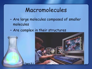
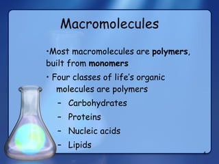
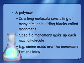
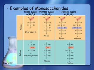
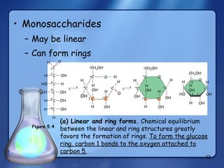
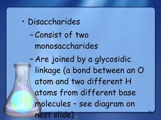
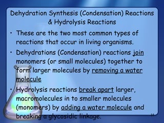
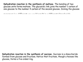
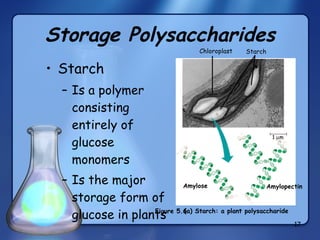
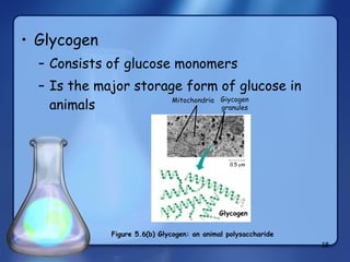
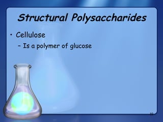
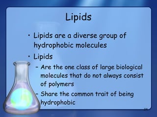
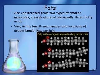
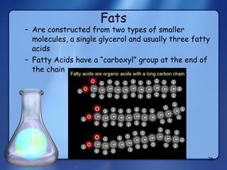
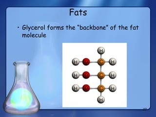
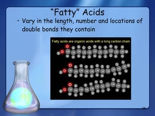
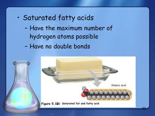
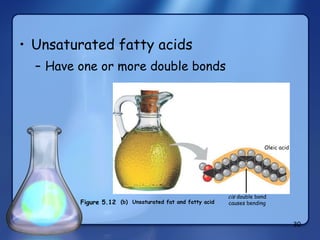
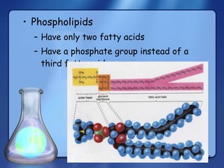
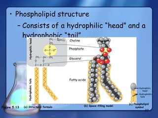
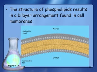
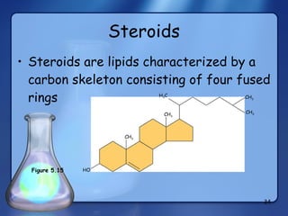
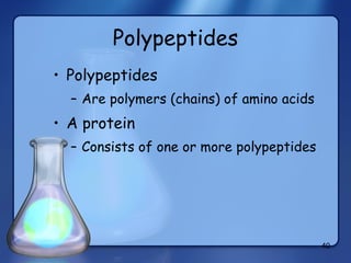
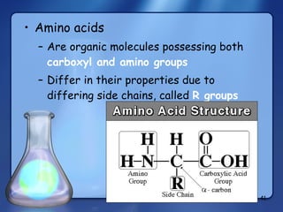
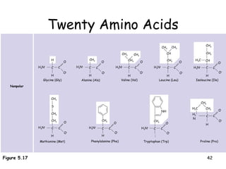
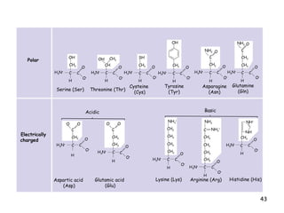
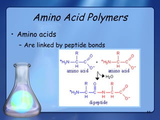
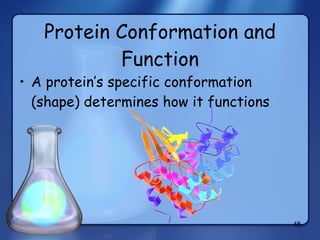
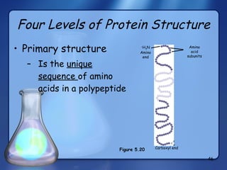
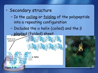
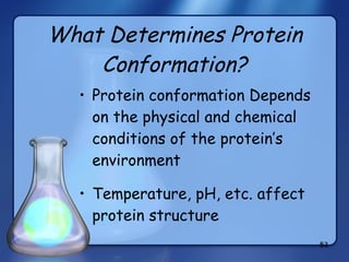
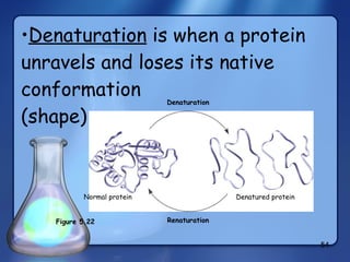
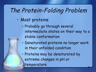
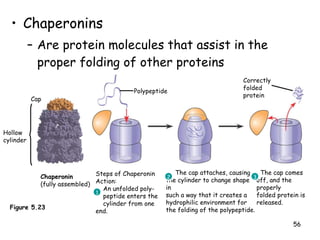
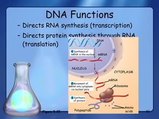
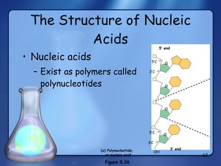
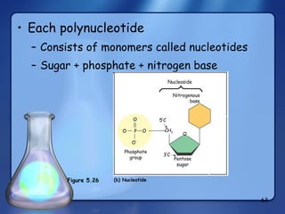
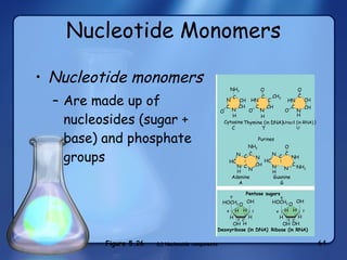
Chemistry of Life Biological MoleculesBiological Molecules.docx
05 macromolecules
1. Chapter 5 The Structure and Function of Macromolecules
2.
3.
4.
5.
6.
7.
8.
9.
10.
11.
12.
13.
14.
15. Dehydration reaction in the synthesis of maltose. The bonding of two glucose units forms maltose. The glycosidic link joins the number 1 carbon of one glucose to the number 4 carbon of the second glucose. Joining the glucose monomers in a different way would result in a different disaccharide. Dehydration reaction in the synthesis of sucrose. Sucrose is a disaccharide formed from glucose and fructose. Notice that fructose, though a hexose like glucose, forms a five-sided ring. H 2 O CH 2 OH Glucose Fructose Sucrose H HO H H OH H OH O H OH CH 2 OH H H O H HO H OH O H CH 2 OH CH 2 OH HO OH H CH 2 OH H OH H O H OH CH 2 OH H HO O H O 1 2 1–2 glycosidic linkage H H HO H H OH H OH O H OH CH 2 OH H O H H OH H OH O H OH CH 2 OH H H 2 O H HO OH H CH 2 OH H OH H O H OH H CH 2 OH H OH H O H OH O 1 4 1– 4 glycosidic linkage Glucose Glucose Maltose OH H
16.
17.
18.
19.
20.
21.
22.
23.
24.
25.
26.
27.
28.
29.
30.
31.
32.
33.
34.
35.
36.
37.
38.
39.
40.
41.
42.
43. O – OH CH 2 C C H H 3 N + O O – H 3 N + OH CH 3 CH C C H O – O SH CH 2 C H H 3 N + C O O – H 3 N + C C CH 2 OH H H H H 3 N + NH 2 CH 2 O C C C O O – NH 2 O C CH 2 CH 2 C C H 3 N + O O – O Polar Electrically charged – O O C CH 2 C C H 3 N + H O O – O – O C CH 2 C C H 3 N + H O O – CH 2 CH 2 CH 2 CH 2 NH 3 + CH 2 C C H 3 N + H O O – NH 2 C NH 2 + CH 2 CH 2 CH 2 C C H 3 N + H O O – CH 2 NH + NH CH 2 C C H 3 N + H O O – Serine (Ser) Threonine (Thr) Cysteine (Cys) Tyrosine (Tyr) Asparagine (Asn) Glutamine (Gln) Acidic Basic Aspartic acid (Asp) Glutamic acid (Glu) Lysine (Lys) Arginine (Arg) Histidine (His)
44.
45.
46.
47.
48.
49.
50. Review of Protein Structure Primary Secondary Tertiary Quaternary + H 3 N Amino end Amino acid subunits helix
51.
52. Fibers of abnormal hemoglobin deform cell into sickle shape. Primary structure Secondary and tertiary structures Quaternary structure Function Red blood cell shape Hemoglobin A Molecules do not associate with one another, each carries oxygen. Normal cells are full of individual hemoglobin molecules, each carrying oxygen 10 m 10 m Primary structure Secondary and tertiary structures Quaternary structure Function Red blood cell shape Hemoglobin S Molecules interact with one another to crystallize into a fiber, capacity to carry oxygen is greatly reduced. subunit subunit 1 2 3 4 5 6 7 3 4 5 6 7 2 1 Normal hemoglobin Sickle-cell hemoglobin . . . . . . Figure 5.21 Exposed hydrophobic region Val Thr His Leu Pro Glul Glu Val His Leu Thr Pro Val Glu