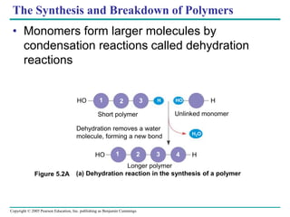
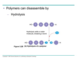
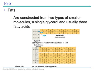
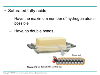
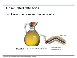
Copyright © 2005 Pearson Education, Inc. publishing as Benjamin Cummings
PowerPoint Lectures for
Biology, Seventh Edition
Neil Campbell and Jane Reece
Lectures by Chris Romero
Chapter 5
The Structure and Function of
Macromolecules
Copyright © 2005 Pearson Education, Inc. publishing as Benjamin Cummings
• Overview: The Molecules of Life
– Another level in the hierarchy of biological
organization is reached when small organic
molecules are joined together
Copyright © 2005 Pearson Education, Inc. publishing as Benjamin Cummings
• Macromolecules
– Are large molecules composed of smaller
molecules
– Are complex in their structures
Figure 5.1
Copyright © 2005 Pearson Education, Inc. publishing as Benjamin Cummings
Concept 5.1: Most macromolecules are
polymers, built from monomers
• Three of the classes of life’s organic
molecules are polymers
– Carbohydrates
– Proteins
– Nucleic acids
Copyright © 2005 Pearson Education, Inc. publishing as Benjamin Cummings
• A polymer
– Is a long molecule consisting of many similar
building blocks called monomers
Copyright © 2005 Pearson Education, Inc. publishing as Benjamin Cummings
The Synthesis and Breakdown of Polymers
• Monomers form larger molecules by
condensation reactions called dehydration
reactions
(a) Dehydration reaction in the synthesis of a polymer
HO H
1 2 3 HO
HO H
1 2 3 4
H
H2O
Short polymer Unlinked monomer
Longer polymer
Dehydration removes a water
molecule, forming a new bond
Figure 5.2A
Copyright © 2005 Pearson Education, Inc. publishing as Benjamin Cummings
• Polymers can disassemble by
– Hydrolysis
(b) Hydrolysis of a polymer
HO 1 2 3 H
HO H
1 2 3 4
H2O
H
HO
Hydrolysis adds a water
molecule, breaking a bond
Figure 5.2B
Copyright © 2005 Pearson Education, Inc. publishing as Benjamin Cummings
The Diversity of Polymers
• Each class of polymer
– Is formed from a specific set of monomers
1 2 3 HO
H
Copyright © 2005 Pearson Education, Inc. publishing as Benjamin Cummings
• Although organisms share the same limited
number of monomer types, each organism is
unique based on the arrangement of
monomers into polymers
• An immense variety of polymers can be built
from a small set of monomers
Copyright © 2005 Pearson Education, Inc. publishing as Benjamin Cummings
• Concept 5.2: Carbohydrates serve as fuel and
building material
• Carbohydrates
– Include both sugars and their polymers
Copyright © 2005 Pearson Education, Inc. publishing as Benjamin Cummings
Sugars
• Monosaccharides
– Are the simplest sugars
– Can be used for fuel
– Can be converted into other organic molecules
– Can be combined into polymers
Copyright © 2005 Pearson Education, Inc. publishing as Benjamin Cummings
• Examples of monosaccharides
Triose sugars
(C3H6O3)
Pentose sugars
(C5H10O5)
Hexose sugars
(C6H12O6)
H C OH
H C OH
H C OH
H C OH
H C OH
H C OH
HO C H
H C OH
H C OH
H C OH
H C OH
HO C H
HO C H
H C OH
H C OH
H C OH
H C OH
H C OH
H C OH
H C OH
H C OH
H C OH
C O
C O
H C OH
H C OH
H C OH
HO C H
H C OH
C O
H
H
H
H H H
H
H H H H
H
H H
C C C C
O
O
O
O
Aldoses
Glyceraldehyde
Ribose
Glucose Galactose
Dihydroxyacetone
Ribulose
Ketoses
Fructose
Figure 5.3
Copyright © 2005 Pearson Education, Inc. publishing as Benjamin Cummings
• Monosaccharides
– May be linear
– Can form rings
H
H C OH
HO C H
H C OH
H C OH
H C
O
C
H
1
2
3
4
5
6
H
OH
4C
6CH2OH 6CH2OH
5C
H
OH
C
H OH
H
2 C
1C
H
O
H
OH
4C
5C
3 C
H
H
OH
OH
H
2C
1 C
OH
H
CH2OH
H
H
OH
HO
H
OH
OH
H
5
3 2
4
(a) Linear and ring forms. Chemical equilibrium between the linear and ring
structures greatly favors the formation of rings. To form the glucose ring,
carbon 1 bonds to the oxygen attached to carbon 5.
OH
3
O H O
O
6
1
Figure 5.4
Copyright © 2005 Pearson Education, Inc. publishing as Benjamin Cummings
• Disaccharides
– Consist of two monosaccharides
– Are joined by a glycosidic linkage
Copyright © 2005 Pearson Education, Inc. publishing as Benjamin Cummings
• Examples of disaccharides
Dehydration reaction
in the synthesis of
maltose. The bonding
of two glucose units
forms maltose. The
glycosidic link joins
the number 1 carbon
of one glucose to the
number 4 carbon of
the second glucose.
Joining the glucose
monomers in a
different way would
result in a different
disaccharide.
Dehydration reaction
in the synthesis of
sucrose. Sucrose is
a disaccharide formed
from glucose and fructose.
Notice that fructose,
though a hexose like
glucose, forms a
five-sided ring.
(a)
(b)
H
HO
H
H
OH H
OH
O H
OH
CH2OH
H
HO
H
H
OH H
OH
O H
OH
CH2OH
H
O
H
H
OH H
OH
O H
OH
CH2OH
H
H2O
H2O
H
H
O
H
HO
H
OH
O
H
CH2OH
CH2OH HO
OH
H
CH2OH
H
OH H
H
HO
OH
H
CH2OH
H
OH H
O
O H
OH
H
CH2OH
H
OH H
O
H
OH
CH2OH
H HO
O
CH2OH
H
H
OH
O
O
1 2
1 4
1–4
glycosidic
linkage
1–2
glycosidic
linkage
Glucose
Glucose Glucose
Fructose
Maltose
Sucrose
OH
H
H
Figure 5.5
Copyright © 2005 Pearson Education, Inc. publishing as Benjamin Cummings
Polysaccharides
• Polysaccharides
– Are polymers of sugars
– Serve many roles in organisms
Copyright © 2005 Pearson Education, Inc. publishing as Benjamin Cummings
Storage Polysaccharides
• Starch
– Is a polymer consisting entirely of glucose
monomers
Copyright © 2005 Pearson Education, Inc. publishing as Benjamin Cummings
– Is the major storage form of glucose in plants
Chloroplast Starch
Amylose Amylopectin
1 m
(a) Starch: a plant polysaccharide
Figure 5.6
Copyright © 2005 Pearson Education, Inc. publishing as Benjamin Cummings
• Glycogen
– Consists of glucose monomers
– Is the major storage form of glucose in animals
Mitochondria Giycogen
granules
0.5 m
(b) Glycogen: an animal polysaccharide
Glycogen
Figure 5.6
Copyright © 2005 Pearson Education, Inc. publishing as Benjamin Cummings
Structural Polysaccharides
• Cellulose
– Is a polymer of glucose
Copyright © 2005 Pearson Education, Inc. publishing as Benjamin Cummings
– Has different glycosidic linkages than starch
(c) Cellulose: 1– 4 linkage of  glucose monomers
H O
O
CH2O
H
H
OH H
H
OH
OH
H
H
HO
4
C
C
C
C
C
C
H
H
H
HO
OH
H
OH
OH
OH
H
O
CH2O
H
H
H
H
OH
OH
H
H
HO
4
OH
CH2O
H
O
OH
OH
HO
4
1
O
CH2O
H
O
OH
OH
O
CH2O
H
O
OH
OH
CH2O
H
O
OH
OH
O O
CH2O
H
O
OH
OH
HO
4
O
1
OH
O
OH OH
O
CH2O
H
O
OH
O OH
O
OH
OH
(a)  and  glucose ring structures
(b) Starch: 1– 4 linkage of  glucose monomers
1
 glucose  glucose
CH2O
H
CH2O
H
1 4 4
1 1
Figure 5.7 A–C
Copyright © 2005 Pearson Education, Inc. publishing as Benjamin Cummings
Plant cells
0.5 m
Cell walls
Cellulose microfibrils
in a plant cell wall Microfibril
CH2OH
CH2OH
OH
O
H
O
O
OH
O
CH2OH
O
O
OH
O
CH2OH OH
OH OH
O
O
CH2OH
O
O
O
H
CH2OH
O
O
O
H
O
O
CH2OH
OH
CH2OH
OH
O
OH OH OH OH
O
OH OH
CH2OH
CH2OH
OH
O
OH CH2OH
O
O
OH CH2OH
OH
 Glucose
monomer
O
O
O
O
O
O
Parallel cellulose molecules are
held together by hydrogen
bonds between hydroxyl
groups attached to carbon
atoms 3 and 6.
About 80 cellulose
molecules associate
to form a microfibril, the
main architectural unit
of the plant cell wall.
A cellulose molecule
is an unbranched 
glucose polymer.
OH
OH
O
O
OH
Cellulose
molecules
Figure 5.8
– Is a major component of the tough walls that
enclose plant cells
Copyright © 2005 Pearson Education, Inc. publishing as Benjamin Cummings
• Cellulose is difficult to digest
– Cows have microbes in their stomachs to
facilitate this process
Figure 5.9
Copyright © 2005 Pearson Education, Inc. publishing as Benjamin Cummings
• Chitin, another important structural
polysaccharide
– Is found in the exoskeleton of arthropods
– Can be used as surgical thread
(a) The structure of the
chitin monomer.
O
CH2O
H
OH
H
H OH
H
NH
C
CH3
O
H
H
(b) Chitin forms the exoskeleton
of arthropods. This cicada
is molting, shedding its old
exoskeleton and emerging
in adult form.
(c) Chitin is used to make a
strong and flexible surgical
thread that decomposes after
the wound or incision heals.
OH
Figure 5.10 A–C
Copyright © 2005 Pearson Education, Inc. publishing as Benjamin Cummings
• Concept 5.3: Lipids are a diverse group of
hydrophobic molecules
• Lipids
– Are the one class of large biological molecules
that do not consist of polymers
– Share the common trait of being hydrophobic
Copyright © 2005 Pearson Education, Inc. publishing as Benjamin Cummings
Fats
• Fats
– Are constructed from two types of smaller
molecules, a single glycerol and usually three
fatty acids
(b) Fat molecule (triacylglycerol)
H H
H H
H
H
H
H
H
H
H
H
H
H
H
H
O
H O H
C
C
C
H
H OH
OH
H
H
H
H
H
H
H
H
H
H
H
H
H
H
H
H
H
C
C
C
C
C
C
C
C
C
C
C
C
C
C
C C
Glycerol
Fatty acid
(palmitic acid)
H
H
H
H
H
H
H
H
H
H
H
H
H
H
H
H
H
H
H
H
H
H
H
H
H
H
H
H
H
H
H
H
H
H
H
H
H
H
H
H
H
H
H
H
H
H
H
H
H
H
H
H
H
H
H
H
H
H
H
H
H
H
H
H
H
H
H
H H H H
H
H
H H
H
H
H
H H H H
H
H
H
H
H
H
H
H
H
H
H
H
H
H
H
H
HO
O
O
O
O
C
C
C C C
C
C
C C C
C
C C C
C
C
C
C C
C
C
C
C
C
C
C
C
C
C
C
C
C
C
C
C
C
C C C
C
C C C
C
C
C
C
C
C
C
C
O
O
(a) Dehydration reaction in the synthesis of a fat
Ester linkage
Figure 5.11
Copyright © 2005 Pearson Education, Inc. publishing as Benjamin Cummings
• Fatty acids
– Vary in the length and number and locations of
double bonds they contain
Copyright © 2005 Pearson Education, Inc. publishing as Benjamin Cummings
• Saturated fatty acids
– Have the maximum number of hydrogen atoms
possible
– Have no double bonds
(a) Saturated fat and fatty acid
Stearic acid
Figure 5.12
Copyright © 2005 Pearson Education, Inc. publishing as Benjamin Cummings
• Unsaturated fatty acids
– Have one or more double bonds
(b) Unsaturated fat and fatty acid
cis double bond
causes bending
Oleic acid
Figure 5.12
Copyright © 2005 Pearson Education, Inc. publishing as Benjamin Cummings
Phospholipids
• Phospholipids
– Have only two fatty acids
– Have a phosphate group instead of a third
fatty acid
Copyright © 2005 Pearson Education, Inc. publishing as Benjamin Cummings
• Phospholipid structure
– Consists of a hydrophilic “head” and
hydrophobic “tails”
CH2
O
P
O O
O
CH2
CH
CH2
O
O
C O C O
Phosphate
Glycerol
(a) Structural formula (b) Space-filling model
Fatty acids
(c) Phospholipid
symbol
Hydrophilic
head
Hydrophobic
tails
–
CH2
Choline
+
Figure 5.13
N(CH3)3
Copyright © 2005 Pearson Education, Inc. publishing as Benjamin Cummings
• The structure of phospholipids
– Results in a bilayer arrangement found in cell
membranes
Hydrophilic
head
WATER
WATER
Hydrophobic
tail
Figure 5.14
Copyright © 2005 Pearson Education, Inc. publishing as Benjamin Cummings
Steroids
• Steroids
– Are lipids characterized by a carbon skeleton
consisting of four fused rings
Copyright © 2005 Pearson Education, Inc. publishing as Benjamin Cummings
• One steroid, cholesterol
– Is found in cell membranes
– Is a precursor for some hormones
HO
CH3
CH3
H3C CH3
CH3
Figure 5.15
Copyright © 2005 Pearson Education, Inc. publishing as Benjamin Cummings
• Concept 5.4: Proteins have many structures,
resulting in a wide range of functions
– Proteins
• Have many roles inside the cell
Copyright © 2005 Pearson Education, Inc. publishing as Benjamin Cummings
• An overview of protein functions
Table 5.1
Copyright © 2005 Pearson Education, Inc. publishing as Benjamin Cummings
• Enzymes
– Are a type of protein that acts as a catalyst,
speeding up chemical reactions
Substrate
(sucrose)
Enzyme
(sucrase)
Glucose
OH
H O
H2O
Fructose
3 Substrate is converted
to products.
1 Active site is available for
a molecule of substrate, the
reactant on which the enzyme acts.
Substrate binds to
enzyme.
2
2
4 Products are released.
Figure 5.16
Copyright © 2005 Pearson Education, Inc. publishing as Benjamin Cummings
Polypeptides
• Polypeptides
– Are polymers of amino acids
• A protein
– Consists of one or more polypeptides
Copyright © 2005 Pearson Education, Inc. publishing as Benjamin Cummings
Amino Acid Monomers
• Amino acids
– Are organic molecules possessing both
carboxyl and amino groups
– Differ in their properties due to differing side
chains, called R groups
Copyright © 2005 Pearson Education, Inc. publishing as Benjamin Cummings
• 20 different amino acids make up proteins
O
O–
H
H3N+
C C
O
O–
H
CH3
H3N+
C
H
C
O
O–
CH3 CH3
CH3
C C
O
O–
H
H3N+
CH
CH3
CH2
C
H
H3N+
CH3
CH3
CH2
CH
C
H
H3N+ C
CH3
CH2
CH2
C
H3N+
H
C
O
O–
CH2
C
H3N+
H
C
O
O–
CH2
NH
H
C
O
O–
H3N+
C
CH2
H2C
H2N C
CH2
H
C
Nonpolar
Glycine (Gly) Alanine (Ala) Valine (Val) Leucine (Leu) Isoleucine (Ile)
Methionine (Met) Phenylalanine (Phe)
C
O
O–
Tryptophan (Trp) Proline (Pro)
H3C
Figure 5.17
S
O
O–
Copyright © 2005 Pearson Education, Inc. publishing as Benjamin Cummings
O–
OH
CH2
C C
H
H3N+
O
O–
H3N+
OH CH3
CH
C C
H
O–
O
SH
CH2
C
H
H3N+
C
O
O–
H3N+ C C
CH2
OH
H H H
H3N+
NH2
CH2
O
C
C C
O
O–
NH2 O
C
CH2
CH2
C C
H3N+
O
O–
O
Polar
Electrically
charged
–O O
C
CH2
C C
H3N+
H
O
O–
O– O
C
CH2
C C
H3N+
H
O
O–
CH2
CH2
CH2
CH2
NH3
+
CH2
C C
H3N+
H
O
O–
NH2
C NH2
+
CH2
CH2
CH2
C C
H3N+
H
O
O–
CH2
NH+
NH
CH2
C C
H3N+
H
O
O–
Serine (Ser) Threonine (Thr)
Cysteine
(Cys)
Tyrosine
(Tyr)
Asparagine
(Asn)
Glutamine
(Gln)
Acidic Basic
Aspartic acid
(Asp)
Glutamic acid
(Glu)
Lysine (Lys) Arginine (Arg) Histidine (His)
Copyright © 2005 Pearson Education, Inc. publishing as Benjamin Cummings
Amino Acid Polymers
• Amino acids
– Are linked by peptide bonds
OH
DESMOSOMES
DESMOSOMES
DESMOSOMES
OH
CH2
C
N
H
C
H O
H OH OH
Peptide
bond
OH
OH
OH
H H
H
H
H
H
H
H
H
H H
H
N
N N
N N
SH
Side
chains
SH
O
O
O O O
H2O
CH2 CH2
CH2 CH2 CH2
C C C C C C
C C
C C
Peptide
bond
Amino end
(N-terminus)
Backbone
(a)
Figure 5.18 (b)
Carboxyl end
(C-terminus)
Copyright © 2005 Pearson Education, Inc. publishing as Benjamin Cummings
Determining the Amino Acid Sequence of a Polypeptide
• The amino acid sequences of polypeptides
– Were first determined using chemical means
– Can now be determined by automated
machines
Copyright © 2005 Pearson Education, Inc. publishing as Benjamin Cummings
Protein Conformation and Function
• A protein’s specific conformation
– Determines how it functions
Copyright © 2005 Pearson Education, Inc. publishing as Benjamin Cummings
• Two models of protein conformation
(a) A ribbon model
(b) A space-filling model
Groove
Groove
Figure 5.19
Copyright © 2005 Pearson Education, Inc. publishing as Benjamin Cummings
Four Levels of Protein Structure
• Primary structure
– Is the unique sequence of amino acids in a
polypeptide
Figure 5.20
–
Amino acid
subunits
+H3N
Amino
end
o
Carboxyl end
o
c
GlyProThrGly
Thr
Gly
Glu
Seu
Lys
Cys
Pro
Leu
Met
Val
Lys
Val
Leu
Asp
AlaVal ArgGly
Ser
Pro
Ala
Gly
lle
Ser
ProPheHisGluHis
Ala
Glu
Val
Val
Phe
Thr
Ala
Asn
Asp
Ser
GlyPro
Arg
Arg
Tyr
Thr
lle
Ala
Ala
Leu
Leu
Ser
Pro
Tyr
Ser
Tyr
Ser
Thr
Thr
Ala
Val
Val
Thr
AsnPro
LysGlu
Thr
Lys
Ser
Tyr
Trp
Lys
Ala
Leu
GluLle Asp
Copyright © 2005 Pearson Education, Inc. publishing as Benjamin Cummings
O C  helix
 pleated sheet
Amino acid
subunits N
C
H
C
O
C N
H
C
O H
R
C N
H
C
O H
C
R
N
H
H
R
C
O
R
C
H
N
H
C
O H
N
C
O
R
C
H
N
H
H
C
R
C
O
C
O
C
N
H
H
R
C
C
O
N
H
H
C
R
C
O
N
H
R
C
H C
O
N
H
H
C
R
C
O
N
H
R
C
H C
O
N
H
H
C
R
C
O
N H
H C R
N H
O
O C N
C
R
C
H O
C
H
R
N H
O C
R
C
H
N H
O C
H C R
N H
C
C
N
R
H
O C
H C R
N H
O C
R
C
H
H
C
R
N
H
C
O
C
N
H
R
C
H C
O
N
H
C
• Secondary structure
– Is the folding or coiling of the polypeptide into a
repeating configuration
– Includes the  helix and the  pleated sheet
H H
Figure 5.20
Copyright © 2005 Pearson Education, Inc. publishing as Benjamin Cummings
• Tertiary structure
– Is the overall three-dimensional shape of a
polypeptide
– Results from interactions between amino acids
and R groups
CH2
CH
O
H
O
C
HO
CH2
CH2 NH3
+ C
-O CH2
O
CH2
S
S
CH2
CH
CH3
CH3
H3C
H3C
Hydrophobic
interactions and
van der Waals
interactions
Polypeptide
backbone
Hyrdogen
bond
Ionic bond
CH2
Disulfide bridge
Copyright © 2005 Pearson Education, Inc. publishing as Benjamin Cummings
• Quaternary structure
– Is the overall protein structure that results from
the aggregation of two or more polypeptide
subunits
Polypeptide
chain
Collagen
 Chains
 Chains
Hemoglobin
Iron
Heme
Copyright © 2005 Pearson Education, Inc. publishing as Benjamin Cummings
• The four levels of protein structure
+H3N
Amino end
Amino acid
subunits
helix
Copyright © 2005 Pearson Education, Inc. publishing as Benjamin Cummings
Sickle-Cell Disease: A Simple Change in
Primary Structure
• Sickle-cell disease
– Results from a single amino acid substitution in
the protein hemoglobin
Copyright © 2005 Pearson Education, Inc. publishing as Benjamin Cummings
• Hemoglobin structure and sickle-cell disease
Fibers of abnormal
hemoglobin
deform cell into
sickle shape.
Primary
structure
Secondary
and tertiary
structures
Quaternary
structure
Function
Red blood
cell shape
Hemoglobin A
Molecules do
not associate
with one
another, each
carries oxygen.
Normal cells are
full of individual
hemoglobin
molecules, each
carrying oxygen




10 m 10 m




Primary
structure
Secondary
and tertiary
structures
Quaternary
structure
Function
Red blood
cell shape
Hemoglobin S
Molecules
interact with
one another to
crystallize into a
fiber, capacity to
carry oxygen is
greatly reduced.
 subunit  subunit
1 2 3 4 5 6 7 3 4 5 6 7
2
1
Normal hemoglobin Sickle-cell hemoglobin
. . .
. . .
Figure 5.21
Exposed
hydrophobic
region
Val Thr
His Leu Pro Glul Glu Val His Leu Thr Pro Val Glu
Copyright © 2005 Pearson Education, Inc. publishing as Benjamin Cummings
What Determines Protein Conformation?
• Protein conformation
– Depends on the physical and chemical
conditions of the protein’s environment
Copyright © 2005 Pearson Education, Inc. publishing as Benjamin Cummings
• Denaturation
– Is when a protein unravels and loses its native
conformation
Denaturation
Renaturation
Denatured protein
Normal protein
Figure 5.22
Copyright © 2005 Pearson Education, Inc. publishing as Benjamin Cummings
The Protein-Folding Problem
• Most proteins
– Probably go through several intermediate
states on their way to a stable conformation
Copyright © 2005 Pearson Education, Inc. publishing as Benjamin Cummings
• Chaperonins
– Are protein molecules that assist in the proper
folding of other proteins
Hollow
cylinder
Cap
Chaperonin
(fully assembled)
Steps of Chaperonin
Action:
An unfolded poly-
peptide enters the
cylinder from one end.
The cap attaches, causing
the cylinder to change shape in
such a way that it creates a
hydrophilic environment for the
folding of the polypeptide.
The cap comes
off, and the properly
folded protein is
released.
Correctly
folded
protein
Polypeptide
2
1
3
Figure 5.23
Copyright © 2005 Pearson Education, Inc. publishing as Benjamin Cummings
• X-ray crystallography
– Is used to determine a protein’s three-
dimensional structure X-ray
diffraction
pattern
Photographic film
Diffracted X-rays
X-ray
source
X-ray
beam
Crystal Nucleic acid Protein
(a) X-ray diffraction pattern (b) 3D computer model
Figure 5.24
Copyright © 2005 Pearson Education, Inc. publishing as Benjamin Cummings
• Concept 5.5: Nucleic acids store and transmit
hereditary information
• Genes
– Are the units of inheritance
– Program the amino acid sequence of
polypeptides
– Are made of nucleic acids
Copyright © 2005 Pearson Education, Inc. publishing as Benjamin Cummings
The Roles of Nucleic Acids
• There are two types of nucleic acids
– Deoxyribonucleic acid (DNA)
– Ribonucleic acid (RNA)
Copyright © 2005 Pearson Education, Inc. publishing as Benjamin Cummings
• DNA
– Stores information for the synthesis of specific
proteins
Copyright © 2005 Pearson Education, Inc. publishing as Benjamin Cummings
– Directs RNA synthesis
– Directs protein synthesis through RNA
1
2
3
Synthesis of
mRNA in the nucleus
Movement of
mRNA into cytoplasm
via nuclear pore
Synthesis
of protein
NUCLEUS
CYTOPLASM
DNA
mRNA
Ribosome
Amino
acids
Polypeptide
mRNA
Figure 5.25
Copyright © 2005 Pearson Education, Inc. publishing as Benjamin Cummings
The Structure of Nucleic Acids
• Nucleic acids
– Exist as polymers called polynucleotides
(a) Polynucleotide,
or nucleic acid
3’C
5’ end
5’C
3’C
5’C
3’ end
OH
Figure 5.26
O
O
O
O
Copyright © 2005 Pearson Education, Inc. publishing as Benjamin Cummings
• Each polynucleotide
– Consists of monomers called nucleotides
Nitrogenous
base
Nucleoside
O
O
O


O P CH2
5’C
3’C
Phosphate
group Pentose
sugar
(b) Nucleotide
Figure 5.26
O
Copyright © 2005 Pearson Education, Inc. publishing as Benjamin Cummings
Nucleotide Monomers
• Nucleotide monomers
– Are made up of nucleosides and phosphate
groups
(c) Nucleoside components
Figure 5.26
CH
CH
Uracil (in RNA)
U
Ribose (in RNA)
Nitrogenous bases
Pyrimidines
C
N
N
C
O
H
NH2
CH
CH
O
C
N
H
CH
HN
C
O
C
CH3
N
HN
C
C
H
O
O
Cytosine
C
Thymine (in DNA)
T
N
HC
N C
C
N
C
CH
N
NH2 O
N
HC
N
H
H
C
C
N
NH
C
NH2
Adenine
A
Guanine
G
Purines
O
HOCH2
H
H H
OH
H
O
HOCH2
H
H H
OH
H
Pentose sugars
Deoxyribose (in DNA) Ribose (in RNA)
OH
OH
CH
CH
Uracil (in RNA)
U
4’
5”
3’
OH H
2’
1’
5”
4’
3’ 2’
1’
Copyright © 2005 Pearson Education, Inc. publishing as Benjamin Cummings
Nucleotide Polymers
• Nucleotide polymers
– Are made up of nucleotides linked by the–OH
group on the 3´ carbon of one nucleotide and
the phosphate on the 5´ carbon on the next
Copyright © 2005 Pearson Education, Inc. publishing as Benjamin Cummings
• The sequence of bases along a nucleotide
polymer
– Is unique for each gene
Copyright © 2005 Pearson Education, Inc. publishing as Benjamin Cummings
The DNA Double Helix
• Cellular DNA molecules
– Have two polynucleotides that spiral around an
imaginary axis
– Form a double helix
Copyright © 2005 Pearson Education, Inc. publishing as Benjamin Cummings
• The DNA double helix
– Consists of two antiparallel nucleotide strands
3’ end
Sugar-phosphate
backbone
Base pair (joined by
hydrogen bonding)
Old strands
Nucleotide
about to be
added to a
new strand
A
3’ end
3’ end
5’ end
New
strands
3’ end
5’ end
5’ end
Figure 5.27
Copyright © 2005 Pearson Education, Inc. publishing as Benjamin Cummings
• The nitrogenous bases in DNA
– Form hydrogen bonds in a complementary
fashion (A with T only, and C with G only)
Copyright © 2005 Pearson Education, Inc. publishing as Benjamin Cummings
DNA and Proteins as Tape Measures of Evolution
• Molecular comparisons
– Help biologists sort out the evolutionary
connections among species
Copyright © 2005 Pearson Education, Inc. publishing as Benjamin Cummings
The Theme of Emergent Properties in the
Chemistry of Life: A Review
• Higher levels of organization
– Result in the emergence of new properties
• Organization
– Is the key to the chemistry of life

chapter_5_the_structure_and_function_of_macromolecules.ppt

  • 1.
    Copyright © 2005Pearson Education, Inc. publishing as Benjamin Cummings PowerPoint Lectures for Biology, Seventh Edition Neil Campbell and Jane Reece Lectures by Chris Romero Chapter 5 The Structure and Function of Macromolecules
  • 2.
    Copyright © 2005Pearson Education, Inc. publishing as Benjamin Cummings • Overview: The Molecules of Life – Another level in the hierarchy of biological organization is reached when small organic molecules are joined together
  • 3.
    Copyright © 2005Pearson Education, Inc. publishing as Benjamin Cummings • Macromolecules – Are large molecules composed of smaller molecules – Are complex in their structures Figure 5.1
  • 4.
    Copyright © 2005Pearson Education, Inc. publishing as Benjamin Cummings Concept 5.1: Most macromolecules are polymers, built from monomers • Three of the classes of life’s organic molecules are polymers – Carbohydrates – Proteins – Nucleic acids
  • 5.
    Copyright © 2005Pearson Education, Inc. publishing as Benjamin Cummings • A polymer – Is a long molecule consisting of many similar building blocks called monomers
  • 6.
    Copyright © 2005Pearson Education, Inc. publishing as Benjamin Cummings The Synthesis and Breakdown of Polymers • Monomers form larger molecules by condensation reactions called dehydration reactions (a) Dehydration reaction in the synthesis of a polymer HO H 1 2 3 HO HO H 1 2 3 4 H H2O Short polymer Unlinked monomer Longer polymer Dehydration removes a water molecule, forming a new bond Figure 5.2A
  • 7.
    Copyright © 2005Pearson Education, Inc. publishing as Benjamin Cummings • Polymers can disassemble by – Hydrolysis (b) Hydrolysis of a polymer HO 1 2 3 H HO H 1 2 3 4 H2O H HO Hydrolysis adds a water molecule, breaking a bond Figure 5.2B
  • 8.
    Copyright © 2005Pearson Education, Inc. publishing as Benjamin Cummings The Diversity of Polymers • Each class of polymer – Is formed from a specific set of monomers 1 2 3 HO H
  • 9.
    Copyright © 2005Pearson Education, Inc. publishing as Benjamin Cummings • Although organisms share the same limited number of monomer types, each organism is unique based on the arrangement of monomers into polymers • An immense variety of polymers can be built from a small set of monomers
  • 10.
    Copyright © 2005Pearson Education, Inc. publishing as Benjamin Cummings • Concept 5.2: Carbohydrates serve as fuel and building material • Carbohydrates – Include both sugars and their polymers
  • 11.
    Copyright © 2005Pearson Education, Inc. publishing as Benjamin Cummings Sugars • Monosaccharides – Are the simplest sugars – Can be used for fuel – Can be converted into other organic molecules – Can be combined into polymers
  • 12.
    Copyright © 2005Pearson Education, Inc. publishing as Benjamin Cummings • Examples of monosaccharides Triose sugars (C3H6O3) Pentose sugars (C5H10O5) Hexose sugars (C6H12O6) H C OH H C OH H C OH H C OH H C OH H C OH HO C H H C OH H C OH H C OH H C OH HO C H HO C H H C OH H C OH H C OH H C OH H C OH H C OH H C OH H C OH H C OH C O C O H C OH H C OH H C OH HO C H H C OH C O H H H H H H H H H H H H H H C C C C O O O O Aldoses Glyceraldehyde Ribose Glucose Galactose Dihydroxyacetone Ribulose Ketoses Fructose Figure 5.3
  • 13.
    Copyright © 2005Pearson Education, Inc. publishing as Benjamin Cummings • Monosaccharides – May be linear – Can form rings H H C OH HO C H H C OH H C OH H C O C H 1 2 3 4 5 6 H OH 4C 6CH2OH 6CH2OH 5C H OH C H OH H 2 C 1C H O H OH 4C 5C 3 C H H OH OH H 2C 1 C OH H CH2OH H H OH HO H OH OH H 5 3 2 4 (a) Linear and ring forms. Chemical equilibrium between the linear and ring structures greatly favors the formation of rings. To form the glucose ring, carbon 1 bonds to the oxygen attached to carbon 5. OH 3 O H O O 6 1 Figure 5.4
  • 14.
    Copyright © 2005Pearson Education, Inc. publishing as Benjamin Cummings • Disaccharides – Consist of two monosaccharides – Are joined by a glycosidic linkage
  • 15.
    Copyright © 2005Pearson Education, Inc. publishing as Benjamin Cummings • Examples of disaccharides Dehydration reaction in the synthesis of maltose. The bonding of two glucose units forms maltose. The glycosidic link joins the number 1 carbon of one glucose to the number 4 carbon of the second glucose. Joining the glucose monomers in a different way would result in a different disaccharide. Dehydration reaction in the synthesis of sucrose. Sucrose is a disaccharide formed from glucose and fructose. Notice that fructose, though a hexose like glucose, forms a five-sided ring. (a) (b) H HO H H OH H OH O H OH CH2OH H HO H H OH H OH O H OH CH2OH H O H H OH H OH O H OH CH2OH H H2O H2O H H O H HO H OH O H CH2OH CH2OH HO OH H CH2OH H OH H H HO OH H CH2OH H OH H O O H OH H CH2OH H OH H O H OH CH2OH H HO O CH2OH H H OH O O 1 2 1 4 1–4 glycosidic linkage 1–2 glycosidic linkage Glucose Glucose Glucose Fructose Maltose Sucrose OH H H Figure 5.5
  • 16.
    Copyright © 2005Pearson Education, Inc. publishing as Benjamin Cummings Polysaccharides • Polysaccharides – Are polymers of sugars – Serve many roles in organisms
  • 17.
    Copyright © 2005Pearson Education, Inc. publishing as Benjamin Cummings Storage Polysaccharides • Starch – Is a polymer consisting entirely of glucose monomers
  • 18.
    Copyright © 2005Pearson Education, Inc. publishing as Benjamin Cummings – Is the major storage form of glucose in plants Chloroplast Starch Amylose Amylopectin 1 m (a) Starch: a plant polysaccharide Figure 5.6
  • 19.
    Copyright © 2005Pearson Education, Inc. publishing as Benjamin Cummings • Glycogen – Consists of glucose monomers – Is the major storage form of glucose in animals Mitochondria Giycogen granules 0.5 m (b) Glycogen: an animal polysaccharide Glycogen Figure 5.6
  • 20.
    Copyright © 2005Pearson Education, Inc. publishing as Benjamin Cummings Structural Polysaccharides • Cellulose – Is a polymer of glucose
  • 21.
    Copyright © 2005Pearson Education, Inc. publishing as Benjamin Cummings – Has different glycosidic linkages than starch (c) Cellulose: 1– 4 linkage of  glucose monomers H O O CH2O H H OH H H OH OH H H HO 4 C C C C C C H H H HO OH H OH OH OH H O CH2O H H H H OH OH H H HO 4 OH CH2O H O OH OH HO 4 1 O CH2O H O OH OH O CH2O H O OH OH CH2O H O OH OH O O CH2O H O OH OH HO 4 O 1 OH O OH OH O CH2O H O OH O OH O OH OH (a)  and  glucose ring structures (b) Starch: 1– 4 linkage of  glucose monomers 1  glucose  glucose CH2O H CH2O H 1 4 4 1 1 Figure 5.7 A–C
  • 22.
    Copyright © 2005Pearson Education, Inc. publishing as Benjamin Cummings Plant cells 0.5 m Cell walls Cellulose microfibrils in a plant cell wall Microfibril CH2OH CH2OH OH O H O O OH O CH2OH O O OH O CH2OH OH OH OH O O CH2OH O O O H CH2OH O O O H O O CH2OH OH CH2OH OH O OH OH OH OH O OH OH CH2OH CH2OH OH O OH CH2OH O O OH CH2OH OH  Glucose monomer O O O O O O Parallel cellulose molecules are held together by hydrogen bonds between hydroxyl groups attached to carbon atoms 3 and 6. About 80 cellulose molecules associate to form a microfibril, the main architectural unit of the plant cell wall. A cellulose molecule is an unbranched  glucose polymer. OH OH O O OH Cellulose molecules Figure 5.8 – Is a major component of the tough walls that enclose plant cells
  • 23.
    Copyright © 2005Pearson Education, Inc. publishing as Benjamin Cummings • Cellulose is difficult to digest – Cows have microbes in their stomachs to facilitate this process Figure 5.9
  • 24.
    Copyright © 2005Pearson Education, Inc. publishing as Benjamin Cummings • Chitin, another important structural polysaccharide – Is found in the exoskeleton of arthropods – Can be used as surgical thread (a) The structure of the chitin monomer. O CH2O H OH H H OH H NH C CH3 O H H (b) Chitin forms the exoskeleton of arthropods. This cicada is molting, shedding its old exoskeleton and emerging in adult form. (c) Chitin is used to make a strong and flexible surgical thread that decomposes after the wound or incision heals. OH Figure 5.10 A–C
  • 25.
    Copyright © 2005Pearson Education, Inc. publishing as Benjamin Cummings • Concept 5.3: Lipids are a diverse group of hydrophobic molecules • Lipids – Are the one class of large biological molecules that do not consist of polymers – Share the common trait of being hydrophobic
  • 26.
    Copyright © 2005Pearson Education, Inc. publishing as Benjamin Cummings Fats • Fats – Are constructed from two types of smaller molecules, a single glycerol and usually three fatty acids (b) Fat molecule (triacylglycerol) H H H H H H H H H H H H H H H H O H O H C C C H H OH OH H H H H H H H H H H H H H H H H H C C C C C C C C C C C C C C C C Glycerol Fatty acid (palmitic acid) H H H H H H H H H H H H H H H H H H H H H H H H H H H H H H H H H H H H H H H H H H H H H H H H H H H H H H H H H H H H H H H H H H H H H H H H H H H H H H H H H H H H H H H H H H H H H H H H H H HO O O O O C C C C C C C C C C C C C C C C C C C C C C C C C C C C C C C C C C C C C C C C C C C C C C C C C C C O O (a) Dehydration reaction in the synthesis of a fat Ester linkage Figure 5.11
  • 27.
    Copyright © 2005Pearson Education, Inc. publishing as Benjamin Cummings • Fatty acids – Vary in the length and number and locations of double bonds they contain
  • 28.
    Copyright © 2005Pearson Education, Inc. publishing as Benjamin Cummings • Saturated fatty acids – Have the maximum number of hydrogen atoms possible – Have no double bonds (a) Saturated fat and fatty acid Stearic acid Figure 5.12
  • 29.
    Copyright © 2005Pearson Education, Inc. publishing as Benjamin Cummings • Unsaturated fatty acids – Have one or more double bonds (b) Unsaturated fat and fatty acid cis double bond causes bending Oleic acid Figure 5.12
  • 30.
    Copyright © 2005Pearson Education, Inc. publishing as Benjamin Cummings Phospholipids • Phospholipids – Have only two fatty acids – Have a phosphate group instead of a third fatty acid
  • 31.
    Copyright © 2005Pearson Education, Inc. publishing as Benjamin Cummings • Phospholipid structure – Consists of a hydrophilic “head” and hydrophobic “tails” CH2 O P O O O CH2 CH CH2 O O C O C O Phosphate Glycerol (a) Structural formula (b) Space-filling model Fatty acids (c) Phospholipid symbol Hydrophilic head Hydrophobic tails – CH2 Choline + Figure 5.13 N(CH3)3
  • 32.
    Copyright © 2005Pearson Education, Inc. publishing as Benjamin Cummings • The structure of phospholipids – Results in a bilayer arrangement found in cell membranes Hydrophilic head WATER WATER Hydrophobic tail Figure 5.14
  • 33.
    Copyright © 2005Pearson Education, Inc. publishing as Benjamin Cummings Steroids • Steroids – Are lipids characterized by a carbon skeleton consisting of four fused rings
  • 34.
    Copyright © 2005Pearson Education, Inc. publishing as Benjamin Cummings • One steroid, cholesterol – Is found in cell membranes – Is a precursor for some hormones HO CH3 CH3 H3C CH3 CH3 Figure 5.15
  • 35.
    Copyright © 2005Pearson Education, Inc. publishing as Benjamin Cummings • Concept 5.4: Proteins have many structures, resulting in a wide range of functions – Proteins • Have many roles inside the cell
  • 36.
    Copyright © 2005Pearson Education, Inc. publishing as Benjamin Cummings • An overview of protein functions Table 5.1
  • 37.
    Copyright © 2005Pearson Education, Inc. publishing as Benjamin Cummings • Enzymes – Are a type of protein that acts as a catalyst, speeding up chemical reactions Substrate (sucrose) Enzyme (sucrase) Glucose OH H O H2O Fructose 3 Substrate is converted to products. 1 Active site is available for a molecule of substrate, the reactant on which the enzyme acts. Substrate binds to enzyme. 2 2 4 Products are released. Figure 5.16
  • 38.
    Copyright © 2005Pearson Education, Inc. publishing as Benjamin Cummings Polypeptides • Polypeptides – Are polymers of amino acids • A protein – Consists of one or more polypeptides
  • 39.
    Copyright © 2005Pearson Education, Inc. publishing as Benjamin Cummings Amino Acid Monomers • Amino acids – Are organic molecules possessing both carboxyl and amino groups – Differ in their properties due to differing side chains, called R groups
  • 40.
    Copyright © 2005Pearson Education, Inc. publishing as Benjamin Cummings • 20 different amino acids make up proteins O O– H H3N+ C C O O– H CH3 H3N+ C H C O O– CH3 CH3 CH3 C C O O– H H3N+ CH CH3 CH2 C H H3N+ CH3 CH3 CH2 CH C H H3N+ C CH3 CH2 CH2 C H3N+ H C O O– CH2 C H3N+ H C O O– CH2 NH H C O O– H3N+ C CH2 H2C H2N C CH2 H C Nonpolar Glycine (Gly) Alanine (Ala) Valine (Val) Leucine (Leu) Isoleucine (Ile) Methionine (Met) Phenylalanine (Phe) C O O– Tryptophan (Trp) Proline (Pro) H3C Figure 5.17 S O O–
  • 41.
    Copyright © 2005Pearson Education, Inc. publishing as Benjamin Cummings O– OH CH2 C C H H3N+ O O– H3N+ OH CH3 CH C C H O– O SH CH2 C H H3N+ C O O– H3N+ C C CH2 OH H H H H3N+ NH2 CH2 O C C C O O– NH2 O C CH2 CH2 C C H3N+ O O– O Polar Electrically charged –O O C CH2 C C H3N+ H O O– O– O C CH2 C C H3N+ H O O– CH2 CH2 CH2 CH2 NH3 + CH2 C C H3N+ H O O– NH2 C NH2 + CH2 CH2 CH2 C C H3N+ H O O– CH2 NH+ NH CH2 C C H3N+ H O O– Serine (Ser) Threonine (Thr) Cysteine (Cys) Tyrosine (Tyr) Asparagine (Asn) Glutamine (Gln) Acidic Basic Aspartic acid (Asp) Glutamic acid (Glu) Lysine (Lys) Arginine (Arg) Histidine (His)
  • 42.
    Copyright © 2005Pearson Education, Inc. publishing as Benjamin Cummings Amino Acid Polymers • Amino acids – Are linked by peptide bonds OH DESMOSOMES DESMOSOMES DESMOSOMES OH CH2 C N H C H O H OH OH Peptide bond OH OH OH H H H H H H H H H H H H N N N N N SH Side chains SH O O O O O H2O CH2 CH2 CH2 CH2 CH2 C C C C C C C C C C Peptide bond Amino end (N-terminus) Backbone (a) Figure 5.18 (b) Carboxyl end (C-terminus)
  • 43.
    Copyright © 2005Pearson Education, Inc. publishing as Benjamin Cummings Determining the Amino Acid Sequence of a Polypeptide • The amino acid sequences of polypeptides – Were first determined using chemical means – Can now be determined by automated machines
  • 44.
    Copyright © 2005Pearson Education, Inc. publishing as Benjamin Cummings Protein Conformation and Function • A protein’s specific conformation – Determines how it functions
  • 45.
    Copyright © 2005Pearson Education, Inc. publishing as Benjamin Cummings • Two models of protein conformation (a) A ribbon model (b) A space-filling model Groove Groove Figure 5.19
  • 46.
    Copyright © 2005Pearson Education, Inc. publishing as Benjamin Cummings Four Levels of Protein Structure • Primary structure – Is the unique sequence of amino acids in a polypeptide Figure 5.20 – Amino acid subunits +H3N Amino end o Carboxyl end o c GlyProThrGly Thr Gly Glu Seu Lys Cys Pro Leu Met Val Lys Val Leu Asp AlaVal ArgGly Ser Pro Ala Gly lle Ser ProPheHisGluHis Ala Glu Val Val Phe Thr Ala Asn Asp Ser GlyPro Arg Arg Tyr Thr lle Ala Ala Leu Leu Ser Pro Tyr Ser Tyr Ser Thr Thr Ala Val Val Thr AsnPro LysGlu Thr Lys Ser Tyr Trp Lys Ala Leu GluLle Asp
  • 47.
    Copyright © 2005Pearson Education, Inc. publishing as Benjamin Cummings O C  helix  pleated sheet Amino acid subunits N C H C O C N H C O H R C N H C O H C R N H H R C O R C H N H C O H N C O R C H N H H C R C O C O C N H H R C C O N H H C R C O N H R C H C O N H H C R C O N H R C H C O N H H C R C O N H H C R N H O O C N C R C H O C H R N H O C R C H N H O C H C R N H C C N R H O C H C R N H O C R C H H C R N H C O C N H R C H C O N H C • Secondary structure – Is the folding or coiling of the polypeptide into a repeating configuration – Includes the  helix and the  pleated sheet H H Figure 5.20
  • 48.
    Copyright © 2005Pearson Education, Inc. publishing as Benjamin Cummings • Tertiary structure – Is the overall three-dimensional shape of a polypeptide – Results from interactions between amino acids and R groups CH2 CH O H O C HO CH2 CH2 NH3 + C -O CH2 O CH2 S S CH2 CH CH3 CH3 H3C H3C Hydrophobic interactions and van der Waals interactions Polypeptide backbone Hyrdogen bond Ionic bond CH2 Disulfide bridge
  • 49.
    Copyright © 2005Pearson Education, Inc. publishing as Benjamin Cummings • Quaternary structure – Is the overall protein structure that results from the aggregation of two or more polypeptide subunits Polypeptide chain Collagen  Chains  Chains Hemoglobin Iron Heme
  • 50.
    Copyright © 2005Pearson Education, Inc. publishing as Benjamin Cummings • The four levels of protein structure +H3N Amino end Amino acid subunits helix
  • 51.
    Copyright © 2005Pearson Education, Inc. publishing as Benjamin Cummings Sickle-Cell Disease: A Simple Change in Primary Structure • Sickle-cell disease – Results from a single amino acid substitution in the protein hemoglobin
  • 52.
    Copyright © 2005Pearson Education, Inc. publishing as Benjamin Cummings • Hemoglobin structure and sickle-cell disease Fibers of abnormal hemoglobin deform cell into sickle shape. Primary structure Secondary and tertiary structures Quaternary structure Function Red blood cell shape Hemoglobin A Molecules do not associate with one another, each carries oxygen. Normal cells are full of individual hemoglobin molecules, each carrying oxygen     10 m 10 m     Primary structure Secondary and tertiary structures Quaternary structure Function Red blood cell shape Hemoglobin S Molecules interact with one another to crystallize into a fiber, capacity to carry oxygen is greatly reduced.  subunit  subunit 1 2 3 4 5 6 7 3 4 5 6 7 2 1 Normal hemoglobin Sickle-cell hemoglobin . . . . . . Figure 5.21 Exposed hydrophobic region Val Thr His Leu Pro Glul Glu Val His Leu Thr Pro Val Glu
  • 53.
    Copyright © 2005Pearson Education, Inc. publishing as Benjamin Cummings What Determines Protein Conformation? • Protein conformation – Depends on the physical and chemical conditions of the protein’s environment
  • 54.
    Copyright © 2005Pearson Education, Inc. publishing as Benjamin Cummings • Denaturation – Is when a protein unravels and loses its native conformation Denaturation Renaturation Denatured protein Normal protein Figure 5.22
  • 55.
    Copyright © 2005Pearson Education, Inc. publishing as Benjamin Cummings The Protein-Folding Problem • Most proteins – Probably go through several intermediate states on their way to a stable conformation
  • 56.
    Copyright © 2005Pearson Education, Inc. publishing as Benjamin Cummings • Chaperonins – Are protein molecules that assist in the proper folding of other proteins Hollow cylinder Cap Chaperonin (fully assembled) Steps of Chaperonin Action: An unfolded poly- peptide enters the cylinder from one end. The cap attaches, causing the cylinder to change shape in such a way that it creates a hydrophilic environment for the folding of the polypeptide. The cap comes off, and the properly folded protein is released. Correctly folded protein Polypeptide 2 1 3 Figure 5.23
  • 57.
    Copyright © 2005Pearson Education, Inc. publishing as Benjamin Cummings • X-ray crystallography – Is used to determine a protein’s three- dimensional structure X-ray diffraction pattern Photographic film Diffracted X-rays X-ray source X-ray beam Crystal Nucleic acid Protein (a) X-ray diffraction pattern (b) 3D computer model Figure 5.24
  • 58.
    Copyright © 2005Pearson Education, Inc. publishing as Benjamin Cummings • Concept 5.5: Nucleic acids store and transmit hereditary information • Genes – Are the units of inheritance – Program the amino acid sequence of polypeptides – Are made of nucleic acids
  • 59.
    Copyright © 2005Pearson Education, Inc. publishing as Benjamin Cummings The Roles of Nucleic Acids • There are two types of nucleic acids – Deoxyribonucleic acid (DNA) – Ribonucleic acid (RNA)
  • 60.
    Copyright © 2005Pearson Education, Inc. publishing as Benjamin Cummings • DNA – Stores information for the synthesis of specific proteins
  • 61.
    Copyright © 2005Pearson Education, Inc. publishing as Benjamin Cummings – Directs RNA synthesis – Directs protein synthesis through RNA 1 2 3 Synthesis of mRNA in the nucleus Movement of mRNA into cytoplasm via nuclear pore Synthesis of protein NUCLEUS CYTOPLASM DNA mRNA Ribosome Amino acids Polypeptide mRNA Figure 5.25
  • 62.
    Copyright © 2005Pearson Education, Inc. publishing as Benjamin Cummings The Structure of Nucleic Acids • Nucleic acids – Exist as polymers called polynucleotides (a) Polynucleotide, or nucleic acid 3’C 5’ end 5’C 3’C 5’C 3’ end OH Figure 5.26 O O O O
  • 63.
    Copyright © 2005Pearson Education, Inc. publishing as Benjamin Cummings • Each polynucleotide – Consists of monomers called nucleotides Nitrogenous base Nucleoside O O O   O P CH2 5’C 3’C Phosphate group Pentose sugar (b) Nucleotide Figure 5.26 O
  • 64.
    Copyright © 2005Pearson Education, Inc. publishing as Benjamin Cummings Nucleotide Monomers • Nucleotide monomers – Are made up of nucleosides and phosphate groups (c) Nucleoside components Figure 5.26 CH CH Uracil (in RNA) U Ribose (in RNA) Nitrogenous bases Pyrimidines C N N C O H NH2 CH CH O C N H CH HN C O C CH3 N HN C C H O O Cytosine C Thymine (in DNA) T N HC N C C N C CH N NH2 O N HC N H H C C N NH C NH2 Adenine A Guanine G Purines O HOCH2 H H H OH H O HOCH2 H H H OH H Pentose sugars Deoxyribose (in DNA) Ribose (in RNA) OH OH CH CH Uracil (in RNA) U 4’ 5” 3’ OH H 2’ 1’ 5” 4’ 3’ 2’ 1’
  • 65.
    Copyright © 2005Pearson Education, Inc. publishing as Benjamin Cummings Nucleotide Polymers • Nucleotide polymers – Are made up of nucleotides linked by the–OH group on the 3´ carbon of one nucleotide and the phosphate on the 5´ carbon on the next
  • 66.
    Copyright © 2005Pearson Education, Inc. publishing as Benjamin Cummings • The sequence of bases along a nucleotide polymer – Is unique for each gene
  • 67.
    Copyright © 2005Pearson Education, Inc. publishing as Benjamin Cummings The DNA Double Helix • Cellular DNA molecules – Have two polynucleotides that spiral around an imaginary axis – Form a double helix
  • 68.
    Copyright © 2005Pearson Education, Inc. publishing as Benjamin Cummings • The DNA double helix – Consists of two antiparallel nucleotide strands 3’ end Sugar-phosphate backbone Base pair (joined by hydrogen bonding) Old strands Nucleotide about to be added to a new strand A 3’ end 3’ end 5’ end New strands 3’ end 5’ end 5’ end Figure 5.27
  • 69.
    Copyright © 2005Pearson Education, Inc. publishing as Benjamin Cummings • The nitrogenous bases in DNA – Form hydrogen bonds in a complementary fashion (A with T only, and C with G only)
  • 70.
    Copyright © 2005Pearson Education, Inc. publishing as Benjamin Cummings DNA and Proteins as Tape Measures of Evolution • Molecular comparisons – Help biologists sort out the evolutionary connections among species
  • 71.
    Copyright © 2005Pearson Education, Inc. publishing as Benjamin Cummings The Theme of Emergent Properties in the Chemistry of Life: A Review • Higher levels of organization – Result in the emergence of new properties • Organization – Is the key to the chemistry of life