Downloaded 14 times
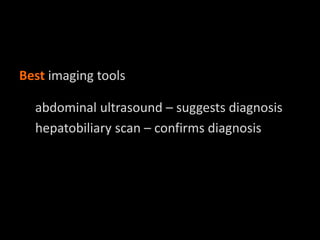
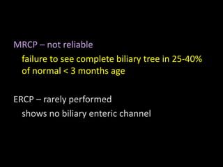
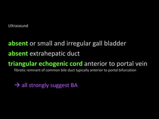
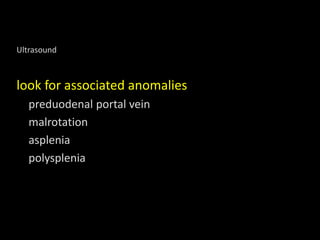
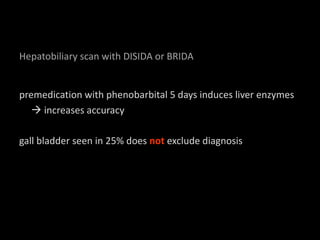
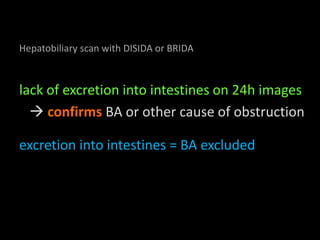
Biliary atresia (BA) is characterized by a hypoplastic or atretic extrahepatic biliary tree, typically presenting as persistent neonatal jaundice from day one. Abdominal ultrasound and hepatobiliary scans are key diagnostic tools, with ultrasound revealing absent or irregular gall bladder and fibrotic remnants, while a lack of intestinal excretion on hepatobiliary scans confirms BA. Additional anomalies may be present, and premedication with phenobarbital can enhance diagnostic accuracy.


![PERI-PROSTHETIC FRACTURE NAIL-PLATE CONSTRUCT [NPC].pptx](https://cdn.slidesharecdn.com/ss_thumbnails/drarunkumardrmohamedashrafperiprostheticfrasturenail-plateconstructnpc-260209164459-7e9d15a1-thumbnail.jpg?width=640&height=640&fit=bounds)


![CTEV [ clubfoot] DR ARUN LAL ,DR MOHAMED ASHRAF travancore medical college k...](https://cdn.slidesharecdn.com/ss_thumbnails/ctevclubfootdrarunlaldrmohamedashraftravancoremedicalcollegekollamkeralaindia-260208063247-18fc466c-thumbnail.jpg?width=640&height=640&fit=bounds)
