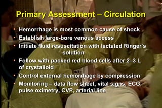
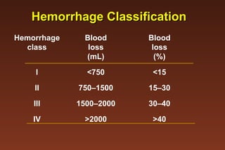
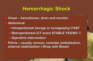
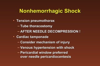
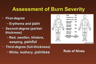
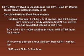
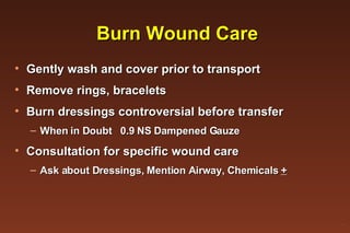
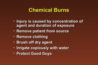
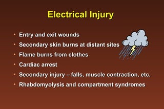
Trauma and Burn Management ® D.F.Naylor, Jr., MD, FACS, FCCS  Staff Intensivist  September 13,2007
Objectives Review initial assessment of the trauma patient Outline treatment of life-threatening injury Discuss use of  radiography to identify injury Outline response to changes in patient’s status Discuss early burn management ®
Trauma Care Principles Simultaneous assessment and treatment through a standardized approach If no improvement or decline in status, start over at primary assessment Early surgical involvement ®
Primary Assessment –  Airway / Breathing Assume cervical spine injury Airway assessment and management  Effects of facial/mandibular fracture Laryngeal/tracheal injury – ecchymosis, hoarseness, edema, subcutaneous air Flail chest from rib fractures Pneumo- / hemothoraces ®
Primary Assessment – Circulation Hemorrhage is most common cause of shock Establish large-bore venous access Initiate fluid resuscitation with lactated Ringer’s  solution  Follow with packed red blood cells after 2–3 L  of crystalloid Control external hemorrhage by compression Monitoring – data flow sheet, vital signs, ECG, pulse oximetry, CVP, arterial line ®
Hemorrhage Classification Hemorrhage Blood Blood class loss loss (mL) (%) I <750 <15 II 750–1500 15–30 III 1500–2000 30–40 IV >2000 >40
Hemorrhagic Shock Chest – hemothorax; drain and monitor Abdominal  Intraperitoneal (lavage or sonography) FAST Retroperitoneal (CT scan) STABLE YES/NO   Operative intervention   Pelvis – usually venous; consider embolization, external stabilization ( Wrap with Sheet) ®
Nonhemorrhagic Shock Tension pneumothorax  Tube thoracostomy AFTER NEEDLE DECOMPRESSION ! Cardiac tamponade  Consider mechanism of injury Venous hypertension with shock Pericardial window preferred over needle pericardiocentesis
Nonhemorrhagic Shock Blunt cardiac injury Consider mechanism of injury ECG nonspecific Cardiac enzymes rarely helpful Monitor at least 4 hours Neurogenic shock Cervical/thoracic spinal cord injury Associated bradycardia, (warm and slow)
Secondary Assessment Identify potentially life-threatening injuries History of event, medical history, drugs, allergies, tetanus immunization  AMPLE Head to toe examination Fully expose patient  Correct and prevent hypothermia Assess for signs of urethral injury FINGER OR TUBE IN EVERY ORFICE ! Neurovascular integrity ®
Secondary Assessment Laboratory data – arterial blood gas, blood counts, electrolytes, coagulation studies, type and cross-match, urinalysis, toxicology, etc Radiograph review  Cervical spine – complete survey  Chest – mediastinal evaluation; tubes/catheters Pelvis – major fractures Cystogram  urethrogram  Skeletal exam
Secondary Assessment CT scan of head CT scan of abdomen if indicated Other issues Nasogastric tube Tetanus prophylaxis Prior Immune Status HyperTet? Antibiotic indications Specialty consultation
Tertiary Assessment Detailed examination to detect all injuries Serial examinations over time  to detect change and occult injuries Return to primary/secondary survey strategies for worsening status Surgical consultation/transfer planning DOCTORS SPEAK TO DOCTORS ! ®
Compartment Syndromes Abdomen  Compromise of venous return due  to high intra-abdominal pressure  Secondary to free blood, fluid,  edema of abdominal contents Evaluate with measure of intrabladder pressure Surgical decompression  ACS = IAH > 25 + Organ Dysfunction
Compartment Syndromes Extremity  Serial examinations Pain, pallor, pulselessness,  paresthesias, paralysis Fasciotomy  Know Sensory only sites  Thumb Web, Great Toe 1st Metatarsal
Burn Injury – Primary Assessment Airway/breathing Upper and lower airway injury Carbon monoxide exposure Bronchoscopy for evaluation Consider early intubation Avoid succinylcholine ®
Burn Injury –  Primary Assessment Circulation Establish intravenous access Crystalloid resuscitation based upon extent and severity of burns Assess for circumferential injury Evaluate for other injuries ®
Assessment of Burn Severity First-degree Erythema and  pain Second-degree  (partial-thickness) Red, swollen, blisters, weeping,  painful Third-degree  (full-thickness) White, leathery,  painless Rule of Nines
Resuscitation –  Burn Shock Primary fluid loss from wound  Secondary nonburn edema SIRS  Principles Avoid excess fluid resuscitation  but maintain organ perfusion Replace components of fluids lost  as well as volume Replace blood as needed ®
Resuscitation – Burn Shock Lactated Ringer’s solution – crystalloid of choice Various formulae for amount and type of crystalloid and colloid resuscitation Parkland formula:  4 mL/kg    % of second- and third-degree burn estimates     body weight in first 24 hrs; deliver one-half calculated volume in first 8 hrs TIme 0  = Time of Injury not arrival in ED Aim for urine output 0.5-1 ml/kg/hr Cautious use of analgesia
80 KG Male involved in Closed space Fire 50 % TBSA  2 nd  Degree Burns arrives instantaneously in ED Calculate Parkland Fluid Requirements Parkland formula:  4 mL/kg    % of second- and third-degree burn estimates     body weight in first 24 hrs; deliver one-half calculated volume in first 8 hrs  SO 4 x 50 x 80 = 16000 ccs/first 24 hours  ONE LITER /hour for 8 hours IF he arrived after an 8 hour transport from OSH— without IVF 8000 ccs + 500 cc’s first hour
Burn Wound Care Gently wash and cover prior to transport Remove rings, bracelets Burn dressings controversial before transfer When in Doubt  0.9 NS Dampened Gauze Consultation for specific wound care  Ask about Dressings, Mention Airway, Chemicals  + ®
Chemical Burns Injury is caused by concentration of  agent and duration of exposure Remove patient from source Remove clothing Brush off dry agent Irrigate copiously with water Protect Good Guys
Electrical Injury Entry and exit wounds Secondary skin burns at distant sites Flame burns from clothes Cardiac arrest Secondary injury –  falls, muscle contraction, etc. Rhabdomyolysis and compartment syndromes
Pediatric Considerations Same general principles as for adults Orotracheal intubation with in-line stabilization Greater risk of injury after cricothyrotomy Diagnostic peritoneal lavage used less frequently   Body surface area/body mass so higher risk  of hypothermia ®
Pediatric Considerations Initial crystalloid bolus 20 mL/kg Hypotension is  late  finding of severe hypovolemia Blood added when crystalloid infusion >40 mL/kg Initial blood transfusion = 10 mL/kg ®
Pediatric Considerations Consider child abuse when discrepancies exist  between history and physical examination Laboratory Skull and skeletal radiographs Fundoscopic exam for hemorrhage WHEN IN DOUBT REPORT IT OUT ! ®
Key Points

Basic Trauma And Burn Management

  • 1.
    Trauma and BurnManagement ® D.F.Naylor, Jr., MD, FACS, FCCS Staff Intensivist September 13,2007
  • 2.
    Objectives Review initialassessment of the trauma patient Outline treatment of life-threatening injury Discuss use of radiography to identify injury Outline response to changes in patient’s status Discuss early burn management ®
  • 3.
    Trauma Care PrinciplesSimultaneous assessment and treatment through a standardized approach If no improvement or decline in status, start over at primary assessment Early surgical involvement ®
  • 4.
    Primary Assessment – Airway / Breathing Assume cervical spine injury Airway assessment and management Effects of facial/mandibular fracture Laryngeal/tracheal injury – ecchymosis, hoarseness, edema, subcutaneous air Flail chest from rib fractures Pneumo- / hemothoraces ®
  • 5.
    Primary Assessment –Circulation Hemorrhage is most common cause of shock Establish large-bore venous access Initiate fluid resuscitation with lactated Ringer’s solution Follow with packed red blood cells after 2–3 L of crystalloid Control external hemorrhage by compression Monitoring – data flow sheet, vital signs, ECG, pulse oximetry, CVP, arterial line ®
  • 6.
    Hemorrhage Classification HemorrhageBlood Blood class loss loss (mL) (%) I <750 <15 II 750–1500 15–30 III 1500–2000 30–40 IV >2000 >40
  • 7.
    Hemorrhagic Shock Chest– hemothorax; drain and monitor Abdominal Intraperitoneal (lavage or sonography) FAST Retroperitoneal (CT scan) STABLE YES/NO  Operative intervention Pelvis – usually venous; consider embolization, external stabilization ( Wrap with Sheet) ®
  • 8.
    Nonhemorrhagic Shock Tensionpneumothorax Tube thoracostomy AFTER NEEDLE DECOMPRESSION ! Cardiac tamponade Consider mechanism of injury Venous hypertension with shock Pericardial window preferred over needle pericardiocentesis
  • 9.
    Nonhemorrhagic Shock Bluntcardiac injury Consider mechanism of injury ECG nonspecific Cardiac enzymes rarely helpful Monitor at least 4 hours Neurogenic shock Cervical/thoracic spinal cord injury Associated bradycardia, (warm and slow)
  • 10.
    Secondary Assessment Identifypotentially life-threatening injuries History of event, medical history, drugs, allergies, tetanus immunization AMPLE Head to toe examination Fully expose patient Correct and prevent hypothermia Assess for signs of urethral injury FINGER OR TUBE IN EVERY ORFICE ! Neurovascular integrity ®
  • 11.
    Secondary Assessment Laboratorydata – arterial blood gas, blood counts, electrolytes, coagulation studies, type and cross-match, urinalysis, toxicology, etc Radiograph review Cervical spine – complete survey Chest – mediastinal evaluation; tubes/catheters Pelvis – major fractures Cystogram  urethrogram Skeletal exam
  • 12.
    Secondary Assessment CTscan of head CT scan of abdomen if indicated Other issues Nasogastric tube Tetanus prophylaxis Prior Immune Status HyperTet? Antibiotic indications Specialty consultation
  • 13.
    Tertiary Assessment Detailedexamination to detect all injuries Serial examinations over time to detect change and occult injuries Return to primary/secondary survey strategies for worsening status Surgical consultation/transfer planning DOCTORS SPEAK TO DOCTORS ! ®
  • 14.
    Compartment Syndromes Abdomen Compromise of venous return due to high intra-abdominal pressure Secondary to free blood, fluid, edema of abdominal contents Evaluate with measure of intrabladder pressure Surgical decompression ACS = IAH > 25 + Organ Dysfunction
  • 15.
    Compartment Syndromes Extremity Serial examinations Pain, pallor, pulselessness, paresthesias, paralysis Fasciotomy Know Sensory only sites Thumb Web, Great Toe 1st Metatarsal
  • 16.
    Burn Injury –Primary Assessment Airway/breathing Upper and lower airway injury Carbon monoxide exposure Bronchoscopy for evaluation Consider early intubation Avoid succinylcholine ®
  • 17.
    Burn Injury – Primary Assessment Circulation Establish intravenous access Crystalloid resuscitation based upon extent and severity of burns Assess for circumferential injury Evaluate for other injuries ®
  • 18.
    Assessment of BurnSeverity First-degree Erythema and pain Second-degree (partial-thickness) Red, swollen, blisters, weeping, painful Third-degree (full-thickness) White, leathery, painless Rule of Nines
  • 19.
    Resuscitation – Burn Shock Primary fluid loss from wound Secondary nonburn edema SIRS Principles Avoid excess fluid resuscitation but maintain organ perfusion Replace components of fluids lost as well as volume Replace blood as needed ®
  • 20.
    Resuscitation – BurnShock Lactated Ringer’s solution – crystalloid of choice Various formulae for amount and type of crystalloid and colloid resuscitation Parkland formula: 4 mL/kg  % of second- and third-degree burn estimates  body weight in first 24 hrs; deliver one-half calculated volume in first 8 hrs TIme 0 = Time of Injury not arrival in ED Aim for urine output 0.5-1 ml/kg/hr Cautious use of analgesia
  • 21.
    80 KG Maleinvolved in Closed space Fire 50 % TBSA 2 nd Degree Burns arrives instantaneously in ED Calculate Parkland Fluid Requirements Parkland formula: 4 mL/kg  % of second- and third-degree burn estimates  body weight in first 24 hrs; deliver one-half calculated volume in first 8 hrs SO 4 x 50 x 80 = 16000 ccs/first 24 hours ONE LITER /hour for 8 hours IF he arrived after an 8 hour transport from OSH— without IVF 8000 ccs + 500 cc’s first hour
  • 22.
    Burn Wound CareGently wash and cover prior to transport Remove rings, bracelets Burn dressings controversial before transfer When in Doubt 0.9 NS Dampened Gauze Consultation for specific wound care Ask about Dressings, Mention Airway, Chemicals + ®
  • 23.
    Chemical Burns Injuryis caused by concentration of agent and duration of exposure Remove patient from source Remove clothing Brush off dry agent Irrigate copiously with water Protect Good Guys
  • 24.
    Electrical Injury Entryand exit wounds Secondary skin burns at distant sites Flame burns from clothes Cardiac arrest Secondary injury –  falls, muscle contraction, etc. Rhabdomyolysis and compartment syndromes
  • 25.
    Pediatric Considerations Samegeneral principles as for adults Orotracheal intubation with in-line stabilization Greater risk of injury after cricothyrotomy Diagnostic peritoneal lavage used less frequently  Body surface area/body mass so higher risk of hypothermia ®
  • 26.
    Pediatric Considerations Initialcrystalloid bolus 20 mL/kg Hypotension is late finding of severe hypovolemia Blood added when crystalloid infusion >40 mL/kg Initial blood transfusion = 10 mL/kg ®
  • 27.
    Pediatric Considerations Considerchild abuse when discrepancies exist between history and physical examination Laboratory Skull and skeletal radiographs Fundoscopic exam for hemorrhage WHEN IN DOUBT REPORT IT OUT ! ®
  • 28.