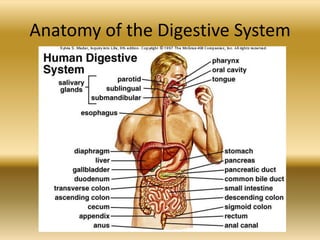
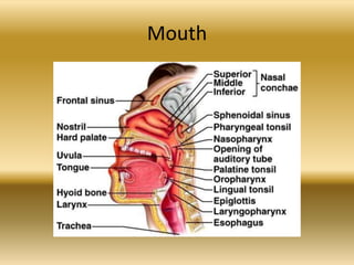
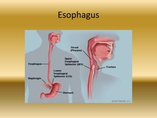
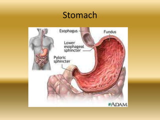
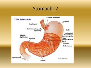
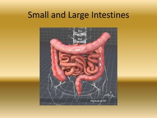
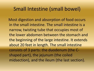
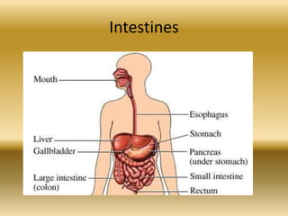
The Digestive System

(Also called Gastrointestinal or
      Alimentary system)
Food We Eat - Healthy
-
-
-
Bottlefeeding
Breastfeeding
Unhealthy Food
-
-
-
-
-
-
-
Animals We Eat
-
-
-
-
-
-
-
Animals We are Eaten by
-
-
-
-
-
-
Being Food
-
Functions
The digestive system begins with the mouth or
  the oral (buccal) cavity, where food enters the
  body, and terminates at the anus where solid
  waste materials leave the body. The functions
  of the digestive system are threefold.
1. It prepares food for absorption
2. It absorbs food into the bloodstream
3. It eliminates solid waste materials
Preparation of Food for Absorption
The ingested food is in a form that cannot reach the cells because of its
   inability to pass through the intestinal mucosa into the
   bloodstream. Therefore, the consumed food must be prepared for
   absorption.
Digestive enzymes are are substances that speed up chemical reactions
   and help in the breakdown (digestion) of complex nutrients:
a) Complex proteins are digested to simpler amino acids;
b) Complex sugars are reduced to simpler sugars, such as glucose;
c) Large fat molecules are broked down to fatty acids and
     triglycerides.
Thus, we can define digestion as a complete process of changing the
     chemical and physical composition of food in order to facilitate
     assimilation of the nourishing ingredients of food by the cells of
     the body.
Absorption of Food into the
              Bloodstream
The digested food must be absorbed into the
  bloodstream by passing through the walls of the small
  intestine. Within the cells, nutrients are catabolized
  (burned) in the presence of oxygen to release energy
  stored in the food. Amino acid nutrients are used to
  anabolize (build) large protein molecules which are
  needed for growth and development of cells. Fatty
  acids and triglycerides are also absorbed through the
  walls of the small intestine but enter lymphatic vessels
  rather than blood vessels. These digested fats
  eventtually enter the bloodstream as lymph vessels
  join blood vessels in the upper chest region.
Elimination of Solid Waste Materials
The third function of the digestive system is to
  eliminate solid waste materials that cannot be
  absorbed into the bloodstream. These waste
  materials are called feces (stools). They are
  concentrated in the large intestine and finally
  pass out of the body through the anus. This
  process is called defecation.
Anatomy of the Digestive System
The Oral Cavity (Mouth)
The digestive system is composed of the gastrointestinal
tract and accessory digestive organs. The gastrointestinal
tract begins at the oral cavity or mouth. Digestion begins in
the mouth, where chemical and mechanical digestion
occurs. Saliva or spit, produced by the salivary glands
(located under the tongue and near the lower jaw), is
released into the mouth. Saliva begins to break down the
food, moistening it and making it easier to swallow. A
digestive enzyme (called amylase) in the saliva begins to
break down the carbohydrates (starches and sugars). One
of the most important functions of the mouth is chewing.
Chewing allows food to be mashed into a soft mass that is
easier to swallow and digest later.
Mouth
Tongue
Tongue
The structures within the oral cavity are cheeks or
bucca and the tongue and its muscles, which
extend across the floor of the mouth. The main
functions of the tongue are manipulation of food
during chewing process, deglutition or
swallowing, speech production, and
determination of taste. The surface of the tongue
has rough elevations; these elevations are taste
buds. The sense organs are called papillae and
are capable of perceiving a variety of flavors
found in food, such as
bitterness, sweetness, saltiness and sourness.
Teeth
Teeth
                    Types of teeth
• The functions of teeth vary, depending on their
  individual shape and size and their location in the
  jaws. The three basic food processing functions
  of the teeth are cutting, holding or grasping, and
  grinding.
• Incisors are single-rooted teeth with a relatively
  sharp thin edge referred to as the incisal edge.
  Located in the front of the mouth, they are
  designed to cut food without the application of
  heavy forces. Central (front teeth) and lateral
  (distal to the centrals) teeth are incisors
-
• Canines, also known as cuspids, are located at
  the corner of the arch. They are designed for
  cutting and tearing foods, which require the
  application of force.
• Premolars are a cross between canines and
  molars. An older term for premolar is
  bicuspid. The pointed cusps hold and grind
  the food. They have a broader surface for
  chewing food. There are two sets of
  premolars in the permanent dentition and NO
  premolars in the primary dentition.
-
• Molars are much larger than premolars. The
  molars have more cusps than other teeth that
  are used to chew or grind up food. There are
  two sets of molars in the primary dentition
  and three sets of molars in the permanent
  dentition.
Tooth
Tooth Structure
Teeth are covered by hard enamel, which gives
  them a white and smooth appearance. The
  enamel is the hardest substance in the body.
  Beneath the enamel is the main structure of the
  tooth, the dentin. It is yellowish and is composed
  of bony tissue which is softer than enamel.
  Dentin is surrounded by a thin layer of modified
  bone called cementum. In the innermost part of
  the tooth is the pulp, a soft, delicate layer, which
  stores the nerves and blood vessels of the tooth.
  The teeth are embedded in pink fleshy tissue
  known as gums, or gingiva.
Tooth_2
Other Structures
• Some of the other structures located within the
  mouth are the hard and soft palates. The hard
  palate lies in the anterrior portion of the roof of
  the mouth, while the soft palate lies in its
  posterior portion. The soft palate forms a
  partition between the mouth and nasopharynx
  and is continuous with the hard palate. Rugae are
  the irregular ridges in the mucous membrane
  covering the anterior portion of the hard palate.
  The entire buccal cavity, like the rest of the
  digesttive tract, is lined with mucous membrane.
-
• After the food is chewed, it is formed into a
  round, sticky mass called a bolus. The bolus is
  pushed by the tongue from the mouth into
  the pharynx. Its downward movement is
  guided into the pharynx or throat by the
  soft, fleshy V-shaped tissue hanging from the
  soft palate, which is called uvula. Uvula also
  functions to aid in speech production.
Pharynx
The pharynx is a muscular tube which serves as a
  common passageway for air from the nasal cavity
  to the larynx (voicebox), as well as for food going
  from the mouth to the esophagus. The pharynx is
  divided into three major sections:
1. The nasopharynx (the throat behind the nose)
2. The oropharynx (the throat behind the moouth)
3. The laryngopharynx (the throat above the
   larynx)
-
• The laryngopharynx is further divided into two
  tubes; one which leads to the lungs, called the
  trachea (wind pipe), and one which leads to
  the stomach, called the esophagus. A small
  flap of tissue, the epiglottis, covers the
  trachea. The main function of the epiglottis is
  to prevent food from entering the
  trachea, thus allowing all food to be
  channeled to the stomach through the
  esophagus
Esophagus
Esophagus
• Anatomically and functionally, the esophagus is
  the least complex section of the digestive tract.
  Its role in digestion is simple: to convey boluses
  of food from the pharynx to the stomach. The
  esophagus begins as an extension of the pharynx
  in the back of the oral cavity. It then courses
  down the neck next to the trachea, through the
  thoracic cavity, and penetrates the diaphragm to
  connect with the stomach in the abdominal
  cavity. It uses rhythmic, wave-like muscle
  movements (called peristalsis) to force food from
  the throat into the stomach.
Stomach
The stomach, which is attached to the end of the
  esophagus by way of the cardiac sphincter, is a stretchy
  sack shaped like the letter J. It is located in the
  abdominal cavity directly below the diaphragm. It has
  three important jobs:

1. To store the food we have eaten
2. To break down the food into a creamy mixture
3. To slowly empty that creamy mixture into the small
   intestine
Stomach
-
The stomach is like a mixer, churning and
mashing together all the small balls of food
that came down the esophagus into smaller
and smaller pieces. It does this with help from
the strong muscles in the walls of the stomach
and gastric juices that also come from the
stomach's walls. These juices contain enzymes
and hydrochloric acid.
Stomach_2
-
Pepsin breaks down protein, rennin curdles
milk, while hydrochloric acid kills bacteria that
might be in the eaten food. The creamy
mixture (digested food) which is emptied into
the small intestine through the pyloric
sphincter is called chyme. The sphincter is
designed to open partially so that large
particles are kept in the stomach for further
mixing and breaking down.
Small and Large Intestines
Small Intestine (small bowel)
Most digestion and absorption of food occurs
in the small intestine. The small intestine is a
narrow, twisting tube that occupies most of
the lower abdomen between the stomach and
the beginning of the large intestine. It extends
about 20 feet in length. The small intestine
consists of 3 parts: the duodenum (the C-
shaped part), the jejunum (the coiled
midsection), and the ileum (the last section).
Intestines
Duodenum - Pancreas
-
• The small intestine has 2 important functions.
  First, the digestive process is completed here
  by enzymes and other substances made by
  intestinal cells, the pancreas, and the liver.
  Glands in the intestine walls secrete enzymes
  that breakdown starches and sugars. The
  pancreas secretes enzymes into the small
  intestine that help breakdown
  carbohydrates, fats, and proteins.
-
The liver produces bile, which is stored in the
gallbladder. Bile helps to make fat molecules
(which otherwise are not soluble in water)
soluble, so they can be absorbed by the body.
Second, the small intestine absorbs the
nutrients from the digestive process. The
inner wall of the small intestine is covered by
millions of tiny fingerlike projections called
villi.
Villi
-
• The villi are covered with even tinier
  projections called microvilli. The combination
  of villi and microvilli increase the surface area
  of the small intestine greatly, allowing
  absorption of nutrients to occur. Undigested
  material travels next to the large intestine.
Large Intestine
The Large Intestine
• The large intestine forms an upside down U
  over the coiled small intestine. It begins at the
  lower right-hand side of the body and ends on
  the lower left-hand side. The large intestine is
  about 5-6 feet long. It has 3 parts: the
  cecum, the colon, and the rectum. The cecum
  is a pouch at the beginning of the large
  intestine.
-
This area allows food to pass from the small
intestine to the large intestine. The colon is
where fluids and salts are absorbed and extends
from the cecum to the rectum. The large intestine
consists of the following sections:
cecum, ascending colon, transverse
colon, descending colon, sigmoid
colon, rectum, anal cana, and anus.
The last part of the large intestine is the
rectum, which is where feces (waste material) are
stored before leaving the body through the anus.
-
• The main job of the large intestine is to
  remove water and salts (electrolytes) from the
  undigested material and to form solid wastes
  that can be excreted. Bacteria in the large
  intestine help to break down the undigested
  materials. The remaining contents of the large
  intestine are moved toward the
  rectum, where feces are stored until they
  leave the body through the anus as a bowel
  movement.
Liver
• The liver is the largest gland, and the largest solid
  organ in the body, weighing some 1.8 kg in men and
  1.3 kg in women. It holds approximately 13% (about
  one pint or 0.57 liter) of our total blood supply at any
  given moment and is estimated to have over 500
  functions. The liver is dark reddish brown in color and
  is divided into two main lobes (the much larger right
  and the smaller left) which are further subdivided into
  approximately 100,000 small lobes, or lobules. It is
  located beneath the diaphragm in the right upper
  quadrant (RUQ) of the abdominal cavity.
Liver
Some of the Liver`s Functions
• processing digested food from the intestine
• controlling levels of fats, amino acids and glucose in the blood
• combating infections in the body
• clearing the blood of particles and infections including bacteria
• neutralizing and destroying drugs and toxins
• manufacturing bile
• storing iron, vitamins and other essential chemicals
• breaking down food and turning it into energy
• manufacturing, breaking down and regulating numerous hormones
  including sex hormones
• making enzymes and proteins which are responsible for most chemical
  reactions in the body, for example those involved in blood clotting and
  repair of damaged tissues (e.g. prothrombin, fibrinogen).
Pancreas
• The pancreas is both an endocrine gland
  (producing several important
  hormones, including insulin, glucagon, and
  somatostatin), as well as an exocrine
  gland, secreting pancreatic juice containing
  digestive enzymes that pass to the small
  intestine. These enzymes help in the further
  breakdown of the carbohydrates, protein, and
  fat in the chyme.
Gallbladder
• The gallbladder is a pear-shaped sac, usually
  stained dark green by the bile it contains. It is
  located in the hollow underside of the liver. Its
  duct, the cystic duct, joins the hepatic duct
  from the liver to form the common bile
  duct, which enters the duodenum. The
  gallbladder receives bile from the liver and
  then concentrates and stores it. It secretes bile
  when the small intestine is stimulated by the
  entrance of fats.
Gallbladder
Finished

The digestive system

  • 1.
    The Digestive System (Alsocalled Gastrointestinal or Alimentary system)
  • 2.
    Food We Eat- Healthy
  • 3.
  • 4.
  • 5.
  • 6.
  • 7.
  • 8.
  • 9.
  • 10.
  • 11.
  • 12.
  • 13.
  • 14.
  • 15.
  • 16.
  • 17.
  • 18.
  • 19.
  • 20.
  • 21.
  • 22.
  • 24.
  • 25.
  • 26.
  • 27.
  • 28.
  • 29.
  • 30.
  • 31.
  • 32.
  • 33.
  • 34.
    Functions The digestive systembegins with the mouth or the oral (buccal) cavity, where food enters the body, and terminates at the anus where solid waste materials leave the body. The functions of the digestive system are threefold. 1. It prepares food for absorption 2. It absorbs food into the bloodstream 3. It eliminates solid waste materials
  • 35.
    Preparation of Foodfor Absorption The ingested food is in a form that cannot reach the cells because of its inability to pass through the intestinal mucosa into the bloodstream. Therefore, the consumed food must be prepared for absorption. Digestive enzymes are are substances that speed up chemical reactions and help in the breakdown (digestion) of complex nutrients: a) Complex proteins are digested to simpler amino acids; b) Complex sugars are reduced to simpler sugars, such as glucose; c) Large fat molecules are broked down to fatty acids and triglycerides. Thus, we can define digestion as a complete process of changing the chemical and physical composition of food in order to facilitate assimilation of the nourishing ingredients of food by the cells of the body.
  • 36.
    Absorption of Foodinto the Bloodstream The digested food must be absorbed into the bloodstream by passing through the walls of the small intestine. Within the cells, nutrients are catabolized (burned) in the presence of oxygen to release energy stored in the food. Amino acid nutrients are used to anabolize (build) large protein molecules which are needed for growth and development of cells. Fatty acids and triglycerides are also absorbed through the walls of the small intestine but enter lymphatic vessels rather than blood vessels. These digested fats eventtually enter the bloodstream as lymph vessels join blood vessels in the upper chest region.
  • 37.
    Elimination of SolidWaste Materials The third function of the digestive system is to eliminate solid waste materials that cannot be absorbed into the bloodstream. These waste materials are called feces (stools). They are concentrated in the large intestine and finally pass out of the body through the anus. This process is called defecation.
  • 38.
    Anatomy of theDigestive System
  • 39.
    The Oral Cavity(Mouth) The digestive system is composed of the gastrointestinal tract and accessory digestive organs. The gastrointestinal tract begins at the oral cavity or mouth. Digestion begins in the mouth, where chemical and mechanical digestion occurs. Saliva or spit, produced by the salivary glands (located under the tongue and near the lower jaw), is released into the mouth. Saliva begins to break down the food, moistening it and making it easier to swallow. A digestive enzyme (called amylase) in the saliva begins to break down the carbohydrates (starches and sugars). One of the most important functions of the mouth is chewing. Chewing allows food to be mashed into a soft mass that is easier to swallow and digest later.
  • 40.
  • 41.
  • 42.
    Tongue The structures withinthe oral cavity are cheeks or bucca and the tongue and its muscles, which extend across the floor of the mouth. The main functions of the tongue are manipulation of food during chewing process, deglutition or swallowing, speech production, and determination of taste. The surface of the tongue has rough elevations; these elevations are taste buds. The sense organs are called papillae and are capable of perceiving a variety of flavors found in food, such as bitterness, sweetness, saltiness and sourness.
  • 43.
  • 44.
    Teeth Types of teeth • The functions of teeth vary, depending on their individual shape and size and their location in the jaws. The three basic food processing functions of the teeth are cutting, holding or grasping, and grinding. • Incisors are single-rooted teeth with a relatively sharp thin edge referred to as the incisal edge. Located in the front of the mouth, they are designed to cut food without the application of heavy forces. Central (front teeth) and lateral (distal to the centrals) teeth are incisors
  • 45.
    - • Canines, alsoknown as cuspids, are located at the corner of the arch. They are designed for cutting and tearing foods, which require the application of force. • Premolars are a cross between canines and molars. An older term for premolar is bicuspid. The pointed cusps hold and grind the food. They have a broader surface for chewing food. There are two sets of premolars in the permanent dentition and NO premolars in the primary dentition.
  • 46.
    - • Molars aremuch larger than premolars. The molars have more cusps than other teeth that are used to chew or grind up food. There are two sets of molars in the primary dentition and three sets of molars in the permanent dentition.
  • 47.
  • 48.
    Tooth Structure Teeth arecovered by hard enamel, which gives them a white and smooth appearance. The enamel is the hardest substance in the body. Beneath the enamel is the main structure of the tooth, the dentin. It is yellowish and is composed of bony tissue which is softer than enamel. Dentin is surrounded by a thin layer of modified bone called cementum. In the innermost part of the tooth is the pulp, a soft, delicate layer, which stores the nerves and blood vessels of the tooth. The teeth are embedded in pink fleshy tissue known as gums, or gingiva.
  • 49.
  • 50.
    Other Structures • Someof the other structures located within the mouth are the hard and soft palates. The hard palate lies in the anterrior portion of the roof of the mouth, while the soft palate lies in its posterior portion. The soft palate forms a partition between the mouth and nasopharynx and is continuous with the hard palate. Rugae are the irregular ridges in the mucous membrane covering the anterior portion of the hard palate. The entire buccal cavity, like the rest of the digesttive tract, is lined with mucous membrane.
  • 51.
    - • After thefood is chewed, it is formed into a round, sticky mass called a bolus. The bolus is pushed by the tongue from the mouth into the pharynx. Its downward movement is guided into the pharynx or throat by the soft, fleshy V-shaped tissue hanging from the soft palate, which is called uvula. Uvula also functions to aid in speech production.
  • 52.
    Pharynx The pharynx isa muscular tube which serves as a common passageway for air from the nasal cavity to the larynx (voicebox), as well as for food going from the mouth to the esophagus. The pharynx is divided into three major sections: 1. The nasopharynx (the throat behind the nose) 2. The oropharynx (the throat behind the moouth) 3. The laryngopharynx (the throat above the larynx)
  • 53.
    - • The laryngopharynxis further divided into two tubes; one which leads to the lungs, called the trachea (wind pipe), and one which leads to the stomach, called the esophagus. A small flap of tissue, the epiglottis, covers the trachea. The main function of the epiglottis is to prevent food from entering the trachea, thus allowing all food to be channeled to the stomach through the esophagus
  • 54.
  • 55.
    Esophagus • Anatomically andfunctionally, the esophagus is the least complex section of the digestive tract. Its role in digestion is simple: to convey boluses of food from the pharynx to the stomach. The esophagus begins as an extension of the pharynx in the back of the oral cavity. It then courses down the neck next to the trachea, through the thoracic cavity, and penetrates the diaphragm to connect with the stomach in the abdominal cavity. It uses rhythmic, wave-like muscle movements (called peristalsis) to force food from the throat into the stomach.
  • 56.
    Stomach The stomach, whichis attached to the end of the esophagus by way of the cardiac sphincter, is a stretchy sack shaped like the letter J. It is located in the abdominal cavity directly below the diaphragm. It has three important jobs: 1. To store the food we have eaten 2. To break down the food into a creamy mixture 3. To slowly empty that creamy mixture into the small intestine
  • 57.
  • 58.
    - The stomach islike a mixer, churning and mashing together all the small balls of food that came down the esophagus into smaller and smaller pieces. It does this with help from the strong muscles in the walls of the stomach and gastric juices that also come from the stomach's walls. These juices contain enzymes and hydrochloric acid.
  • 59.
  • 60.
    - Pepsin breaks downprotein, rennin curdles milk, while hydrochloric acid kills bacteria that might be in the eaten food. The creamy mixture (digested food) which is emptied into the small intestine through the pyloric sphincter is called chyme. The sphincter is designed to open partially so that large particles are kept in the stomach for further mixing and breaking down.
  • 61.
    Small and LargeIntestines
  • 62.
    Small Intestine (smallbowel) Most digestion and absorption of food occurs in the small intestine. The small intestine is a narrow, twisting tube that occupies most of the lower abdomen between the stomach and the beginning of the large intestine. It extends about 20 feet in length. The small intestine consists of 3 parts: the duodenum (the C- shaped part), the jejunum (the coiled midsection), and the ileum (the last section).
  • 63.
  • 64.
  • 65.
    - • The smallintestine has 2 important functions. First, the digestive process is completed here by enzymes and other substances made by intestinal cells, the pancreas, and the liver. Glands in the intestine walls secrete enzymes that breakdown starches and sugars. The pancreas secretes enzymes into the small intestine that help breakdown carbohydrates, fats, and proteins.
  • 66.
    - The liver producesbile, which is stored in the gallbladder. Bile helps to make fat molecules (which otherwise are not soluble in water) soluble, so they can be absorbed by the body. Second, the small intestine absorbs the nutrients from the digestive process. The inner wall of the small intestine is covered by millions of tiny fingerlike projections called villi.
  • 67.
  • 68.
    - • The villiare covered with even tinier projections called microvilli. The combination of villi and microvilli increase the surface area of the small intestine greatly, allowing absorption of nutrients to occur. Undigested material travels next to the large intestine.
  • 69.
  • 70.
    The Large Intestine •The large intestine forms an upside down U over the coiled small intestine. It begins at the lower right-hand side of the body and ends on the lower left-hand side. The large intestine is about 5-6 feet long. It has 3 parts: the cecum, the colon, and the rectum. The cecum is a pouch at the beginning of the large intestine.
  • 71.
    - This area allowsfood to pass from the small intestine to the large intestine. The colon is where fluids and salts are absorbed and extends from the cecum to the rectum. The large intestine consists of the following sections: cecum, ascending colon, transverse colon, descending colon, sigmoid colon, rectum, anal cana, and anus. The last part of the large intestine is the rectum, which is where feces (waste material) are stored before leaving the body through the anus.
  • 72.
    - • The mainjob of the large intestine is to remove water and salts (electrolytes) from the undigested material and to form solid wastes that can be excreted. Bacteria in the large intestine help to break down the undigested materials. The remaining contents of the large intestine are moved toward the rectum, where feces are stored until they leave the body through the anus as a bowel movement.
  • 73.
    Liver • The liveris the largest gland, and the largest solid organ in the body, weighing some 1.8 kg in men and 1.3 kg in women. It holds approximately 13% (about one pint or 0.57 liter) of our total blood supply at any given moment and is estimated to have over 500 functions. The liver is dark reddish brown in color and is divided into two main lobes (the much larger right and the smaller left) which are further subdivided into approximately 100,000 small lobes, or lobules. It is located beneath the diaphragm in the right upper quadrant (RUQ) of the abdominal cavity.
  • 74.
  • 75.
    Some of theLiver`s Functions • processing digested food from the intestine • controlling levels of fats, amino acids and glucose in the blood • combating infections in the body • clearing the blood of particles and infections including bacteria • neutralizing and destroying drugs and toxins • manufacturing bile • storing iron, vitamins and other essential chemicals • breaking down food and turning it into energy • manufacturing, breaking down and regulating numerous hormones including sex hormones • making enzymes and proteins which are responsible for most chemical reactions in the body, for example those involved in blood clotting and repair of damaged tissues (e.g. prothrombin, fibrinogen).
  • 76.
    Pancreas • The pancreasis both an endocrine gland (producing several important hormones, including insulin, glucagon, and somatostatin), as well as an exocrine gland, secreting pancreatic juice containing digestive enzymes that pass to the small intestine. These enzymes help in the further breakdown of the carbohydrates, protein, and fat in the chyme.
  • 78.
    Gallbladder • The gallbladderis a pear-shaped sac, usually stained dark green by the bile it contains. It is located in the hollow underside of the liver. Its duct, the cystic duct, joins the hepatic duct from the liver to form the common bile duct, which enters the duodenum. The gallbladder receives bile from the liver and then concentrates and stores it. It secretes bile when the small intestine is stimulated by the entrance of fats.
  • 79.
  • 80.