This book provides an overview of surgical bariatric procedures for weight loss and treating obesity-related diseases. It describes the most common bariatric surgery procedures performed worldwide and examines how they have evolved over time in Italy and internationally. For each procedure, it discusses indications, surgical techniques, potential complications, and outcomes related to weight loss and comorbidities. It also covers the problem of weight regain after surgery and discusses different types of revisional surgeries to address this issue.


![1
1L. Angrisani (Ed), Bariatric and Metabolic Surgery,
Updates in Surgery
DOI: 10.1007/ 978-88-470-3944-5_1, © Springer-Verlag Italia 2017
A. Vitiello (*)
Department of Clinical Medicine and Surgery, University of Naples Federico II
Naples, Italy
e-mail: antoniovitiello_@hotmail.it
1History of Obesity Surgery in Italy
Vincenzo Pilone, Ariola Hasani, Giuliano Izzo, Antonio Vitiello,
and Pietro Forestieri
1.1 Epidemiology of Obesity in Italy
Overweight and obesity rates are constantly increasing in industrialized
countries. In 2013, according to statistical data, more than one out of ten Italian
adults (11.3%) is obese, while 34.5% of the population is overweight [1].
However, the latest data show that the proportion of overweight adults has only
mildly increased since the early 2000s and the rate has been stabilizing in recent
years. In this context, southern regions have a higher prevalence of obesity; for
example, the obese population in Puglia represents 13.6% compared with 9%
in Lombardia, and the overweight population is 39.2% in Campania compared
with 30% in Trentino-Alto Adige [1].
1.2 Early Years of Bariatric Surgery
Bariatric surgery in Italy began in early 1970, a time when obesity was still
considered worldwide as being the consequence of an inappropriate lifestyle and
not a serious multifactorial disease. In 1972 in Milan, Montorsi [2–5] performed
the first jejunoileal bypass (JIB) following a long period of research on obesity
and its related pathologies. He was a pioneer not only as a bariatric surgeon but
as a physician, since he understood that a multidisciplinary approach was the
only effective way to achieve success in the treatment of obese patients. In the
same period intense bariatric research took place in different Italian institutions
by different groups: Montorsi and Doldi in Milan, Battezzati and Scopinaro in](https://image.slidesharecdn.com/bariatricandmetabolicsurgery-180301214153/85/Bariatric-and-metabolic-surgery-16-320.jpg)
![2 V. Pilone et al.
Genoa, Mazzeo and Forestieri in Naples, Morino and Toppino in Turin, Grassi
and Santoro in Rome, Vecchioni and Baggio in Verona, and Vassallo in Pavia. The
initial experience with JIB showed good outcomes with acceptable compliance
but also unsatisfactory weight loss and catastrophic results such as liver failure,
bypass enteritis, and excessive weight loss with severe malnutrition requiring
reintervention. Media and medical societies firmly opposed this surgery, inducing
some bariatric surgeons to abandon the practice and others to find new solutions.
In Genoa in 1973, Scopinaro [6–9] began his first series of JIB and at the same
time ideated and experimented with a new procedure on animals – biliopancreatic
diversion (BPD) – performed for the first time on humans on 12 May 1976.
The procedure consisted of a distal gastrectomy with a long-limb Roux-en-Y
reconstruction and an enteroenteric anastomosis performed in the terminal ileum.
BPD was conceived in an attempt to avoid complications associated with JIB,
which were primarily due to the presence of the long blind loop, non-selective
malabsorption, and intestinal adaptation syndrome. What Scopinaro observed
on animals was then confirmed in patients: BPD seemed to solve the primary
problems associated with JIB. Scopinaro represents a milestone in the history
of bariatric surgery worldwide, not only as a surgeon but also for his important
studies on intestinal physiology, which allowed better comprehension of intestinal
absorption. Different techniques were developed as variations or simplifications
of the Scopinaro procedure, thus confirming that BPD still represents one of the
most effective bariatric procedures, even after 40 years. Mazzeo and Forestieri
[10] in Naples performed the first series of JIB in 1974, at a time when many
authors reported weight regain likely due to the alimentary reflux in the excluded
loop. In an attempt to solve this side effect, Forestieri [11] ideated the end-to-
side jejunoileostomy with an antireflux valve system, the successful outcomes
of which were presented at the Biennial World Congress of the International
College of Surgeons in Athens, Greece, in 1976. This modification resulted in
extensive application worldwide, and Forestieri [12, 13] was the first Italian
surgeon cited in the history of the evolution of bariatric surgery, published by
Buchwald [14]. In Turin in 1975, Morino began his bariatric experience with the
JIB, and after inconsistent results, in 1978, he adopted the Roux-en-Y gastric
bypass and, in 1983, Mason’s vertical gastroplasty. In Rome, after evaluating the
outcomes of his own extensive experience with JIB, Santoro was one of the first
authors to describe postoperative adaptation syndrome and bypass intolerance
syndrome [15–18].
In 1979, the School of Montorsi performed the first biliointestinal bypass
in Italy in an attempt to reduce the effects of bacterial overgrowth in the blind
loop and malabsorption of bile salts. In 1990, Doldi definitively adopted the
biliointestinal bypass as the standard procedure in obese patients who were
candidates for malabsorptive procedures.
In Bologna, in 1991, Amenta and Cariani [19–21] began their bariatric
experience with Mason’s vertical gastroplasty, and later in 1996, they adopted
the laparoscopic Roux-en-Y gastric bypass (LRYGB) procedure. After constant](https://image.slidesharecdn.com/bariatricandmetabolicsurgery-180301214153/85/Bariatric-and-metabolic-surgery-17-320.jpg)
![31 History of Obesity Surgery in Italy
research and clinical activity, in 2002, they introduced a modification to
preserve the possibility of endoscopically and radiologically evaluating the
excluded gastroenteric tract: the Roux-en-Y gastric bypass on vertical banded
gastroplasty (Amenta-Cariani), which is still the standard procedure in their
center for treating obesity. In 1997, in Pavia, Vassallo [22] introduced an
evolution of BPD: BPD coupled with transitory gastroplasty, which preserves the
duodenal bulb. The gastroplasty is transitory due to the use of a biodegradable
polydioxanone (PDS) band.
The enthusiastic bariatric activity and the need to gather and share experiences
led Italian bariatric surgeons create the Italian Society of Bariatric Surgery
(SICOB) in Genoa in 1991 and, with Carlo Vassallo, to the institution of the first
School of Bariatric Surgery, entrusted to the Associazione Chirurghi Ospedalieri
Italiani (ACOI). SICOB is one of five founding societies of the International
Federation for the Surgery of Obesity and Metabolic Disorders (IFSO) and the
first bariatric society in the world to add the concept of metabolic surgery to its
name, changing it to Society of Bariatric and Metabolic Surgery in 2007.
1.3 The Beginning of Laparoscopy
Italians surgeons have always been pioneers in the surgical treatment of obesity
and weight-related diseases. In the early 1990s, they began proposing and
adopting several endoscopic and laparoscopic procedures. In 1993, for the first
time worldwide, Catona [23] placed a silicone gastric band laparoscopically;
the same year, Favretti [24] performed the first laparoscopic adjustable gastric
banding (LAGB), which allowed placement of the posterior aspect of the band
in the thickness of the mesogastrium, thus creating an extremely small (virtual)
anterior gastric pouch. This perigastric intervention was the initial gold standard
technique for LAGB. Later, a different approach – the pars flaccida technique
– gained popularity, since it is more effective in preventing slippage and other
complications after band placement [25, 26].
In the mid-1990s, many other bariatric centers began their experience with
LAGB, which rapidly became the most frequently performed gastric bypass
procedure in Italy. Satisfactory results of LAGB on specific patients, such as the
superobese, those with low body mass index (BMI), and the elderly individuals,
were accomplished and the results published before they were reported by other
countries. The Italian Group for Lap-Band still leads international guidelines and
perspectives due to the extensive knowledge accumulated over the past 15 years.
In 1995, Catona [27] performed the first videolaparoscopic vertical banded
gastroplasty (LVBG), and in 2002, Morino [28] published a series of 250 cases
showing that LVBG was an effective and safe procedure in morbidly obese
patients, providing good weight loss with a low morbidity rate and minimum
discomfort. However, in superobese patients, LVBG was questionable, and](https://image.slidesharecdn.com/bariatricandmetabolicsurgery-180301214153/85/Bariatric-and-metabolic-surgery-18-320.jpg)
![4 V. Pilone et al.
more complex procedures were taken into account. As with open surgery, the
laparoscopic approach allowed the creation of a calibrated transgastric window
using a circular stapler and the fashioning of a linear gastric pouch along the
lesser curve using a linear stapler. The operation was completed by positioning a
polypropylene band at the distal part of the gastric pouch. In 2001, Forestieri et al.
[29, 30] demonstrated that success following use of the BioEnterics Intragastric
Balloon (BIB) in patients undergoing LAGB was predictive of weight loss after
banding (BIB test). Success of adjustable banding in Italy and other industrialized
countries was definitely due to the feasibility of using the laparoscopic approach.
On the other hand, the diffusion of laparoscopy in bariatric surgery was certainly
induced by the satisfactory outcomes of LAGB. However, it did not take long
for Italian surgeons to begin performing more advanced procedures using a
minimally invasive approach.
1.4 The Modern Era
At the beginning of the third millennium, the extensive knowledge gained
regarding surgical treatment of obesity and the laparoscopic experience with
restrictive procedures also induced many surgeons to perform laparoscopically
procedures that were more complex than LAGB. Several centers began
performing LRYGB at approximately the same time (it is indeed difficult to
establish who was the first). In 2007, Angrisani et al. [31] were the first to report
their 5-year outcomes with LRYGB, which resulted in better weight loss and
a reduced number of failures compared with LAGB, despite the significantly
longer operative time and possible life-threatening complications.
Italian bariatric surgeons have also proposed and performed laparoscopic
modifications of the traditional gastric bypass technique. In June 2001, Lesti [32]
designed and performed the first laparoscopic gastric bypass with fundectomy
and exploration of the remnant stomach. The idea was to remove the gastric
fundus and create a passage between the pouch and the remnant stomach,
which can therefore be investigated endoscopically. At the same time, Furbetta
[33] designed a new procedure: the functional gastric bypass (FGB). In this
technique, a gastric band is positioned around the upper part of the stomach, with
the addition of a hand-sewn side-to-side gastroenterostomy between the gastric
pouch and the small bowel in the form of an omega loop. Inflation or deflation
of the band allows activation or deactivation of the bypass. In 2006, Parini [34]
et al. published their outcomes with robotic Roux-en-Y gastric bypass using the
Da Vinci robot-assisted approach. The authors found that the performance of
gastrojejunostomy anastomosis using the robot is easier and the results more
certain than with the same laparoscopic procedure, because it is performed with
the help of a tridimensional view and restored hand–eye coordination.](https://image.slidesharecdn.com/bariatricandmetabolicsurgery-180301214153/85/Bariatric-and-metabolic-surgery-19-320.jpg)
![51 History of Obesity Surgery in Italy
The first experience with laparoscopic malabsorptive surgery was published
by Scopinaro [35, 36] in 2002, who described the technique and reported early
results of laparoscopic biliopancreatic diversion (LBPD). In 2003, the same
authors described in detail their experience with 42 patients using a retrocolic
submesocolic approach to create a gastroenteroanastomosis.
The biliopancreatic diversion with duodenal switch (BPD-DS) [37, 38] was
initially performed with a two-stage approach, creating a “sleeve” resection of
the stomach as the first step. This laparoscopic sleeve gastrectomy (LSG) was
intended to reduce operative risk (American Society of Anesthesiologists score)
in superobese patients undergoing bariatric surgery. In 2006, Basso et al. [39, 40]
were the first to publish their experience showing that LSG alone represented a
safe and effective procedure to achieve marked weight loss as well as significantly
reduce major obesity-related comorbidities. The authors found that using this
approach caused a reduction of ghrelin, thus providing a metabolic effect as well.
As for LAGB, in the early period of laparoscopy, the effectiveness and feasibility
of LSG induced many centers to prefer this procedure over LRYGB and LBPD.
The ability of Italian bariatric surgeons to foresee new and promising
procedures is demonstrated by the recent success of the laparoscopic mini-gastric
bypass (LMGB). This new intervention, following a similar trend in the United
States, has raised doubt concerning the risk of determining biliary gastritis and
cancer of the gastric pouch in the long term. In June 2012, despite skepticism,
LMGB was approved in Italy by SICOB, and a multicenter retrospective study
claiming its effectiveness has already been carried out [41]. Although bariatric
surgery in Italy is continuously moving toward new frontiers, we cannot find
a better way to conclude this brief history than citing the godfather of this
discipline, Nicola Scopinaro: “Only the long experience, culture, dedication
of professionals who really do this surgery with the only aim of giving these
unfortunate patients hope for the future can guarantee the correct use of bariatric
operations.”
References
1. ISTAT - Istituto Nazionale di Statistica (2014) Condizioni di salute e ricorso ai servizi
sanitari. Anno 2013. Parte seconda - Fattori di rischio e prevenzione. 2.2 Sovrappeso e
obesità (Tavole 2.2.1–2.2.5) http://www.istat.it/it/files/2014/12/tavoledicembre.zip?title=La
+salute+e+il+ricorso+ai+servizi+sanitari+-+29%2Fdic%2F2014+-+Tavole.zip
2. Montorsi W, Doldi SB (1981) Surgical treatment of massive obesity: our experience with
jejuno-ileal bypass. World J Surg 5:801–806
3. Doldi SB, Montorsi W (1984) Jejuno-ileal latero-lateral bypass. Defects of mechanical
sutures. Presse Med 13:1571–1572 [Article in French]
4. Montorsi W, Doldi SB, Klinger R, Montorsi F (1986) Surgical therapy for morbid obesity.
Int Surg 71:84–86
5. Doldi SB, Lattuada E, Zappa MA et al (1998) Biliointestinal bypass: another surgical option.
Obes Surg 8:566–569](https://image.slidesharecdn.com/bariatricandmetabolicsurgery-180301214153/85/Bariatric-and-metabolic-surgery-20-320.jpg)
![6 V. Pilone et al.
6. Scopinaro N, Gianetta E, Berretti B, Caponnetto A (1976) A case of severe obesity treated
jejunoileal bypass: 1-year clinical course. Minerva Chir 31:341–359
7. Scopinaro N, Gianetta E, Civalleri D et al (1979) The biliopancreatic bypass for functional
surgical treatment of obesity. Minerva Med 70:3537–3547
8. Scopinaro N, Gianetta E, Civalleri D et al (1979) Bilio-pancreatic bypass for obesity: I. An
experimental study in dogs. Br J Surg 66:613–617
9. Scopinaro N, Gianetta E, Civalleri D et al (1979) Bilio-pancreatic bypass for obesity: II.
Initial experience in man. Br J Surg 66:618–620
10. Mazzeo F, Forestieri P, De Luca L (1977) Risultati del bypass digiuno-ileale termino-laterale.
Atti del III Congresso Nazionale dell’Unione Italiana contro l’Obesità. Società Editrice
Universo, Roma
11. Forestieri P, De Luca L, Mazzeo F, Scrocca A (1976) End-to-side jejuno-ileal bypass in the
treatment of gross obesity: a modified Payne technique. Abstracts from Proceedings of the
XX Biennial World Congress of the International College of Surgeons
12. Forestieri P, De Luca L, Bucci L, Mazzeo F (1977) Surgical treatment of high degree obesity.
Our own criteria to choose the appropriate type of jejuno-ileal bypass. A modified Payne
technique. Chirurgia Gastroenterologica 11:401–405
13. Forestieri P, Formisano C, Bucci L et al (1984) Surgical therapy of severe obesity: results and
complications. Our experience with termino-lateral jejuno-ileal bypass (personal method).
Minerva Chir 39:1307–1314 [Article in Italian]
14. Buchwald H (2014) The evolution of metabolic/bariatric surgery. Obes Surg 24:1126–1135
15. Grassi G, Cantarelli I, Dell’Osso A (1975) Intestinal bypass in the treatment of severe
obesity. Personal experience. Chirurgie 101:920–927 [Article in French]
16. Santoro E, Allegri C, Ciaraldi F (1977) A new technique of jejuno-ileal anastomosis for the
treatment of morbid obesity. Surg Italy 7:126–132
17. Santoro E, Allegri C, Garofalo A (1980) Special gastric bypass in associated diabetes and
obesity. Chirurgia Generale 1:167–169
18. Santoro E et al (1984) Gastroplastiche e by pass gastro-enterici nella cura chirurgica della
grande obesità. G Chir 5:124–127
19. Cariani S, Amenta E (2007) Three-year results of Roux-en-Y gastric bypass-on-vertical
banded gastroplasty: an effective and safe procedure which enables endoscopy and X-ray
study of the stomach and biliary tract. Obes Surg 17:1312–1318
20. Cariani S, Palandri P, Della Valle E et al (2008) Italian multicenter experience of Roux-
en-Y gastric bypass on vertical banded gastroplasty: four-year results of effective and safe
innovative procedure enabling traditional endoscopic and radiographic study of bypassed
stomach and biliary tract. Surg Obes Relat Dis 4:16–25
21. Cariani S, Agostinelli L, Leuratti L et al (2009) Roux en-Y gastric bypass on vertical
banded gastroplasty (variante Amenta-Cariani): risultati a 5 anni di follow-up. Osp Ital
Chir 15:421–431
22. Vassallo C, Negri L, Della Valle A et al (1997) Biliopancreatic diversion with transitory
gastroplasty preserving duodenal bulb: 3 years’ experience. Obes Surg 7:30–33
23. Catona, A, Gossenberg M, La Manna A, Mussini G (1993) Laparoscopic gastric banding:
preliminary series. Obes Surg 3:207–209
24. Favretti F, Cadiere GB, Segato G et al (1995) Laparoscopic adjustable gastric banding (LAP-
BAND): technique and results. Obes Surg 5:364–371
25. Angrisani L, Lorenzo M, Esposito G et al (1997) Laparoscopic adjustable silicone gastric
banding: preliminary results of the University of Naples experience. Obes Surg 7:19–21
26. Morino M, Toppino M, Garrone C (1997) Disappointing long-term results of laparoscopic
adjustable silicone gastric banding. Br J Surg 84:868–869
27. Catona A, Gossenberg M, Mussini G et al (1995) Videolaparoscopic vertical banded
gastroplasty. Obes Surg 5:323–326
28. Morino M, Toppino M, Bonnet G et al (2002) Laparoscopic vertical banded gastroplasty for
morbid obesity: assessment of efficacy. Surg Endosc 16:1566–1172](https://image.slidesharecdn.com/bariatricandmetabolicsurgery-180301214153/85/Bariatric-and-metabolic-surgery-21-320.jpg)
![9
L. Busetto (*)
Center for the Study and the Integrated Management of Obesity, Department of Medicine,
University Hospital of Padua
Padua, Italy
e-mail: luca.busetto@unipd.it
L. Angrisani (Ed), Bariatric and Metabolic Surgery,
Updates in Surgery
DOI: 10.1007/ 978-88-470-3944-5_2, © Springer-Verlag Italia 2017
2Current Indications to Bariatric Surgery in
Adult, Adolescent, and Elderly Obese Patients
Luca Busetto, Paolo Sbraccia, and Ferruccio Santini
2.1 Introduction
Indications for obesity surgery were for the first time formalized in 1991 [1].
Since then and until recently, indications remained substantially unchanged
worldwide. In recent years, however, the accrual of new data on the efficacy
and safety of obesity surgery in patients not originally included in the first
indications, coupled with the growing burden of obesity epidemics and the
still unmet need for nonsurgical weight loss strategies, opened the way to
several attempts to revise original criteria. In this chapter, previous and novel
guidelines for bariatric surgery are revised in the context of new clinical and
epidemiologic data.
2.2 Bariatric Surgery in Adults
The prevalence of obesity in adults is increasing worldwide. According to the
World Health Organization (WHO) Global Database on Body Mass Index (BMI),
39% of adults (age ≥18 years) were overweight and 13% were obese in 2014 [2].
Prevalence of obesity varies greatly across the WHO regions, being much more
prevalent in the Americas, in Europe, and in the eastern Mediterranean region.
The global prevalence of obesity has nearly tripled since 1980 [2], configuring
an unprecedented “epidemic” for a noncommunicable disease. An even greater
increase has occurred for the most severe forms of obesity. Whereas the general
prevalence of obesity (BMI >30 kg/m2
) doubled in the last 15 years of the](https://image.slidesharecdn.com/bariatricandmetabolicsurgery-180301214153/85/Bariatric-and-metabolic-surgery-23-320.jpg)
![10 L. Busetto et al.
twentieth century in the USA, the prevalence of morbid obesity (BMI >40 kg/m2
)
had a four-fold increase and the prevalence of superobesity (BMI >50 kg/m2
) had
a six-fold increase [3].
As stated, indications for obesity surgery were for the first time formalized in
1991, at the very beginning of the obesity epidemics, when obesity surgery had
a very limited diffusion and was still in an early stage of development. The 1991
guidelines were formalized by an expert consensus conference endorsed by the
US National Institutes of Health (NIH) and contained a statement on criteria for
patient selection [1]. The guidelines, purely based on expert opinion, indicated
bariatric surgery in morbidly obese patients fulfilling the following criteria:
• BMI >40 kg/m2
(or BMI >35 kg/m2
with comorbid conditions)
• age groups from 18 to 60 years
• obesity lasting >5 years
• patients who failed to lose weight or to maintain long-term weight loss despite
appropriate nonsurgical medical care
• patient willingness to participate in a postoperative multidisciplinary
treatment program.
Comorbid conditions for which patients with BMI 35–40 kg/m2
could be
indicated to bariatric surgery were not clearly specified in the 1991 guidelines.
However, they were generally considered as conditions significantly contributing
to morbidity and mortality in obese patients and in which surgically induced
weight loss is expected to improve the disorder (such as metabolic disease,
cardiorespiratory disease, disabling joint disease, and others).
Contraindications for bariatric surgery reported in the 1991 document [1],
and constantly confirmed thereafter, can be summarized as follows:
• absence of a period of identifiable medical management
• patients unable to participate in prolonged medical follow-up
• psychotic disorders, severe depression, and personality and eating disorders
• alcohol abuse and/or drug dependencies
• diseases threatening life in the short term
• patients unable to care for themselves and have no adequate family or social
support.
Despite the fact that the 1991 indications were not supported by any evidence-
based result at the time of their release, they subsequently proved to be clinically
reasonable according to results obtained in long-term controlled studies. The most
important long-term study in bariatric surgery is the Swedish Obese Subjects
(SOS) study, a controlled trial that compared the outcome of 2000 patients who
underwent bariatric surgery by various techniques with that of a matched control
group that received conventional treatment [4]. In the surgery group, the average
10-year weight loss from baseline stabilized at 16.1%, whereas in controls, the
average weight during the observation period increased by 1.6%. This substantial
difference in weight loss was associated with significant differences in relevant
clinical outcomes. Cumulative overall mortality in the surgery group was 34%
lower than that observed in controls [5], the incidence of fatal and nonfatal first-](https://image.slidesharecdn.com/bariatricandmetabolicsurgery-180301214153/85/Bariatric-and-metabolic-surgery-24-320.jpg)
![112 Current Indications to Bariatric Surgery in Adult, Adolescent, and Elderly Obese Patients
time cardiovascular events was 33% lower [6], the number of first-time cancers
was 42% lower in women [7], and the incidence of new cases of diabetes mellitus
(DM) was 83% lower [4]. In patients already having type 2 DM at enrollment,
the DM remission rate 2 years after surgery was 16.4% in controls and 72.3%
in the surgery group [8]. Despite the fact that type 2 DM tends to relapse over
time in >50% of surgical patients having short-term remission, the cumulative
incidence of microvascular and macrovascular complications was, respectively,
56% and 32% lower in the surgical group than in the control group [8].
The general contents of the NIH 1991 guidelines have been repeatedly and,
until recently, confirmed in several international documents (ACC/AHA/TOS
2013; NICE 2014; IFSO-EC/EASO 2014) [9–11], with only minimal changes
and specifications. In particular, according to the National Institute for Health
and Clinical Excellence (NICE) 2014 guidelines, recognized failure of a previous
nonsurgical treatment program may not be strictly required in patients with
extremely high BMI (>50 kg/m²) [10].As for BMI criterion, it is important to note
that a documented previous high BMI should be considered, meaning that weight
loss as a result of intensified preoperative treatment is not a contraindication for
the planned bariatric surgery, even if patients reach a BMI below that required
for surgery. Furthermore, bariatric surgery is indicated in patients who exhibited
substantial weight loss following a conservative treatment program but started
to regain weight [11].
The first attempt at opening the way to bariatric surgery in some patients
having a BMI below the usual boundaries for indication was in patients with type
2 DM. This significant and still debated step was stimulated by accumulating
evidences about the efficacy and safety of modern bariatric surgery in diabetic
patients with mild obesity (BMI 30–35 kg/m2
). In particular, groups of patients
with these characteristics were included in some of the randomized, controlled,
clinical trials comparing bariatric surgery and conventional treatment in obese
patients with type 2 DM. First, Dixon et al. randomized obese patients (BMI 30–
40 kg/m2
) with recently diagnosed type 2 DM to gastric banding or conventional
therapy with a focus on weight loss. At 2-year follow-up, remission of DM was
achieved in 73% patients in the surgical group and 13% in the conventional-
therapy group [12]. More recently, Schauer et al. randomized obese patients
(BMI 27–43 kg/m2
) with uncontrolled type 2 DM to receive either intensive
medical therapy alone or intensive medical therapy plus gastric bypass or sleeve
gastrectomy in the STAMPEDE (Surgical Treatment and Medications Potentially
Eradicate Diabetes Efficiently) trial. The primary endpoint was a glycated
hemoglobin (HbA1c) level of ≤6.0%. At 3 years, the target was achieved in 5% of
patients in the medical-therapy group compared with 38% of those in the gastric-
bypass group and 24% of those in the sleeve-gastrectomy group. Both weight
loss and glycemic control were greater in the surgical groups than in the medical-
therapy group [13]. Finally, Ikramuddin et al. randomized obese diabetic patients
(BMI 30–40 kg/m2
) to receive intensive medical management or gastric bypass
plus an intensive lifestyle-medical management protocol. The primary endpoint](https://image.slidesharecdn.com/bariatricandmetabolicsurgery-180301214153/85/Bariatric-and-metabolic-surgery-25-320.jpg)
![12 L. Busetto et al.
was a composite goal of HbA1c ≤7.0%, low-density lipoprotein cholesterol ≤100
mg/dL, and systolic blood pressure ≤130 mmHg. At 12 months, 49% of patients
in the gastric bypass group and 19% in the lifestyle-medical management
group achieved the composite goal [14]. The results observed in these small
randomized trials have been confirmed in several prospective and retrospective
studies specifically dedicated to the application of bariatric surgery in diabetic
patients with BMI <35 kg/m2
[15, 16]. A direct comparison among these studies
is difficult because of substantial differences in inclusion criteria, primary
procedures, and definition of therapeutic goals. However, the overall message
is a confirmation of the superiority of bariatric surgery over medical therapy in
producing an improvement of metabolic control and/or achieving remission of
type 2 DM in patients with mild obesity, without substantial differences with
respect to results observed in patients with more severe obesity forms.
The first official position in favor of the use of bariatric surgery in patients
with type 2 DM and mild obesity was held by the International Diabetes
Federation (IDF) in 2011 [17]. The IDF suggested that bariatric surgery should
be considered in diabetic patients with BMI 30–35 kg/m2
when DM cannot be
adequately controlled by optimal medical regimen, especially in the presence
of other major cardiovascular disease risk factors [17]. More recently, the
2013 clinical practice guidelines of the American Association of Clinical
Endocrinologists, the Obesity Society, and the American Society for Metabolic
and Bariatric Surgery, suggested that a bariatric procedure may be offered to
patients with BMI 30–34.9 kg/m2
and with DM or metabolic syndrome [18].
Taking into account the common observation of a higher probability of DM
remission after surgery in patients with a shorter DM history, the NICE 2014
obesity guidelines suggested bariatric surgery in patients with mild obesity and
recent-onset type 2 DM [10]. Finally, application of bariatric surgery to diabetic
patients with BMI 30–35 kg/m2
is permitted on an individual basis in the recent
Interdisciplinary European Guidelines on Metabolic and Bariatric Surgery [11]
and the European Guidelines for Obesity Management in Adults [19]. However,
it should be noted that this opening to the application of bariatric surgery in
diabetic patients with a BMI level below the traditional limits for surgery is
not uniformly accepted. In particular, the 2014 American Diabetes Association
(ADA) Standards of Medical Care in Diabetes [20] confirmed that although
small trials have shown glycemic benefit of bariatric surgery in patients with type
2 DM and BMI 30–35 kg/m2
, there is currently insufficient evidence to generally
recommend surgery in patients with BMI <35 kg/m2
). Particular emphasis has
been posed regarding the lack of long-term data demonstrating net benefit in this
particular group of patients [18–20].
Apart from type 2 DM, a case in favor of the use of obesity surgery has been
raised also for patients with mild obesity suffering from other severe obesity-
related health problems. The superiority of bariatric surgery over a lifestyle-
medical management program in inducing weight loss and improving comorbid
conditions has been demonstrated in nondiabetic patients with moderate obesity](https://image.slidesharecdn.com/bariatricandmetabolicsurgery-180301214153/85/Bariatric-and-metabolic-surgery-26-320.jpg)
![132 Current Indications to Bariatric Surgery in Adult, Adolescent, and Elderly Obese Patients
(BMI 30–35 kg/m2
) by a small randomized, controlled trial with a 2-year
follow-up [21]. On the basis of these results, the Clinical Issue Committee of
the American Society for Metabolic and Bariatric Surgery recommended that
for patients with BMI 30–35 kg/m2
who do not achieve substantial and durable
weight and comorbidity improvement with nonsurgical methods, bariatric
surgery should be an available option [22].
The question of the eventual inclusion of patients with mild obesity in surgical
treatment protocols should be viewed in the context of the present criticism to
the pivotal role of BMI levels in guiding therapeutic decisions. The simple use
of BMI can be misleading in clinical practice, taking into account that BMI
calculation is only a proxy for fat-mass measurement and that the relationships
between BMI levels and the occurrence of obesity-related comorbidities is
imprecise. An effort in favor of a better characterization or phenotyping of obese
patients, well beyond simple BMI levels, is urgently advocated [23]. In this
context, a recent position statement from the International Federation for the
Surgery of Obesity and Metabolic Disorders regarding bariatric surgery in class
I obesity highlighted the inadequacy of the simple BMI value as an indicator of
the clinical state and comorbidity burden in the obese patient [24]. The document
emphasized the common clinical observation that patients with relatively low
BMI values may have a comorbidity burden similar to or greater than patients
with more severe obesity and concluded that denial of bariatric surgery to
obese patients with BMI 30–35 kg/m2
suffering from severe comorbidities and
not achieving weight control with nonsurgical therapy does not appear to be
clinically justified [24]. However, it should be emphasized that long-term results
describing the risk/benefit ratio of bariatric surgery in patients with moderate
obesity (with or without DM) are not available; therefore, potential risks related
to excessive weight loss should be considered with caution in this category of
patients.
2.3 Bariatric Surgery in Adolescents
Obesity trends in children and adolescents mimicked trends of the obesity
epidemic observed in adults, and the alarming prevalence of obesity has been
observed at young ages in several countries worldwide. In this age group, the
aggressive campaign against obesity and unhealthy dietary pattern seems to have
achieved initial positive results. Among US children and adolescents aged 2–19
years, obesity prevalence stabilized between 2003 and 2004 and 2011 and 2012
overall (−0.2 percentage points), with a significant decrease among 2- to 5-year-
old US children (−5.5 percentage points) [25]. Data from other countries have
also shown a decline or stabilization of obesity levels in children. Despite this
encouraging progress, the global situation remains alarming. In 2011–2012, the
prevalence of obesity in the United States was 16.9% in individuals 2 to 19](https://image.slidesharecdn.com/bariatricandmetabolicsurgery-180301214153/85/Bariatric-and-metabolic-surgery-27-320.jpg)
![14 L. Busetto et al.
years [25]. In Italy, 22.2% of children in primary school were overweight, and
10.6% were obese in 2012, with even worst figures in the southern regions of
the country [26]. Obesity epidemics in children and adolescents substantially
challenged pediatric medicine, which is now facing complications once typical
only of adulthood: insulin resistance, type 2 DM, dyslipidemia, nonalcoholic
fatty liver disease, metabolic syndrome, hypertension [27]. These complications
are associated in children and adolescents with cardiovascular events, cancer,
and premature death, as in adults [27]. Obese children are also at higher risk
of precocious puberty, polycystic ovary syndrome, sleep apnea, orthopedic
complications, and psychological and social disturbances [28]. Finally, obese
children have a higher probability of becoming obese adults, thus fueling the
current epidemic of obesity and related diseases [28].
The NIH 1991 guidelines did not suggest the use of bariatric surgery in
the severely obese population <18 year old [1], and young patients have had
limited access to the procedure for many years. However, under pressure of the
dramatic increase in obesity in young people, bariatric surgery for adolescents
has been progressively increasing, with results undergoing careful and complete
review [29], including a randomized controlled trial. O’Brien et al. compared
bariatric surgery (gastric banding) with a lifestyle intervention program in a
small group of adolescents 14–18 years of age and BMI >35 kg/m2
. The authors
confirmed the superiority of bariatric surgery at 2-year follow-up in terms of
weight loss and improvement in comorbidities and quality of life (QoL) [30].
The efficacy and safety of bariatric surgery in adolescents was recently tested
in the Teen-Longitudinal Assessment of Bariatric Surgery (Teen-LABS) study, a
prospective clinical and laboratory study of teenagers undergoing gastric bypass
and sleeve gastrectomy at five centers in the United States. At 3 years after the
procedure, mean weight decreased by 27%, with significant improvements in
cardiometabolic health and weight-related QoL [31].
The paucity of reliable data regarding the efficacy and safety of bariatric
surgery in children and adolescents resulted in more stringent indication criteria
than those applied to adults. According to the Interdisciplinary European
Guidelines on Metabolic and Bariatric Surgery [11], and in agreement with the
recommendations of a consensus document of American pediatricians [32], in
adolescents with severe obesity, bariatric surgery can be considered if the patient
meets the following conditions:
• BMI >40 kg/m2
(or 99.5th percentile for respective age) and at least one
comorbidity
• followed at least 6 months of organized weight-reducing attempts in a
specialized center
• shows skeletal and developmental maturity
• is capable of committing to comprehensive medical and psychological evalu-
ation before and after surgery
• is willing to participate in a postoperative multidisciplinary treatment
program.](https://image.slidesharecdn.com/bariatricandmetabolicsurgery-180301214153/85/Bariatric-and-metabolic-surgery-28-320.jpg)
![152 Current Indications to Bariatric Surgery in Adult, Adolescent, and Elderly Obese Patients
However, on the base of new knowledge, it now seems reasonable to move
the indications for bariatric surgery in adolescents closer to those used in adults.
Recently proposed selection criteria are as follows [29]:
• BMI >35 kg/m2
and serious comorbidities (type 2 DM, moderate or severe
obstructive sleep apnea [apnea-hypopnea index (AHI) >15 events/h),
pseudotumor cerebri, and severe steatohepatitis]
• BMI >40 kg/m2
and another comorbidity [mild obstructive sleep apnea
(AHI ≥5 events/h), hypertension, insulin resistance, glucose intolerance,
dyslipidemia, impaired QoL or activities of daily living]
• Tanner stage IV or V (unless severe comorbidities indicate bariatric surgery
earlier)
• skeletal maturity of at least 95% of estimated growth
• ability to understand what dietary and physical activity changes will be re-
quired for optimal postoperative outcomes
• evidence of mature decision making, with appropriate understanding of
potential risks and benefits of surgery
• evidence of appropriate social support without evidence of abuse or neglect
• appropriate treatment of possible coexisting psychiatric conditions (depres-
sion, anxiety, or binge-eating disorder)
• evidence that family and patient have the ability and motivation to comply
with recommended treatments pre- and postoperatively, including consistent
use of micronutrient supplements; evidence may include a history of reliable
attendance at office visits for weight management and compliance with other
medical needs.
The procedures for which there is enough evidence to recommend bariatric
surgery for adolescents are gastric banding [30], gastric bypass [31], and sleeve
gastrectomy [31]. It is usually recommended that the procedure be performed
in highly specialized centers with extensive multidisciplinary experience and
pediatric surgical skills [11].
2.4 Bariatric Surgery in the Elderly
The prevalence of obesity in the elderly is increasing in Western countries. Data
from the National Health and Nutrition Examination Survey (NHANES 2011–
2012) showed that in the US population, the prevalence of obesity in people >60
years was 32% in men and 38% in women [25]. The association between obesity
and morbidity (hypertension, dyslipidemia, glucose intolerance, type 2 DM,
cardiovascular diseases) in younger adults is maintained in the older population
[33]. Moreover, obesity is now recognized as an important disability factor in
the elderly [34].
The NIH 1991 guidelines did not suggest the use of bariatric surgery in
severely obese individuals >60 years [1]. However, those guidelines were](https://image.slidesharecdn.com/bariatricandmetabolicsurgery-180301214153/85/Bariatric-and-metabolic-surgery-29-320.jpg)
![16 L. Busetto et al.
written in a time when obesity surgery was in a very early stage of development,
was mostly conducted as open surgery, and had a relatively high surgical risk.
Moreover, the problem of obesity in the elderly was not appreciated at that time.
The advent of the laparoscopic approach reduced the risk and greatly improved
postsurgery recovery. On the other hand, the increase in life span coupled with
advances in modern medicine considerably increased the number of patients with
a very long lifespan. Preventing obesity-related disability in this large population
has become one of the major challenges in obesity treatment in several countries
[33]. Over the same period, some initial experiences with bariatric surgery in
elderly patients began to report satisfactory results [35–40]. Generally, these
studies were conducted in patients between 60 and 70 years old who were in
good clinical and physical condition. The studies reported a slightly greater
incidence of postoperative complications and lower weight loss compared with
younger patients yet displaying advantages in terms of improvement or remission
of comorbid conditions and amelioration of functional autonomy and QoL.
Ultimately, bariatric surgery can be considered for patients >60 years and
who have indications similar to those applied in the younger adult patient after a
careful individual estimate of risks and benefits and with the primary aim being
the potential improvement in QoL and the patient’s functional status [11, 33].
2.5 Conclusions
In conclusion, available data confirm the safety and efficacy of bariatric surgery
in adult patients with severe obesity. Technical progress in bariatric surgery and
growing scientific evidence now suggest that surgery could be a valid therapeutic
options in patients for which it was not originally indicated, such those with mild
obesity but severe obesity-related health burden, obese adolescents, and obese
elderly patients with good functional status and long life expectancy.
References
1. National Institutes of Health (1991) Gastrointestinal surgery for severe obesity. National
Institutes of Health Consensus Development Conference Draft Statement. Obes Surg
1:257–265
2. World Health Organization (2015) Obesity and overweight. Fact sheet No 311. http://www.
who.int/topics/obesity/en
3. Sturm R (2003) Increases in clinically severe obesity in the United States, 1986-2000. Arch
Intern Med 163:2146–2148
4. Sjöström L (2013) Review of the key results from the Swedish Obese Subjects (SOS) trial –
a prospective controlled intervention study of bariatric surgery. J Intern Med 273 219–234
5. Sjöström L, Narbro K, Sjöström CD et al (2007) Swedish Obese Subjects Study: effects of
bariatric surgery on mortality in Swedish obese subjects. N Engl J Med 357:741–752](https://image.slidesharecdn.com/bariatricandmetabolicsurgery-180301214153/85/Bariatric-and-metabolic-surgery-30-320.jpg)
![19
A. Santonicola (*)
Gastrointestinal Unit, Department of Medicine and Surgery, University of Salerno
Salerno, Italy
e-mail: antonellasantonicola83@gmail.com
L. Angrisani (Ed), Bariatric and Metabolic Surgery,
Updates in Surgery
DOI: 10.1007/ 978-88-470-3944-5_3, © Springer-Verlag Italia 2017
3Bariatric Surgery Worldwide
Luigi Angrisani, Giampaolo Formisano, Antonella Santonicola,
Ariola Hasani, and Antonio Vitiello
3.1 Introduction
The obesity epidemic represents one of the main challenges for modern
medicine. Data from the World Health Organization (WHO) estimates that more
than 10% of the world’s adult population is obese. Over the past decade, several
studies have proved that bariatric surgery is the gold standard for the treatment
of morbid obesity and weight-related comorbidities and is far more effective
than nonsurgical interventions [1, 2]. However, only a small percentage of
obese people actually undergo surgical treatment. Bariatric surgery has indeed
evolved over time: several procedures have been developed over years, and
some of them have already been abandoned. The choice of a specific bariatric
procedure has been generally influenced by different factors, such as published
results, worldwide trends, local conditions, and surgical-team experience. Four
worldwide surveys of bariatric surgery have been published [3–6], which offer
snapshots of the evolution of this discipline around the world. Recently, we
reported an overview describing the number and types of procedures performed
worldwide in 2013 [7], along with trends of the most common procedures over
the 2003–2013 decade. Our analysis showed that Roux-en-Y gastric bypass
(RYGB) was the most frequently performed procedure, sleeve gastrectomy (SG)
experienced a steep increase (+37% from 2003 to 2013), while the popularity of
adjustable gastric banding (AGB) significantly declined.
In 2014, the International Federation for the Surgery of Obesity and
Metabolic Disorders (IFSO) endorsed a new survey, the aim of which was not
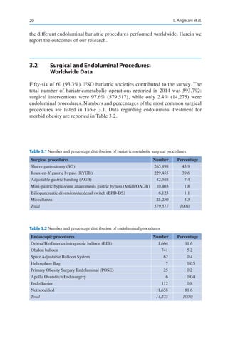
only to update the previous study but to provide a comprehensive overview of](https://image.slidesharecdn.com/bariatricandmetabolicsurgery-180301214153/85/Bariatric-and-metabolic-surgery-33-320.jpg)
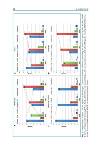
![233 Bariatric Surgery Worldwide
decrease of 3.9, 3, and 11% was observed for RYGB in USA/Canada, Europe,
and Asia/Pacific, respectively. In Latin/South America, RYGB still represents
the most popular procedure, while SG decreased by 2.9%. AGB plateaued in
the American regions and declined in Europe and Asia/Pacific (–7.5 and –4.7%,
respectively). BPD-DS plateaued in three of four IFSO chapters (USA/Canada,
Asia/Pacific, Latin/South America), whereas a slight decrease was registered in
Europe (–0.7%). MGB/OAGB increased in the Asia/Pacific region only (+2.7%);
it plateaued in Latin/South America and slightly decreased in Europe (–0.7%).
No data on MGB/OAGB were reported by USA/Canada. Data on regional trends
are summarized in Fig. 3.2.
3.5 Discussion and Conclusions
Over recent years, different endoluminal procedures – Orbera intragastric balloon,
BioEnterics intragastric balloon (BIB), Obalon balloon, SpatzAdjustable Balloon
System, Heliosphere Bag, Primary Obesity Surgery Endoluminal (POSE) weight-
loss procedure, StomaphyX, Apollo Overstitch Endosurgery, EndoBarrier – have
gained popularity among bariatric surgeons in the attempt to fill the gap between
medical and surgical treatment for borderline patients [8]. A total of 14,275
endoluminal procedures were reported in 2014, but since they have not been
analyzed, it is not yet possible to determine a trend. However, they represent
an evolving field of bariatric surgery, either as primary or revision procedures,
and it is very likely that they will become more popular in the coming years;
therefore, specific analysis is mandatory in future studies.
In order to optimize data collection, we added a specific section to the enquiry
form of our previous survey [7], asking for endoscopic techniques. Moreover,
we chose the definition “mini-gastric bypass/one anastomosis gastric bypass”
(MGB/OAGB), as suggested by other authors [9, 10], in order to avoid data loss
due to the high heterogeneity of definitions. This survey also provides short-term
trend, from 2011 to 2014, of MGB/OAGB, the first experience on which was
published by Rutledge in 2001 [11], and which then spread around the world,
with some authors claiming to prove its efficacy and safety [12]. Worldwide,
MGB/OAGB analysis reveals that this intervention increased only in Asia/
Pacific and plateaued in all the other areas.
A 23.6% increase in bariatric/metabolic procedures was reported from 2013
to 2014, which may have been caused by the higher response rate (93.3 vs.
90.7%) compared with the previous survey [7]. Therefore, better reporting rather
than a real increase may partially explain this result.
From 2003 to 2013, SG continuously gained success in all IFSO chapters,
and in 2014, it was the most performed procedure globally, overcoming RYGB.
As we hypothesized in our previous study [7], the easier surgical technique of
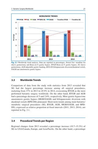
SG compared with RYGB, together with the promising long-term outcomes [13,](https://image.slidesharecdn.com/bariatricandmetabolicsurgery-180301214153/85/Bariatric-and-metabolic-surgery-37-320.jpg)
![24 L. Angrisani et al.
14], could explain these findings. Analysis of regional trends shows that SG is
the most common bariatric procedure in all regions except Latin/South America.
In that area SG declined and RYGB remains the most performed intervention.
In conclusion, the current IFSO survey indicates that in 2014, there was a
universal increase in bariatric surgery, and SG definitely replaced RYGB as the
preferred intervention. Also bariatric endoluminal procedures have been reported
consistently.
References
1. Picot J, Jones J, Colquitt JL et al (2009) The clinical effectiveness and cost-effectiveness of
bariatric (weight loss) surgery for obesity: a systematic review and economic evaluation.
Health Technol Assess 13:1–190, 215–357, iii-iv
2. Colquitt JL, Pickett K, Loveman E, Frampton GK (2014) Surgery for weight loss in adults.
Cochrane Database Syst Rev 8:CD003641
3. Scopinaro N (1998) The IFSO and obesity surgery throughout the world. Obes Surg 8:3–8
4. Buchwald H, Williams SE (2004) Bariatric surgery worldwide 2003. Obes Surg
14:1157–1164
5. Buchwald H, Oien DM (2009) Metabolic/bariatric surgery worldwide 2008. Obes Surg
19:1605–1611
6. Buchwald H, Oien DM (2011) Metabolic/bariatric surgery worldwide. Obes Surg 23:427–436
7. Angrisani L, Santonicola A, Iovino P et al (2015) Bariatric surgery worldwide 2013. Obes
Surg 25:1822–1832
8. Mathus-Vliegen EM (2014) Endoscopic treatment: the past, the present and the future. Best
Pract Res Clin Gastroenterol 28:685–702
9. Rutledge R (2014) Naming the mini-gastric bypass. Obes Surg 24:2173
10. Carbajo MA, Luque-de-León E (2015) Mini-gastric bypass/one-anastomosis gastric bypass–
standardizing the name. Obes Surg 25:858–859
11. Rutledge R (2001) The mini-gastric bypass: experience with the first 1,274 cases. Obes Surg
11:276–280
12. Georgiadou D, Sergentanis TN, Nixon A et al (2014) Efficacy and safety of laparoscopic
mini gastric bypass. A systematic review. Surg Obes Relat Dis 10:984–991
13. Diamantis T, Apostolou KG, Alexandrou A et al (2014) Review of long-term weight loss
results after laparoscopic sleeve gastrectomy. Surg Obes Relat Dis 10:177–183
14. Angrisani L, Santonicola A, Hasani A et al (2015) Five-year results of laparoscopic sleeve
gastrectomy: effects on gastroesophageal reflux disease symptoms and co-morbidities. Surg
Obes Relat Dis pii:S1550-7289(15)00855-2](https://image.slidesharecdn.com/bariatricandmetabolicsurgery-180301214153/85/Bariatric-and-metabolic-surgery-38-320.jpg)
![25
4
L. Angrisani (Ed), Bariatric and Metabolic Surgery,
Updates in Surgery
DOI: 10.1007/ 978-88-470-3944-5_4, © Springer-Verlag Italia 2017
N. Di Lorenzo (*)
Department of Experimental Medicine and Surgery, University of Rome Tor Vergata
Rome, Italy
e-mail: nicola.di.lorenzo@uniroma2.it
Evolution of Bariatric Surgery in Italy: Results
of the National Survey
Nicola Di Lorenzo, Giuseppe Navarra, Vincenzo Bruni,
Ida Camperchioli, and Luigi Angrisani
4.1 Introduction
Over the last few decades, the number of overweight and obese individuals in-
creased worldwide and became a major public health challenge in high-, middle-,
and low-income countries. Overall, 31.8% of the Italian adult population – 39.8%
men, 24.4% women – is overweight (body mass index, BMI ≥25 kg/m2
and <30
kg/m2
) and 8.9% – 8.5% men, 9.4% women – is obese (BMI ≥30 kg/m2
) [1].
While governments, national health systems, and scientific societies draw
strategies to battle the obesity epidemic, the disappointing long-term efficacy of
conventional weight reduction treatments has contributed to the steep increase
in the number of bariatric procedures performed worldwide [2–4]. Undoubtedly,
bariatric surgery is the best available approach by which to achieve and maintain
significant weight loss over the long term, together with a better quality of life,
improvement in or remission of comorbidities, and a significant reduction in
overall mortality [5–7]. Several surgical options are available: some have been
proved to be safe and efficient; some are still investigational. The choice of
a specific procedure depends on specific local conditions, such as a patient’s
alimentary disorders and comorbidities and the experience of surgical staff [8].
According to data from the annual survey of the International Federation for the
Surgery of Obesity and Metabolic Disease (IFSO), the total number of metabolic/
bariatric procedures performed worldwide progressed form 340,768 in 2011 to
468,609 in 2013 [8, 9].
In Italian hospitals belonging to the National Health system – as in some other
countries aiming at zero mortality related to being overweight or obese [10] –](https://image.slidesharecdn.com/bariatricandmetabolicsurgery-180301214153/85/Bariatric-and-metabolic-surgery-39-320.jpg)
![26 N. Di Lorenzo et al.
bariatric surgery is indicated in patients with BMI >40 kg/m2
or BMI >35 kg/m2
with significant comorbidities in case of failure of nonsurgical treatments over
an extended period, by previous psychological evaluation of the patient, and
according to eligibility criteria established by the guidelines of the National
Institutes of Health Consensus Development Conference Statement [11].
4.2 Creation and Evolution of the SICOB
The Italian Society for Obesity Surgery and Metabolic Diseases (SICOB) is a
scientific community composed of Italian specialists battling obesity – including
surgeons, psychologists and psychiatrists, nutritionists, and dietitians – with
the purpose of improving the art and science of bariatric and metabolic surgery
by continually increasing the quality and safety of care and treatment of obese
people, providing educational and support programs for surgeons and integrated
health professionals, and monitoring the number, type, safety, and long-term
outcome of surgeries through the use of a national register.
The Society started its activity as the Italian Group of Bariatric Surgeons
(GICO) in 1990 and became SICOB in 1995, with different targets [12]. Among
them were to:
• promote and improve treatment of obesity and metabolic diseases through a
multidisciplinary approach
• promote scientific research in this field
• regularly define and upgrade guidelines
• provide educational and support programs for surgeons and integrated health
professionals
• become the recognized authority on bariatric and metabolic surgery
• monitor and certify number, type, safety, and long-term outcome of surgeries
through the use of a national register
• serve professional needs of its members.
In the attempt to recognize the quality of treatment offered by bariatric centers
in Italy, three levels of SICOB certified centers have been identified:
1. Excellence centers, which must perform at least four different surgical
procedures recognized by SICOB, including redo surgery, with at least 100
surgical procedures/year.
2. Accredited centers, which must perform at least three different surgical
procedures and no less than 50 surgical procedures/year.
3. Associated centers, which must perform at least two different surgical
procedures with at least 25 surgical procedures/years.
All levels share the following features:
• they follow the same patient selection standards
• they have a multidisciplinary group
• they record all surgical activity on the national register](https://image.slidesharecdn.com/bariatricandmetabolicsurgery-180301214153/85/Bariatric-and-metabolic-surgery-40-320.jpg)
![274 Evolution of Bariatric Surgery in Italy: Results of the National Survey
• they provide patient follow-up >50%, wholly recorded on the national register
• they have an available intensive care unit in the hospital.
4.3 Creation and Improvement of the SICOB Register
The SICOB register was created in January 1996 to record clinical data related
to bariatric surgery in Italy [13].
The register holds data on the number of patients, is updated regularly,
and allows a reliable comparison between surgical procedures using the same
comparative method. Since 1996, three kinds of register have been designed:
The first (1996–2003) had 50 registry contributors and recorded 10,250
interventions (Table 4.1) that comprised: adjustable silicon gastric banding
(ASGB) (43.3%), vertical banded gastroplasty (VBG) (33.2%), biliopancreatic
diversion (BPD) (17%), Roux-en-Y gastric bypass (RYGB) (4.2%), BioEnterics
intragastric balloon (BIB) (1.7%), nonadjustable gastric banding (NAGB) (0.5%),
and other techniques (0.1%). In this first period, results in terms of percentage of
excess weight loss (%EWL) at 5-year follow-up were 69.3% after BPD, 59.8%
after VBG, and 39.9% after ASGB. There were early complications with 16.4% of
RYGB, 10.6% of BPD, 8.2% of BIB, 7.8% of VBG, 2% of ASGB patients. Late
complications with reinterventions occurred in 9.4% of ASGB (7% due to major
complications), 5.3% of BPD, 3.4% of VBG, and 2.6% of RYGB procedures.
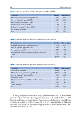
From 2004 to 2006, an online database was available, providing real-time
updates, mandatory fields, avoidance of missing data, and improved data quality
and processing efficiency. A total of 5975 surgical procedures were recorded
on this database (Table 4.2): ASGB were performed in 57% of cases, RYGB in
21.5%, VBG in 9.7%, BPD in 7.3%, BIB in 2.6%, and sleeve gastrectomy (SG)
in 1.9% of cases. Results in terms of %EWL at 5 years were 65% after BPD,
57.7% after RYGB, 57.3% after VBG, and 39.1% after ASGB; %EWL at 9-year
follow-up were 66% after BPD, 55.2% after RYGB, 51.2% after ASGB, and
50.3% after VBG.
Table 4.1 Bariatric procedures recorded in Italy Between 1996 and 2003
Procedures Number Percentage
Adjustable silicon gastric banding (ASGB) 4437 43.3
Vertical banded gastroplasty (VBG) 3405 33.2
Biliopancreatic diversion (BPD) 1,741 17.0
Roux-en-Y gastric bypass (RYGB) 427 4.2
Bioenteric intragastric balloon (BIB) 175 1.7
Nonadjustable gastric banding (NAGB) 54 0.5
Others 11 0.1
Total 10,250 100.0](https://image.slidesharecdn.com/bariatricandmetabolicsurgery-180301214153/85/Bariatric-and-metabolic-surgery-41-320.jpg)
![294 Evolution of Bariatric Surgery in Italy: Results of the National Survey
As is clearly shown by collected data, the key objective of the national
register – to accumulate sufficient data to allow a comprehensive report on
outcomes following bariatric surgery – has been met. Over the last few years,
more procedures have been recorded, new centers have begun uploading their
caseloads, and long-term follow-up data are available for more patients. At
present, the register allows constant monitoring of bariatric surgery in Italy,
not just in terms of number of procedures performed but especially in terms
of outcomes. It renders SICOB, de facto, the recognized authority on bariatric
surgery, since it is the only scientific body in Italy to contain data not only on
the safety but also on long-term outcomes, such as %EWL and comorbidity
remission.
4.4 Results of the National Survey
Thanks to data extracted from the national register, SICOB recently released
data of a national survey on bariatric surgery in Italy [14]. From 1996 to the
present, SICOB centers grew from 53 to 108: 56 centers (51.9%) are in the north,
24 (22.2%) in the center, 21 (19.4%) in the south, and 7 (6.5%) on the islands.
Excellence centers have grown from 28% in 2011 to 37% in 2015, while centers
performing <50 procedures decreased from 48% to 33%. Bariatric surgeries
performed have increased from 5974 procedures in 2008 to 11,435 in 2015, with
>95% of the them being performed laparoscopically over the last five years.
Data on type of surgery performed between 2008 and 2015 show a clear drop
in bands, down from >50% to 21%; a limited decrease in gastric bypass, down
to 16.6% from 23.6%, which is compensated by the number of mini-gastric
bypasses performed in 2015 (870; 7.6% of cases). During the same period, the
most striking data is the explosion of sleeve gastrectomies performed, jumping
from 8.9% to 48.5% of cases.
The reason for these changes could be related to suboptimal long-term results
after gastric bandings and a limited but still present number of cases of weight
regain. At present, sleeve gastrectomy is by far the most popular procedure
because it is quick, efficient, and can be converted to duodenal switch (DS) or
RYGB in case of weight regain.
References
1. Gallus S, OdoneA, LugoAet al (2013) Overweight and obesity prevalence and determinants
in Italy: an update to 2010. Eur J Nutr 52:677–685
2. Lecube A, de Hollanda A, Calañas A et al (2015) Trends in bariatric surgery in Spain in
the twenty-first century: baseline results and 1-month follow-up of the RICIBA, a national
registry. Obes Surg [Epub ahead of print] doi:10.1007/s11695-015-2001-3](https://image.slidesharecdn.com/bariatricandmetabolicsurgery-180301214153/85/Bariatric-and-metabolic-surgery-43-320.jpg)
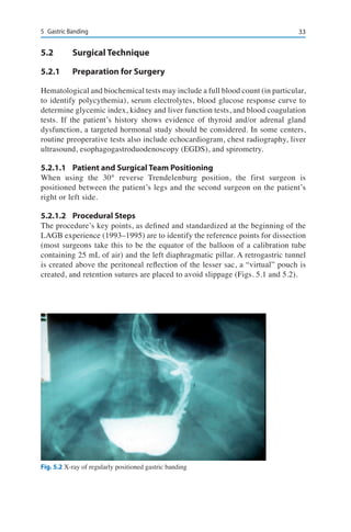
![32 M. De Luca et al.
knowledge of the band’s physical specifications and compliance with a detailed
adjustment protocol [1–4]. It is also important to consider the physiological
behavior of the implanted gastric band, particularly during swallowing and
gastric-pouch emptying. Activating the satiety mechanism requires patient
understanding of the importance of eating slowly and an awareness of when to
stop eating. Patients should be trained to eat only when they are hungry and to eat
until they are not hungry – not until they are full. Behavioral compliance includes
choosing foods of the right texture and a good balance of macronutrients, as
well as avoiding energy-dense snacks. Attention to all these elements of care is
essential if suboptimal results are to be avoided [4, 5].
Fig. 5.1 Gastric band implanted (a), virtual pouch (b), and gastrogastric sutures (c)
a c
b](https://image.slidesharecdn.com/bariatricandmetabolicsurgery-180301214153/85/Bariatric-and-metabolic-surgery-46-320.jpg)
![355 Gastric Banding
(perforations and slippage). Once the lesser pars flaccida of the lesser omentum
has been divided up to the extragastric vagal fibers (which should be preserved),
the caudate lobe of the liver and the right diaphragmatic pillar become
visible. This is the starting point for the blunt dissection toward the angle of
His, remaining in front of the plane of the diaphragmatic pillars, exactly as in
fundoplication procedures for gastroesophageal reflux disease or hiatal hernia
repair. Introduction and positioning of the band, band locking, sutures, port
positioning, and band adjustment is the same as in the perigastric technique.
5.2.4 Pars Flaccida versus Perigastric Technique
It is possible to use a combination of the two previous techniques, especially in
the case of visceral obesity, to avoid too much tissue being included in the band
and consequent risk of early gastric stenosis. The dissection starts according to
the pars flaccida approach and, once the tunnel is created, shifts to the perigastric
technique. An anteroposterior perigastric opening along the lesser curvature
is created close the equator of the calibration tube balloon, and the tip of the
calibration tube is then grasped and pulled through. Therefore, the band is
positioned from the angle of His to the perigastric window. Band introduction,
positioning, and locking; sutures; port positioning; and band adjustment are the
same than in perigastric technique [6].
5.3 Results
5.3.1 Weight Loss
In their systematic review, Buchwald et al. found a mean percentage of excess
weight loss (%EWL) of 47.5% for patients who underwent bariatric surgery
[7]. Tice et al. reported 48% EWL at 1 year [8]. A recent report on a large series
of gastric bands from the UK [9] reported results on 2356 primary gastric
band procedures. Mean excess body mass index (BMI) loss at 1, 2, 3, and 5
years was 43.97 ± 27.4%, 51.8 ± 37.41%, 49.7 ± 36.88%, and 52.6 ± 41.74%,
respectively [9].
Several studies have compared weight loss between the gastric band and
Roux-en-Y gastric bypass (RYGB), and results suggest that although after RYGB
initial weight loss was greater, after 2–5 years there was no significant difference
in %EWL. These finding are consistent with a systematic review by O’Brien et
al., who found that mean %EWL for standard gastric bypass was higher than for
gastric banding at year 1 and 2 but was not statistically different at years 3–7.
Note that this was primarily attributed to fading of the effect of RYGB, whereas
weight loss with the band remains relatively stable [10].](https://image.slidesharecdn.com/bariatricandmetabolicsurgery-180301214153/85/Bariatric-and-metabolic-surgery-49-320.jpg)
![36 M. De Luca et al.
5.3.2 Type 2 Diabetes Mellitus
Buchwald et al. described a gradation of effects for diabetes resolution following
different surgical techniques. Regarding gastric banding, their systematic review
reported 56.7% of patients achieving diabetes resolution and 80% achieving
resolution or improvement [11].
5.3.3 Mortality
In the Swedish Obese Subject (SOS) Study, the adjusted 10-year mortality rate
was significantly (31%) lower than in the nonsurgical group, and most surgical
patients were treated with gastric banding. A study comparing LAGB versus
nonsurgical treatment showed a statistically significant 60% reduction in total
mortality in favor of the LAGB group at a mean follow-up of 5.7 and 7.2 years,
respectively [12]. In addition, Peeters et al. compared the mortality rate in 1468
morbidly obese patients treated by gastric banding with 5960 patients from
an established population-based control group. They found that the surgically
treated group were 73% less likely to die of their disease than those in the control
group [13].
5.4 Complications (According to Clavien-Dindo Classification)
LAGB surgery is not without complications, but these occur on a smaller scale
and have a much lower risk profile compared with other methods currently used
in obesity surgery [14, 15].
5.4.1 Gastric Perforation (Grade IIIb)
The stomach may be perforated during surgery (0.2–0.8% of cases), mainly during
creation of the retrogastric tunnel. This step can be difficult in patients with very
high BMI, visceral obesity, and in men. Gastric perforation is characterized by
free leakage of gastric contents into the peritoneum. Confirmation is provided
by a methylene blue test. If the perforation is detected during surgery, and if it
occurs in a site distant from the band, some surgeons have repaired the stomach
laparoscopically and placed the band successfully. If exposure is not satisfactory,
it is advisable to postpone band placement, suture the stomach wall, drain the area,
and place a nasogastric tube in situ. If the perforation is detected postoperatively
and gross contamination has already occurred, causing peritonitis, the band must
be removed, the gastric wall (possibly) sutured, and drainage performed with a
nasogastric tube.](https://image.slidesharecdn.com/bariatricandmetabolicsurgery-180301214153/85/Bariatric-and-metabolic-surgery-50-320.jpg)
![38 M. De Luca et al.
terms of weight loss and always requires band removal. Causes of erosion can
be a combination of small, undetected injuries to the gastric wall during surgery;
necrosis due to pressure of the band; and access port infection. Some authors
believe that first the access port becomes infected and the infection then travels
along the tubing to the band, causing erosion. However, most surgeons believe
that access port infection is almost always a late manifestation of erosion. An
upper GI X-ray series and consequent esophagogastric devascularization and
splenectomy (EGDS) are diagnostic. Treatment consists of band removal (5%)
via laparoscopy or orally by endoscopy, especially if the band is contained
completely within the gastric lumen. If endoscopic removal is contemplated,
general anesthesia is strongly recommended [16, 17].
5.4.6 Gastric Necrosis (Grade IVa)
Gastric necrosis (0.1%) means necrosis of the upper gastric pouch and may occur
early in the postoperative period or later, when it is likely to be the result of a
long-term undetected stomach slippage/pouch dilatation, both of which increase
pressure on the gastric wall, thereby decreasing blood supply to the fundus. The
theoretical link between stomach slippage and necrosis is precisely why stomach
slippage must be considered a surgical emergency. An upper GI X-ray series and
consequent EGDS are diagnostic.
Treating gastric necrosis consists of exploratory laparoscopy or laparotomy,
the methylene blue test, gastric suture, gastric resection, or nasogastric tube and
drainage.
5.4.7 Tubing/Port Access System (Grade IIIa)
The port is an essential component of the band system, and its placement requires
careful attention. The tubing/port access system can be linked to design features
at the interface between the access port and the tubing and in part to the method
of port placement. Port inversion (flipped port) and leakage are the most frequent
problems. Treatment consists of port repositioning or replacement, and in most
cases, surgery can be performed under local anaesthesia (3%).
Widely differing complication rates are reported in the literature [18]. This is
likely to be partly attributable to the surgical technique deployed, but primarily
to the quality of the after-care and follow-up.
5.5 Conclusions
Whereas further refinement of surgical technique may reduce complication
rates, it is unlikely to improve the 50–60% EWL rate, which is such a consistent](https://image.slidesharecdn.com/bariatricandmetabolicsurgery-180301214153/85/Bariatric-and-metabolic-surgery-52-320.jpg)

![41
6
L. Angrisani (Ed), Bariatric and Metabolic Surgery,
Updates in Surgery
DOI: 10.1007/ 978-88-470-3944-5_6, © Springer-Verlag Italia 2017
E. Soricelli (*)
Department of Surgical Sciences, Sapienza University of Rome
Rome, Italy
e-mail: emanuele.soricelli@uniroma1.it
Sleeve Gastrectomy
Emanuele Soricelli, Giovanni Casella, Alfredo Genco,
and Nicola Basso
6.1 Introduction
Sleeve gastrectomy (SG) was performed for the first time in 1988 by Hess and
Hess as part of a hybrid malabsorptive procedure, the biliopancreatic diversion
with duodenal switch (BPD-DS) [1]. Unlike the original Scopinaro biliopancreatic
diversion (BPD), which consisted of a horizontal subtotal gastrectomy with a
gastroileal anastomosis, the BPD-DS combined a vertical gastrectomy, namely
SG, with an end-to-end suprapapillary duodenoileal anastomosis. The rational
was to maintain a proper gastric restriction, avoiding the occurrence of marginal
ulcers at the gastroileal anastomosis, the incidence of which was considerably
high after BPD [2].
Several studies show that BPD-DS was as effective as the Scopinaro BPD
in terms of weight loss, moreover, the malabsorption-related side-effects, such
as diarrhea, number of daily stools, vomiting, bone pain, and lack of serum
vitamins and minerals, were less severe after BPD-DS than after BPD because
of the longest common channel’s length in the former (100 cm vs. 50 cm,
respectively) [3]. In 2000, Ren et al. demonstrated the feasibility of BPD-DS
with a laparoscopic approach [4]; however, in high-risk, superobese patients,
it was affected by a high incidence of complications and mortality. In order to
reduce the overall surgical risk, Regan et al. proposed splitting the procedure into
two surgical stages: laparoscopic SG (LSG) in the first stage, and BPD-DS after
an average 11-month interval [5].
The good results of LSG, as a first stage, in terms of weight loss and
resolution of comorbidities and the great compliance of patients, encouraged
spreading of this procedure. Moreover, a mounting number of published studies
supported the effectiveness of LSG as a sole operation [6]. As a consequence,](https://image.slidesharecdn.com/bariatricandmetabolicsurgery-180301214153/85/Bariatric-and-metabolic-surgery-55-320.jpg)
![42 E. Soricelli et al.
in 2009, the American Society for Metabolic and Bariatric Surgery (ASMBS)
issued a position statement recommending LSG as an approved primary bariatric
procedure [7].
At first, LSG was classified as a restrictive procedure, since its weight-
loss effectiveness was entirely attributed to reduction of the gastric capacity.
However, it soon became evident that significant modifications of gastrointestinal
hormones play a preeminent role. Changes in ghrelin (GHR), glucagon-like
peptide-1 (GLP-1), and peptide tyrosine-tyrosine (PYY), induced by the gastric
resection, are of paramount importance in weight loss and glucose homeostasis
effects of the procedure [8].
At present LSG is the second most performed bariatric procedure after
Roux-en-Y gastric bypass (RYGB), and it has the fastest increment rate in the
last decade [9]. The common perception of LSG as a safe and easy-to-perform
procedure has had a major role in its noteworthy worldwide spread. Because
of the lack of gastrointestinal anastomosis and the short operative time, many
surgeons consider LSG an ideal option to start a novel bariatric activity. This
might represent a boomerang effect, because LSG entails some key technical
points that require adequate training: dissection of the stomach from the
spleen, and complete detachment of the posterior gastric wall from the anterior
aspect of the pancreas and diaphragmatic crura are necessary to perform an
adequate fundectomy, which is of utmost importance for both the restrictive
and the hormonal effects of LSG. Furthermore, the postoperative course can be
affected by life-threatening complications such as gastric leak, the management
of which requires a specific experience and should be performed in dedicated
institutions.
6.2 Surgical Technique
The laparoscopic technique was standardized by Ren et al. in 2000 [4].
However, during the following years, several details have been modified. In
this chapter, we emphasize technical points in light of our own experience of
~900 cases since 2002.
6.2.1 Preparation for Surgery
In all patients, preoperative workup included history and physical examination,
routine laboratory tests, esophagogastroduodenoscopy, abdominal ultrasonogra-
phy, nutritional and psychiatric evaluation, and additional examinations and/or
consultations when indicated. The day before surgery, subcutaneous low-molec-
ular-weight heparin (LMWH) is administered.](https://image.slidesharecdn.com/bariatricandmetabolicsurgery-180301214153/85/Bariatric-and-metabolic-surgery-56-320.jpg)
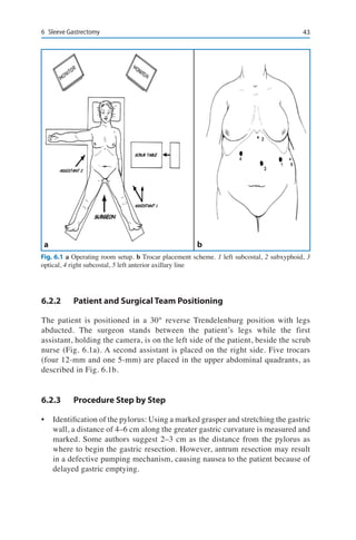
![44 E. Soricelli et al.
• Skeletonization of the greater curvature: By means of radiofrequency or
ultrasound energy devices, the gastrocolic ligament is dissected close to
the gastric wall starting at the median third of the greater curvature, where
the ligament is thin. The dissection proceeds downward to the antrum until
the 4- to 6-cm mark is reached. In this area, access to the omental bursa
might be difficult because of the frequent presence of adhesions between the
posterior wall of the stomach and the anterior aspect of the pancreas. Then,
skeletonization proceeds upward to the angle of His. Attachments with the
upper pole of the spleen should be divided carefully in order to avoid splenic
injury or bleeding from short gastric vessels.
• Complete mobilization of the fundus and posterior gastric wall: In this phase,
the surgeon can move his or her left hand from the right subcostal trocar to
the subxiphoid trocar to facilitate the approach to the gastroesophageal area.
The dissection of the fundus is completed when clear exposure of the left
diaphragmatic pillar is obtained (Fig. 6.2a). If the Belsey’s fat pad on the
anterior aspect of the gastroesophageal junction is redundant, its resection
may be useful to properly expose the area where the stapler will be placed.
The posterior gastric wall must be completely freed from adhesions. When
present, posterior gastric vessels should be divided, as they can lessen
compliance of the stomach during resection. At the end of mobilization, the
left gastric vessels and left crus are clearly exposed, and the stomach can be
easily moved on this axis – like the page of a book (Fig. 6.2b). The proper
accomplishment of these surgical steps is of primary importance in order to
achieve a complete fundectomy. Since GHR secretion is predominant in the
gastric fundus, its ablation plays a main role in the LSG mechanism of action
[10, 11]. On the other hand, accurate mobilization of the gastric fundus and
gastroesophageal junction entails the division of the short gastric vessels and,
when present, the posterior gastric artery and phrenic branches [12]. This
might hamper the blood supply in this area, favoring the onset of gastric
leaks, which occur quite uniformly at the uppermost part of the suture line.
During this surgical step, risks (leak) and benefits (functional result) should
be carefully weighted.
• Inspection of the hiatal area and possible hiatoplasty: Enlarged hiatus and
hiatal hernias must be identified. A hiatal orifice with a diameter >3 cm
is considered abnormal (Fig. 6.2c). A macroscopically evident fingerprint
indentation of the diaphragm just above the esophageal emergence should
be considered suspicious for the presence of a hiatal hernia, indicating
dissection of the hiatal area. This can be easily approached from the left, as
the fundus has been previously mobilized. When present, the hernia sac and
gastroesophageal fat pad are dissected and reduced within the abdominal
cavity. A posterior hiatoplasty is performed by approaching the right and
the left diaphragmatic pillars with two or three interrupted nonabsorbable
sutures (Fig. 6.2d).](https://image.slidesharecdn.com/bariatricandmetabolicsurgery-180301214153/85/Bariatric-and-metabolic-surgery-58-320.jpg)
![456 Sleeve Gastrectomy
• Orogastric tube insertion: This tube is inserted by the anesthesiologist and
pushed down, possibly through the pylorus. It is then placed against the
lesser curvature in order to calibrate the resection. In our clinical practice,
we use a 48-Fr bougie, although in the literature the use of tube sizes from
30- to 60-Fr is reported [13]. Literature data correlate the smaller size
of the bougie to a higher incidence of gastric leaks. Furthermore, a clear
relationship between bougie size and postoperative weight loss is lacking
[14–18]. In our experience, the residual capacity of the gastric remnant
does not depend on bougie size but mainly on the degree of countertraction
exerted on the stomach walls when resecting and on accurate dissection of
the gastric fundus [19].
• Gastric resection and staple-line reinforcement: This step is performed
using a linear stapler applied alongside the calibrating bougie. The height
of the cartridges must be chosen according to gastric-wall thickness, since
it decreases from the antrum to the corpus and fundus. We prefer a staple
height of 4.4 or 4.1 mm near the antrum and 3.8 or 3.5 mm on the corpus
and fundus. In revision surgery cases, the use of higher staples might be
advisable because of the presence of thick scar tissue. Before closing and
a
c
b
d
Fig. 6.2 a Dissection of the gastric fundus from the anterior aspect of the left pillar. b Left gastric
vessels are visualized together with the left pillar. c Enlarged hiatus. d Posterior hiatoplasty with
nonabsorbable stitches. LP left pillar, GF gastric fundus, LGV left gastric vessels, VN vagus nerve,
RP right pillar](https://image.slidesharecdn.com/bariatricandmetabolicsurgery-180301214153/85/Bariatric-and-metabolic-surgery-59-320.jpg)
![46 E. Soricelli et al.
firing the stapler, the anterior and posterior gastric walls should be stretched
homogeneously by two graspers placed exactly at the greater curve (Fig.
6.3a). At the incisura angularis, stretching is somewhat loosened to avoid
functional strictures. The last cartridge is fired 1–2 cm away from the angle
of His (Fig. 6.3b) so that the staple line does not fall within the critical area
[12]. The staple line is reinforced by buttressing with absorbable polymer
membrane (Seamguard, Gore) and meticulously checked for bleeding spots,
which can be managed by using hemostatic clips or stitches. After moving
the stomach specimen away from the left subcostal space, the final end of the
membrane is fixed with two nonabsorbable sutures to the left pillar to avoid
sliding of the stomach tubule into the mediastinum (Fig. 6.4b). A nasogastric
tube is positioned in the gastric remnant, and a methylene blue dye test is
routinely performed to check for complete sealing of the staple line and to
evaluate the residual gastric capacity, usually 60–80 mL.
• Specimen extraction: The specimen is extracted by grabbing its distal end
with a grasper. It is easily brought out of the abdominal cavity through the
slightly enlarged right subcostal access. Care must be taken not to open
Fig. 6.3 a Homogeneous countertraction of both anterior and posterior gastric walls by means
of two graspers placed exactly at the greater curvature. b The last staple is fired 1–2 cm from the
angle of His. c The caudal tip of the resected stomach is extracted, using a grasper, through the
slightly enlarged right subcostal access. d Careful extraction of the specimen without retrieval
bags or endoloop
a
c
b
d](https://image.slidesharecdn.com/bariatricandmetabolicsurgery-180301214153/85/Bariatric-and-metabolic-surgery-60-320.jpg)
![476 Sleeve Gastrectomy
the specimen during these manoeuvres (Fig. 6.3c, d). No retrieval bags or
endoloops are needed [20]. A gauze soaked in povidone-iodine solution
(betadine) is left for 1–2 min at the retrieval site to avoid wound infection.
Drains are not routinely placed, and the nasogastric tube is removed at the
end of the procedure.
• Additional procedure: When gallbladder stones are present, cholecystectomy
is routinely performed at completion of the LSG procedure. The same trocars
are used. Occasionally, in complicated cases, an additional 5-mm trocar is
added 5-cm laterally to the right subcostal trocar.
6.2.4 Postoperative Management
Patients are mobilized on the same day of the operation and maintained with
intravenous fluid therapy, proton pump inhibitors (PPIs), and analgesics.
LMWH is administered subcutaneously 6 h after surgery and continued for 2
weeks. Short-term antibiotic therapy is added. Upper gastrointestinal contrast
(Gastrografin) study is performed on the second postoperative day. Afterward,
patients are put on a liquid diet and discharged on the fourth postoperative day.
Soft diets with mashed and soft foods are prescribed for 4 weeks after surgery.
One month after surgery, patients resume normal diet with the advice of adding
one type of food at a time; meat may take longer to be tolerated. Five small meals
a day are suggested. Postoperative follow-up is performed at 1, 3, 6, 12, 18, and
24 months after the operation and annually thereafter. Controls involve physical
examination, blood tests (including vitamin B1
, B12
, folate, and serum iron,
calcium, and vitamin D levels), upper gastrointestinal contrast (first month and
first year), and liver ultrasound (sixth month). Endoscopic check is mandatory 2
years after the operation in all patients. Oral PPIs and ursodeoxycholic acid for 6
months, and multivitamin tablets for 1 year, are prescribed.
6.3 Results
6.3.1 Weight Loss
Short- to midterm outcomes of LSG in terms of weight loss are very good.
According to the 2012 Fourth International Consensus Summit, accounting for
46,133 LSG performed by 130 surgeons worldwide, the percentage of excess
weight loss (%EWL) at 1, 2, 3, 4, and 5 years was 59.3%, 59.0%, 54.7%, 52.3%,
and 52.4%, respectively [21]. However, since LSG was approved as a stand-
alone bariatric procedure only in 2009, long-term results from large series are
lacking. In a recent review, Diamantis et al. reported on the 5-year results of
nine studies enrolling 258 patients overall, with a mean %EWL of 62.3% [22].](https://image.slidesharecdn.com/bariatricandmetabolicsurgery-180301214153/85/Bariatric-and-metabolic-surgery-61-320.jpg)
![48 E. Soricelli et al.
Consistently, Sieber et al. showed a percentage of excess body mass index loss
(%EBMIL) of 57.4% in their series of 54 patients 5 years after LSG [23]. These
results seem to be maintained at a follow-up of ≥6 years; Eid et al. reported on a
%EWL of 46% in 21 patients 8 years after LSG [14], while Sarela et al. showed
a mean %EWL of 69% in 13 patients at a follow-up of 8 years or more [24].
We recently published the long-term results of our monocentric series of 148
patients with at least 6 years of follow-up; %EWL at 6 years was 67.3% and, a
%EWL >50% (success rate) was achieved in 83.1% of patients. At a follow-up of
≥7 years, results were not significantly different (%EWL 65.7%; success rate 81%),
confirming that the weight loss effect of this procedure is maintained over time [19].
6.3.2 Effect on Comorbidities
LSG is associated with a high rate of resolution of type 2 diabetes mellitus
(T2DM) and other obesity-related comorbidities, such as arterial hypertension
(AH) and obstructive sleep apnea (OSA) [25]. In the ASMBS 2009 position
statement, accounting for 754 patients, T2DM remission ranged from 14 to
100%, AH from 15 to 93%, and OSA from 39 to 100% [7].
Weight loss and comorbidity resolution positively affect secondary cardiac
structural and hemodynamic changes, referred to as obesity cardiomyopathy,
including an increase in left-ventricular (LV) wall thickness, mass, and diameters,
with systolic and diastolic dysfunction [26–29]. In our experience, LSG patients
showed a significant change in LV shape in terms of mass, geometry, and diastolic
function. These modifications were related to weight loss and to improvement of
the metabolic syndrome, resulting in a significant reduction of the Framingham
Risk Score [30].
Concerning T2DM, in several studies, 60–80% of diabetic obese patients
undergoing LSG achieve remission of their pathology [31–34]. These results
compared very favorably with those obtained after an intensive medical regimen
[35] and were not statistically different from those after RYGB [36, 37]. The
effectiveness of LSG on T2DM remission seems to be related to the functional
reserve of β cells; in fact, diabetes postoperative duration >10 years, low
C-peptide levels, and need for insulin therapy to control glycemia, are negative
prognostic factors [38, 39]. In our series, T2DM remission occurred in 100% of
patients with DM duration <10 years and in 31% with DM duration >10 years
[38]. The beneficial action of LSG on T2DM occurs very early: Peterli et al.
reported a significant modification of GLP-1 and PYY plasma levels 7 days after
the procedure [40]. These results have been confirmed in a study by our group,
which showed a significant increase of insulin secretion and sensitivity, plasma
PYY, and GLP-1 just 72 h postoperatively, before the ingestion of any food [41].
At the same time, GHR values were significantly lower than those before the](https://image.slidesharecdn.com/bariatricandmetabolicsurgery-180301214153/85/Bariatric-and-metabolic-surgery-62-320.jpg)
![496 Sleeve Gastrectomy
operation. Since GLP-1 and PYY are cosecreted from L cells in the small bowel
in response of food ingestion, an intrinsic neurohormonal effect of LSG was
suggested to explain these early changes. A gastric hypothesis was formulated
[41], postulating that the diminished hydrochloric acid production induced by
the significant reduction of oxyntic cell mass stimulates the vagally innervated
antral mucosa, left intact by LSG, to secret gastrin-releasing peptide and, as a
consequence, GLP-1 and PYY, without any food ingestion.
The long-term antidiabetic effects of LSG are not well documented due to
the novelty of the procedure. In a small series of patients, at 5-year follow-up,
remission was present in 87.8% of cases [42]. In our experience of 65 obese and
diabetic patients submitted to LSG, remission was present in 57 patients (87%) and
amelioration in 7 patients (10%) at a mean follow-up of 63 months (unpublished
data). Most important, once remission was achieved, it was maintained in all
cases except two, although weight regain occurred in six patients.
6.4 Complications (According to Clavien-Dindo Classification)
The postoperative mortality rate varies from 0.1 to 0.5% [21, 43, 44]. Early diag-
nosis is the most important factor to ensure a positive solution of complications,
the management of which is often challenging and should be accomplished in
bariatric centers by dedicated medical teams.
6.4.1 Bleeding (Grades II–IIIb)
Bleeding (1.1–8.7%) occurs most frequently within the first 24–48 h and almost
always into the abdominal cavity, while it rarely determines hematemesis or
melena [43, 45, 46]. More commonly, it originates from the staple line, in
which case it increases the risk of gastric leak. Other sites of bleeding are the
gastroepiploic and the short gastric vessels, which are divided during stomach
mobilization, and trocar accesses. Hepatic or splenic injuries may cause severe
postoperative bleeding if not recognized and managed intraoperatively. Once
the hemodynamic parameters are stable, computed tomography (CT) scan is
mandatory to define the bleeding site and quantify the hemoperitoneum. In
case of hemorrhage from the staple line, CT images show a hematoma close
to the gastric remnant. Bleeding can be self-limiting, requiring only blood
transfusions, or it can be managed by interventional radiology. In case of
massive uncontrolled hemorrhage, open or laparoscopic surgical exploration is
mandatory. Suture line reinforcement has significantly reduced the occurrence
of this complication [47].](https://image.slidesharecdn.com/bariatricandmetabolicsurgery-180301214153/85/Bariatric-and-metabolic-surgery-63-320.jpg)
![50 E. Soricelli et al.
6.4.2 Staple-Line Leak (Grades IIIa–IV)
Staple-line leak (0–7%) represents the most frequent complication after LSG,
and it may be life-threatening. In most cases, leaks occur early, during the first
postoperative week; late leaks (from the 8th to the ~40th postoperative day) are
less frequent [13, 44].
Ninety percent of leaks occur just below the gastroesophageal junction
(Fig. 6.4a). Both pathophysiological and technical factors seem to play a role
in the development of gastric leaks after LSG. The former are represented by
high intragastric pressure, sliding of the gastric tubule in the low-pressure
mediastinum, lower thickness of the gastric fundus wall, and presence of a
critical area of vascularization on the left side of the cardias region, where the
oesophageal and gastric arterial systems are bordering [12]. Technical aspects
include small bougie size (<36 F) and injury to the gastric wall during hemostasis
or dissecting manoeuvers.
Practical rules are:
1. The staple line should not involve the critical area of vascularization,
remaining 1–2 cm lateral to the angle of His (Fig. 6.3b).
2. The end of the staple line should be fixed to the left diaphragmatic pillar with
two nonabsorbable sutures (Fig. 6.4b).
3. Countertraction should be somewhat loosened at the incisura angularis while
resecting the stomach in order to avoid functional stenosis, which results in
increased intragastric pressure (Fig. 6.4a).
The management of leaks is challenging, requiring a multidisciplinary
approach, and it should be performed in experienced bariatric institutions.
Timely diagnosis is the most important prognostic factor. Therefore, patients are
advised to immediately contact the bariatric team in case of strange or unusual
symptoms of any kind.
In our experience, operative treatment is reserved only for patients with
hemodynamic instability and signs of acute peritonitis. Peritoneal toilet and proper
drainage are recommended. Attempts to repair the fistula are contraindicated
because of the high incidence of recurrence and the risk of adding further severe
complications. In most cases, staple-line leaks can be successfully managed by
percutaneous CT-guided drainage, alone or in combination with stent placement
and enteral nutrition (Fig. 6.4c), without surgical intervention [48]. Unsuccessful
control of the leak may require total gastrectomy or creation of a Roux limb.
Staple-line reinforcement seems to reduce the incidence of postoperative
complications. To date, different reinforcement options have been proposed,
such as oversewing the staple line with a running or inverting absorbable suture,
buttressing the staple line with absorbable materials (bovine pericardium strips
or porcine small intestine submucosa), and applying fibrin glue or hemostatic
agents to the staple line. Routine reinforcement of the staple line, regardless of
type, has been demonstrated to significantly reduce the incidence of bleeding.
Data from a recent review, accounting for 8,920 patients, showed that buttressing](https://image.slidesharecdn.com/bariatricandmetabolicsurgery-180301214153/85/Bariatric-and-metabolic-surgery-64-320.jpg)
![516 Sleeve Gastrectomy
the staple line with absorbable polymer membrane is more effective than other
reinforcement methods for preventing gastric leaks [49].
6.4.3 Stenosis (Grades II–IIIb)
Stenosis (0.2–4%) usually it occurs at the transition between corpus and antrum
of the gastric tubule, at the incisura angularis. It can be transient and related
to transient dysmotility of the gastric muscular layers in this area, or it can be
caused by incorrect orientation of the stapler during resection, resulting in an
organic stenosis with long-lasting dysphagia and/or vomiting. A twisted sleeve
may also cause symptomatic stenosis. An upper gastrointestinal contrast study
is indicated to confirm gastric outlet obstruction (Fig. 6.4d). Endoscopy has
both a diagnostic and therapeutic value. Repeated endoscopic dilations are
the first approach, while placing endoscopic stents should be considered as an
alternative solution.
Fig. 6.4 a Upper gastric leak in patient with organic stenosis at the incisura angularis and dilation
of the prestenosis segment. b Final end of the buttressing membrane is fixed to the left pillar by two
nylon stitches (arrows). c Management of gastric leak using abdominal drainage, stent placement,
and enteral nutrition. d Functional stenosis at the middle third of the gastric tubule. LP left pillar,
BM buttressing membrane, EN enteral nutrition
a
c
b
d](https://image.slidesharecdn.com/bariatricandmetabolicsurgery-180301214153/85/Bariatric-and-metabolic-surgery-65-320.jpg)
![52 E. Soricelli et al.
In case of persistence of symptoms with nutrition problems, reoperation
should be considered. Conversion to RYGB is the treatment of choice [50].
Laparoscopic seromyotomy of the stenotic tract (stricturoplasty) has also been
proposed [51].
6.4.4 Gastroesophageal Reflux Disease (Grades II–IIIb)
The relationship between LSG and gastroesophageal reflux disease (GERD) is
still a matter of discussion (varying from 0 to 30%). While in some published
series postoperative improvement of GERD symptoms has been reported, in
others, worsening has been noted [52–55]. The rationale of these conflicting data
might be ascribed to the coexistence of different pathophysiological mechanisms,
which could promote/worsen or improve GERD symptoms. The former entail
lower esophageal sphincter (LES) malfunction due to section of sling fibers,
sliding of the stomach tubule into the mediastinum determining diminished
intraluminal pressure in the cardiac segment, increased intraluminal pressure
in the gastric remnant, and delayed emptying of the stomach in case of mid-
gastric stenosis of the lumen. The latter are represented by accelerated gastric
emptying and reduced acid secretion. The presence of a hiatal hernia could also
be associated with an increased risk of postoperative GERD development or
worsening. Data concerning the effectiveness of hiatoplasty on GERD after LSG
are not conclusive. However, in the Fourth International Consensus Summit on
LSG held in 2012, there was general agreement that when a hiatal hernia is
present, it should be repaired at the time of the bariatric procedure [21].
In a recent endoscopic survey of our patients submitted to LSG with a
3- to 5-year follow-up, a 15% incidence of peptic esophageal lesions and of
nondysplastic Barrett’s esophagus was found, with no correlation between the
severity of reflux symptoms and the degree of esophageal lesions (unpublished
data). These lesions were highly responsive to full-dose therapy with PPIs. For
this reason, a careful postoperative follow-up schedule including an endoscopy
within 2 years from the operation is recommended to our patients.
In patients complaining of reflux symptoms and not responsive to PPI therapy,
conversion to RYGB is a valid option.
References
1. Hess DS, Hess DW (1998) Biliopancreatic diversion with a duodenal switch. Obes Surg
8:267–282
2. Scopinaro N, Gianetta E, Pandolfo N et al (1976) Bilio-pancreatic bypass. Proposal and
preliminary experimental study of a new type of operation for the functional surgical
treatment of obesity. Minerva Chir 31:560–566
3. Marceau P, Hould FS, Simard S et al (1998) Biliopancreatic diversion with duodenal switch.
World J Surg 22:947–954](https://image.slidesharecdn.com/bariatricandmetabolicsurgery-180301214153/85/Bariatric-and-metabolic-surgery-66-320.jpg)
![57
7
L. Angrisani (Ed), Bariatric and Metabolic Surgery,
Updates in Surgery
DOI: 10.1007/ 978-88-470-3944-5_7, © Springer-Verlag Italia 2017
C. Giardiello (*)
General, Emergency and Metabolic Surgery Unit, Department of Surgery and Obesity Center,
Pineta Grande Hospital
Castelvolturno, Italy
e-mail: cristiano.giardiello@pinetagrande.it
Roux-en-Y Gastric Bypass
Cristiano Giardiello, Pietro Maida, and Michele Lorenzo
7.1 Introduction
Bariatric surgery is actually considered the only therapeutic option in patients
with morbid obesity who are affected by life-threatening comorbidities.
Although a wide range of surgical bariatric options are offered, at this time,
>50% of patients undergo a procedure based on the laparoscopic Roux-en-Y
gastric bypass scheme [1].
In 1966, Mason and Ito first described the technique of laparotomic Roux-
en-Y gastric bypass for treating morbid obesity [2]. In 1994, Wittgrove et al.
reported the technique, and their preliminary experience with the laparoscopic
Roux-en-Y gastric bypass (LRYGB) [3]. Since then, bariatric surgeons
worldwide have made several modifications to the original technique in
order to improve results and decrease complications. These technical changes
were not accepted after randomized prospective studies but on the basis of
the experience of individual bariatric surgeons and mutual collaboration with
colleagues. Moreover, the biggest controversy regarding this procedure is the
lack of clarity regarding its mechanism of action and its profound metabolic
effect [4]. Variations in technical setup and operative procedures are without
significant effects in term of safety and weight loss and mainly depend on
surgeons’ preferences (Table 7.1).
In this chapter, we describe two different technical approaches to LRYGB,
both characterized by the absence of mesenteric section: the reverse technique
and the double-loop technique.](https://image.slidesharecdn.com/bariatricandmetabolicsurgery-180301214153/85/Bariatric-and-metabolic-surgery-70-320.jpg)
![58 C. Giardiello et al.
7.2 Surgical Technique
7.2.1 Preoperative Patient Preparation
Appropriate preoperative surgical preparation is essential for the procedure.
Intragastric balloon or liver shrinkage diet prior surgery is recommended, with
the objective of reducing liver size and improving intraoperative visualization
[5]. Prophylactic broad-spectrum intravenously administered antibiotics are
routinely given at anesthesia induction and continued for several days, according
to patient condition. Thromboprophylaxis is also done routinely using low-
molecular-weight heparin preoperatively and then for 1–2 weeks postoperatively.
Lower-limb pneumatic compression is also applied intraoperatively.
7.2.2 Patient and Surgeon Positioning
Patients are placed in the reverse Trendelenburg position with the split-leg
approach. Usually, the operating surgeon stands between the patient’s legs, with
assistants on either side of the patient.
7.2.3 Pneumoperitoneum and Trocar Positioning
A 15- to 18-mmHg pneumoperitoneum is created by inserting a Veress needle
into the left midclavicular line just below the costal margin. Five trocars are
usually inserted: The first (T1) is inserted in the xiphoumbilical line 15 cm
Table 7.1 Laparoscopic Roux-en-Y gastric bypass: areas of surgical technical variation
Areas of surgical technical variation
1. Alimentary limb position a. Retrogastric or antegastric
b. Retrocolic or antecolic
2. Length of alimentary and biliopancreatic limb a. Alimentary limb
– 100 cm
– 150 cm
–200 cm
b. Biliopancreatic limb
– 25 cm
– 50 cm
– 100 cm
3. Anastomosis technique a. Hand sewn
b. Circular stapler
– endoesophageal assistance
– endogastric assistance
c. Linear stapler](https://image.slidesharecdn.com/bariatricandmetabolicsurgery-180301214153/85/Bariatric-and-metabolic-surgery-71-320.jpg)
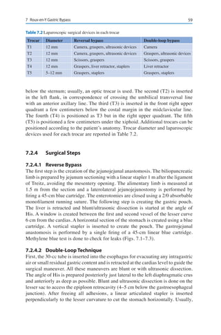
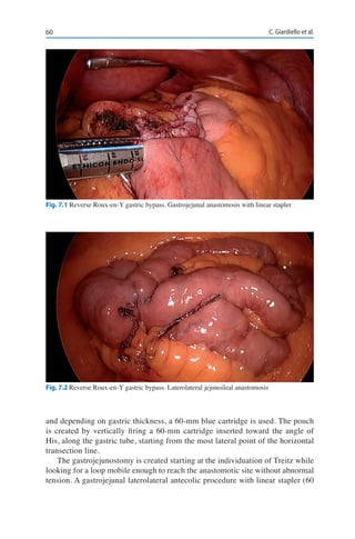
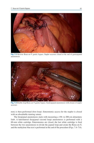
![62 C. Giardiello et al.
7.3 Complications (According to Clavien-Dindo Classification)
After LRYGB, several significant and potentially catastrophic complication can
occur (Table 7.3). The overall complication rate, reported in a recent systematic
review with meta-analysis, is ~21% (12–33%) [6]. The reoperation rate is 3–20%
and mortality rate ~0.38% (<30 days) and 0.72% (>30 days). Early complications
Fig. 7.5 Double-loop Roux-en-Y gastric bypass. On the left the jejunal loop still unsectioned, on
the right the gastrojejunal anastomosis
Fig. 7.6 Double-loop Roux-en-Y gastric bypass. Jejunal stumps](https://image.slidesharecdn.com/bariatricandmetabolicsurgery-180301214153/85/Bariatric-and-metabolic-surgery-75-320.jpg)
![637 Roux-en-Y Gastric Bypass
tend to be related to technical issue; late complications tend to include metabolic
or nutritional problems. Clavien-Dindo classification is inserted to compare
outcome data among different centers [7].
7.3.1 Gastrointestinal Bleeding (Grade IIa)
The rate of gastrointestinal bleeding after gastric bypass is 0.8–4.4%. This
complication can start from anywhere in the gastrointestinal tract, and it may be
clinically expressed by hematemesis, melena, or intraperitoneal bleeding. These
manifestations are usually accompanied by early tachycardia and tachypnea,
abdominal pain, or distension. The gastrojejunal anastomosis is considered
the most frequently involved site after LRYGB. A conservative treatment with
intravenously applied liquid infusion and blood transfusion is usually therapeutic
following minimal blood loss. In massive hemorrhages, a laparoscopic or
laparotomic attempt is mandatory [6, 8–10].
7.3.2 Leak (Grade III)
Anastomotic and staple-line leak can result in high morbility rates. Fistulas,
peritonitis, abscess formation, sepsis, and multiorgan failure can be all observed
as life-threatening consequences after a leak. Leak occurs more frequently in
gastrojejunal anastomosis and occurs less frequently in the staple-line margin.
Intraoperative diagnosis is usually performed using the methylene blue test.
Postoperatively, a leak must be suspected by the clinical presence of tachycardia
(>120 bpm), tachypnea, fever, abdominal pain, hypotension, and hiccup. Upper
gastrointestinal contrast studies with gastrografin are usually able to precisely
reveal the presence and site of a leak. Therapeutic approach can be conservative,
with a nasogastric tube, fluids, and antibiotics; and endoscopic stent is the most
frequently performed therapeutic option, and only in unsuccessful cases is
Table 7.3 Laparoscopic Roux-en-Y gastric bypass: incidence and timing of main complication
presentation
Complication Rate (%) Timinga
Bleeding 0.8–4.4 Early
Leak 1–2 Early/Intermediate
Obstructionb
1–5 Late
Ulcers 4 Late
Strictures 5 Late
a
Early, <1 week; intermediate, 1 week–1 month; late, >1 month
b
Regarding reversal bypass and double-loop bypass; in the authors’ experience, this complication
is absent](https://image.slidesharecdn.com/bariatricandmetabolicsurgery-180301214153/85/Bariatric-and-metabolic-surgery-76-320.jpg)
![64 C. Giardiello et al.
laparoscopic closure of the defect indicated. Only in rare cases with unsuccessful
conservative, endoscopic, and laparoscopic attempts a total gastrectomy is
mandatory. Staple line reinforcement is proven to be effective for preventing this
complication [6, 11–13].
7.3.3 Bowel Occlusion (Grade IIIb)
Intestinal occlusion in bariatric surgery is considered a complication specifically
related to the LRYGB. The rate of this complication is 1.5–5%, and most cases
were diagnosed during the first 12 months after surgery. Then, the incidence slowly
decreased until 42–48 months postoperatively. This complication, in particular,
occurs more frequently in the laparoscopic than in the laparotomic approach.
This fact is probably linked to the absence of mesenteric opening without scares.
The bowel can move freely into the abdomen without provoking obstruction or
entering new open mesenteric spaces (Petersen space). In the double-loop or
reverse bypass technique, the mesenterium is not open, and this complication is
never observed. The clinical presentation of bowel obstruction in patients who
underwent LRYGB is different than in nonobese patients. Due to the small volume
of the gastric pouch, vomit volume is scarce, while vague abdominal discomfort
or colic are more frequently observed. The diagnosis is usually made by CT scan
with gastrografin, and the only therapy is surgical [6, 14, 15].
7.3.4 Stricture (Grades IIIa, IIIb)
The anastomotic stricture rate after LRYGB is 5% and occurs primarily at the
site of the gastrojejunal anastomosis. This complication is usually related to
marginal ulcer scar and tension and ischemia on anastomotic stumps. The 21-
mm circular stapler is now considered in the pathogenesis of this complication.
Usually, the clinical presentation of anastomotic stricture is characterized by
nausea, vomiting, and dysphagia one or more months after surgery. The gold
standard of gastrojejunal anastomosis treatment is endoscopic dilatation. Only
in unsuccessful cases is it necessary to perform a laparoscopic revision. Rarely a
stricture is localized near the jejunojejunal anastomosis. Clinical presentation is
not substantially different, and a laparoscopic revision must be performed [6, 16].
7.4 Results
LRYGB results in significant early weight loss, which is partially maintained after
longer follow-up. Most data come from a meta-analysis of Buchwald et al., which
reports 62% excess weight loss (%EWL) and 0.5% 30-day mortality rate [17].](https://image.slidesharecdn.com/bariatricandmetabolicsurgery-180301214153/85/Bariatric-and-metabolic-surgery-77-320.jpg)
![657 Roux-en-Y Gastric Bypass
Most patients are expected to lose >50% of their excess weight. Weight loss
is maximal at 2 years (32%) and declines during the following years (25%).
Compared with conservative treatment, the Swedish Obesity Study (SOS)
demonstrated better results in operated patients than in patients with dietetic
treatment or who underwent vertical banded gastroplasty or laparoscopic
adjustable gastric banding [18]. These results were confirmed in randomized
controlled trials, as described by Angrisani et al., who compared LRYGB and
laparoscopic adjustable gastric banding (LAGB) [19]. Long-term comparative
results with laparoscopic sleeve gastrectomy (LSG) are lacking. Technically
different approaches to gastric bypass do not seem to be considerably different
in terms of weight loss and comorbidities, as observed for mini-gastric bypass,
reverse bypass, and the double-loop technique.
Improvement in obesity-related comorbidities is considered strictly
proportional to weight loss [17], and some observations regarding improvement
in comorbidities have recently been related to the type of bariatric procedure
[17, 20, 21]. This is the case with type 2 diabetes mellitus and gastroesophageal
reflux disease. In these conditions, a specific role for RYGB was discovered,
which is probably linked to the new anatomy of the digestive tract after surgery.
Gastric bypass appears to have superior effects on type 2 diabetes remission to
other restrictive procedures, such as LAGB or LSG. These effects are mainly
related to duodenal exclusion, or switch. Buchwald et al., in a meta-analysis
study, showed that diabetes improved in 80%, 57%, and 95% of patients treated
with gastric bypass, gastric banding, or duodenal switch, respectively [17]. Lee
et al., in a randomized controlled trial, studied the effects of RYGB and LSG
in patients with a body mass index (BMI) of 25–35 kg/m2
[22]. They observed
that diabetic patients who underwent RYGB achieved greater disease remission
than LSG patients (93 vs. 43%). More recently, the STAMPEDE (Surgical
Treatment and Medications Potentially Eradicate Diabetes Efficiently) trial
reported 3-year follow-up results comparing poorly controlled diabetic
patients who underwent RYGB, LSG, or medical treatment [23]. Glycated
hemoglobin (HbA1c) <6.0% was observed in 51% of patients who underwent
RYGB, 37% of those treated with LSG, and 24.5% of those treated medically.
The Diabetes Surgery Randomized Controlled Trial demonstrated that, in
patients with BMI 30–39.9 kg/m2
, RYGB resulted in HBA1c <7%, low-density
lipoprotein cholesterol <100 mg/dL, systolic blood pressure <130 mmHg in
49%, while similar results were achieved in only 19% of patients under medical
treatment [24]. Adams et al., in two different trials, demonstrated a significant
improvement in major cardiovascular and metabolic risk factors after RYGB
compared with more invasive procedures [25, 26].
Nonalcoholic fatty liver disease (NAFLD) is common in morbidly obese
patients. In the majority of patients with this disease who underwent RYGB resulted
in a reduction of steatosis grade, liver inflammation, and fibrosis [27]. Moreover,
the real role of bariatric surgery in patients with this disease is still a matter of
concern, and NAFLD alone in not currently considered an indication for RYGB.](https://image.slidesharecdn.com/bariatricandmetabolicsurgery-180301214153/85/Bariatric-and-metabolic-surgery-78-320.jpg)
![69L. Angrisani (Ed), Bariatric and Metabolic Surgery,
Updates in Surgery
DOI: 10.1007/ 978-88-470-3944-5_8, © Springer-Verlag Italia 2017
8
M. De Luca (*)
Department of Surgery, Montebelluna Treviso Hospital
Montebelluna, Italy
e-mail: nnwdel@tin.it
Mini-Gastric Bypass/One Anastomosis
Gastric Bypass
Maurizio De Luca, Emilio Manno, Mario Musella, and Luigi Piazza
8.1 Introduction
The concept of a gastric bypass, consisting of one anastomosis, was first
introduced by Mason in 1967 [1]. In this early configuration, the gastric pouch
was very high, short, and had a horizontal shape, exposing the esophageal
mucosa to caustic alkaline bile reflux coming from the jejunal loop (Fig. 8.1). It
was therefore abandoned soon. Nevertheless, the spread of laparoscopic surgery
led Rutledge to return to this concept by introducing in 1997 a different version
of a single anastomosis gastric bypass, which he named the mini-gastric bypass
(MGB). It consisted of a laterolateral anastomosis between a long-sleeved gastric
pouch starting at the level of the crow’s foot and a jejunal loop approximately
180–250 cm distal from the duodenal ligament of Treitz [2] (Fig. 8.2). A technical
variation was then proposed in 2005 by a Spanish group with the definition of
one anastomosis gastric bypass (OAGB) [3]. Since then, other names, such as
single anastomosis gastric bypass (SAGB) or omega loop gastric bypass (OLGB)
have been proposed to define this same technique [4, 5]. This confusion led a
group of surgeons especially experienced with this technique to suggest in 2013
the name mini-gastric bypass/one anastomosis gastric bypass (MGB/OAGB) to
define this surgery [6].
When presented in 2001, MGB/OAGB raised several doubts due to the
predictable high rate of two worrisome complications: bile reflux in the short
term, and gastric-pouch cancer over the long term [7]. These were attributed
to the proposed technique, which comprised a single-loop anastomosis, thus
resembling the Billroth II reconstruction following subtotal gastrectomy.
However, it must be remarked that although some bariatric surgeons still
confound the two techniques, MGB/OAGB from a technical point of view is](https://image.slidesharecdn.com/bariatricandmetabolicsurgery-180301214153/85/Bariatric-and-metabolic-surgery-81-320.jpg)
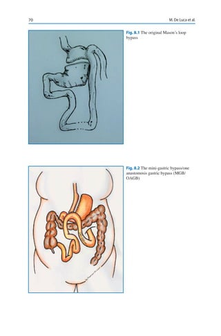
![718 Mini-Gastric Bypass/One Anastomosis Gastric Bypass
definitely not the old loop gastric bypass proposed by Mason in 1967 [1]. The
differences between the two techniques are well depicted in Figs. 8.1 and 8.2.
Furthermore, high rates of bile reflux, often expected by opponents, have been
rarely reported by MGB/OAGB performers and does not exceed 2% of all
operated patients [4, 7]. Conversely, an interesting systematic review showed the
risk of esophagogastric cancer to be extremely low following bariatric surgery
and to be unknown following MGB/OAGB [8].
MGB/OAGB appears extremely effective in reducing obesity-related
comorbidities, offering a good quality of life with a very acceptable complication
rate [9]. Although not officially recognized in the USA as a bariatric procedure,
the trend in the use of MGB/OAGB in Europe and Asia Pacific is rapidly growing
[10], placing it as the most frequently performed procedure following sleeve
gastrectomy (SG), Roux-en-Y gastric bypass (RYGB), and adjustable gastric
banding (AGB).
8.2 Technique
MGB/OAGB is routinely performed with a standard five-port laparoscopic
technique. Patients are placed in the reverse Trendelenburg position with legs
spread. At the beginning of the operation, the surgeon stands between the
patient’s legs in order to prepare the phrenogastric ligament then, in some cases,
moves to the right side of the patient. The monitor is at the head of the operating
table and in some cases on the left side of the patient. The operation consists of
two steps: a long-sleeved gastric tube along the lesser curvature and a Billroth
type II loop gastrojejunostomy with a 180- to 250-cm afferent limb.
8.2.1 Long and Narrow Gastric Pouch
The first trocar – for the camera – is placed in the midpoint between the xiphoid and
the umbilicus. The second trocar is placed in the right hypochondrium, the third
is inserted in the left hypochondrium, symmetrical to the previous one, the fourth
just under the xiphoid process, and the fifth in the right quadrant at mid-clavicular
line on the same level of the camera [11]. A long gastric tube is the cornerstone
of this procedure, placing the single anastomosis of a loop bypass away from the
esophagocardial junction and thereby avoiding the problem of bile esophagitis that
occurs with the original Mason’s loop gastric bypass [1]. The gastric tube should
be constructed as long and narrow as possible and is usually created by applying
one horizontal 45-mm roticulator linear stapler at the angle of the lesser curvature,
just above the left branch of the crow’s foot. Multiple vertical 60-mm roticulator
linear staple cartridges are placed upward to the angle of His and calibrated along
a 32- to 36-Fr bougie, similar to the vertical part of a SG [5]. A 28- to 36-Fr bougie](https://image.slidesharecdn.com/bariatricandmetabolicsurgery-180301214153/85/Bariatric-and-metabolic-surgery-83-320.jpg)
![72 M. De Luca et al.
is strongly recommended for controlling the width of the tube. A narrow tube may
help avoid weight regain and may also reduce acid production and so decrease the
risk of marginal ulcer [4]. Although some data indicate that a running absorbable
seromuscular-seromuscular invagination suture protects against leakage [3], no
reinforcement was routinely done on the staple line.
8.2.2 Gastrojejunostomy
At this point, the surgeon begins the second part of the procedure, moving the
omentum upward. Sectioning of the greater omentum into a bivalve is rarely
needed. The jejunum is then identified at the ligament of Treitz and measured
with a graded grasper to 180–250 cm from the Treitz ligament according to
the patient’s preoperative body mass index (BMI), previous operations, and
alimentary behavior. The proximal limb should be always placed on the patient’s
left side and distal limb on the right to avoid torsion of the intestinal mesentery
[4]. The original Rutledge technique is an end-to-side anastomosis, but most
surgeons prefer a side-to-side technique, with the afferent limb higher than the
efferent loop so as to form an isoperistalsis conduit [2, 4, 5]. Some surgeons
have proposed placing an anchoring suture for the afferent limb on the staple
line of the gastric pouch, with 6 to 10 sutures acting as a valve to inhibit bile
reflux [3, 7]. An antecolic terminolateral gastrojejunostomy is performed using
a posterior 45-mm Roticulator linear stapler and an anterior running suture or a
continuous manual suture with an adsorbable suture. The anastomosis is created
with a size of 1.5–3 cm, which is wider than for the RYGB because the restriction
is provided by the narrow-sleeved tube rather than the small anastomosis used in
RYGB [4]. Anastomosis is checked by intraoperative methylene blue test. Some
authors introduce a nasogastric tube into the efferent loop, and some place a
drain close to the anastomosis until the second postoperative day.
8.3 Results
8.3.1 Weight Loss and Weight-Related Disease
Rutledge reported a percentage of excess weight loss (%EWL) of 77% at 24
months in a series of 1274 cases [2]. Carbajo et al. reported a %EWL of 80%
at 24 months considering a series of 209 patients [3]. Noun et al. considered
1000 patients who underwent OAGB with a mean of 5-year follow-up; %EWL
was 72.5%. The 50%EWL was achieved for 95% of patients at 18 months and
for 89.8% at 60 months. Operative time was 89 min, and length of hospital stay
was 1.85 days for primary surgery and 2.35 for revision surgery [11]. Bruzzi
et al. reported a 5-year postoperative percentage of excess body mass index](https://image.slidesharecdn.com/bariatricandmetabolicsurgery-180301214153/85/Bariatric-and-metabolic-surgery-84-320.jpg)
![738 Mini-Gastric Bypass/One Anastomosis Gastric Bypass
loss (%EBMIL) of 71.5 ± 26.5% in 175 patients who underwent to OAGB.
Postoperative gastrointestinal quality of life index (GIQLI) score of the treatment
group was significantly higher than the preoperative score of the control group
(110.3 ± 17.4 vs. 92.5 ± 15.9; p<0.001) [9]. Musella and the Italian Multicenter
Study Group reported a %EWL of 77% considering 974 OAGB patients and a
5-year follow-up rate of 83.9% [7].
Lee et al. compared weight loss following RYGB patients with 40.5%
preoperative BMI and OAGB patients with 41.1% preoperative BMI. At the fifth
postoperative year, the %EWL was 58.7% for the RYGB group and 64.9% for
the OAGB group. The residual excess weight <50% at 2 years was achieved in
75% of the RYGB group and 95% of the OAGB group. Operative morbidity was
higher in RYGB group (20% vs. 7.5%). The authors evaluated hospital stay, which
was longer in RYGB group (6.9 vs. 5.5 day) and cumulative dose of analgesic
medication, which was larger in the RYGB group (3.4 vs. 2.0 doses). A significant
improvement in obesity-related clinical parameters and complete resolution of
metabolic syndrome in both groups were noted. GIQLI increased significantly
without any significant difference between groups [12]. The same authors then
evaluated two groups of patients – RYGB and OAGB – after 10 years of experience
and with a follow-up of 1–10 years. Major complications were higher for the
RYGB group (3.2% vs. 1.8%). At 5 years postoperatively, RYGB and OAGB
patients had a BMI of 27.7 and 29.2, kg/m2
respectively; %EWL was 60.1% vs.
72.9%. There were no significant differences in GIQLI score at 5 years between
groups [13]. Quan et al. conducted a comprehensive literature search by retrieving
the databases of PubMed, EMBASE, and the Cochrane Library and identified 16
studies for systematic review and 15 articles for meta-analysis. They compared
OAGB with laparoscopic adjustable gastric banding (LAGB), laparoscopic SG
(LSG), and laparoscopic RYGB (LRYGB). OAGB showed significantly higher
weight loss compared to LAGB, LSG, and LRYGB [14].
8.3.2 Type 2 Diabetes Mellitus and Comorbidities
Lee et al., in a retrospective study of 443 patients with a BMI >35 kg/m2
and
12-month follow-up (100% of patients) evaluated fasting plasma glucose (FPG)
and glycated hemoglobin (HbA1c). FPG reached normal range in 89.5% of
patients and HbA1c lowered to <7% in 76.5% of patients; 90% of type 2 diabetes
mellitus (T2DM) patients came off their medication, and there was a significant
improvement in lipid levels [15]. In a retrospective study of 62 patients with a
very low BMI (23–35 kg/m2
) and 24-month follow-up (100% of patients), the
same authors recorded that BMI ranged from 30.1 to 23 kg/m2
, FPG from 195 to
106 mg/dL, and HbA1c from 9.7% to 5.9%; 55% of patients were off their T2DM
medication at the final follow-up [16]. In the meta-analysis and systematic review
by Quan et al., OAGB showed comparable or better results regarding T2DM
remission versus LAGB, LSG, and LRYGB [14].The Italian Multicenter Study](https://image.slidesharecdn.com/bariatricandmetabolicsurgery-180301214153/85/Bariatric-and-metabolic-surgery-85-320.jpg)
![74 M. De Luca et al.
Group reported T2DM remission of 84.4% and hypertension resolution of 87.5%
in 974 OAGB patients at 60-month follow-up [7]. Finally, a recent multicenter
European study [17] has shown the efficacy of both MGB/OAGB and LSG in
determining T2DM remission at 12 months. This paper suggested that T2DM
remission is independent from weight loss, with MGB/OAGB outperforming
LSG on univariate analysis.
8.4 Complications (According to Clavien-Dindo Classification)
8.4.1 Early complications
8.4.1.1 Gastrointestinal bleeding (Grades IIIa, IIIb)
Gastrointestinal anastomosis bleeding should be suspected in the presence
of hemodynamic instability and decrease of hemoglobin levels if no signs of
abdominal bleedings are present. Most cases are treated by endoscopy with
adrenaline injection, clips, and stitches at the bleeding source. Chevallier et
al. reported only one case of gastrointestinal bleeding from the anastomosis in
>1000 patients treated endoscopically [5].
8.4.1.2 Leak (Grade IIIb)
Gastrointestinal leak is probably the most fearsome complication in MGB/
OAGB. Due to the presence of a high flux of bile coming from the afferent
loop, anastomosis leak often causes a clinical onset of choleperitoneum that
requires immediate surgical revision. Surgical strategy depends on the extent
of the defect. In the presence of a large defect, conversion to RYGB could be
the best choice. If the defect is small, primary closure can give good results. In
addition to gastrointestinal anastomosis, leakage coming from the gastric pouch
and gastric remnant have been described [7], with an incidence ranging from 0.6
to 1.8% [18].
8.4.1.3 Abdominal Bleeding (Grades II–IIIb)
The most frequent cause of abdominal bleeding is bleeding from trocar insertion
sites. Frequency ranges from 0.1 to 2.5% [5]. Bleeding following spleen injury
is very rare.
8.4.1.4 Peritonitis (Grade IIIb)
Peritonitis is caused by small-bowel mobilization using the grasper during the
laparoscopic procedure and always requires surgical revision. The strategy is
related to the distance from the gastrointestinal anastomosis. Conversion to
RYGB is indicated if the perforation is close to the gastrointestinal junction [5].
Primary closure and drainage is recommended in all the other cases.](https://image.slidesharecdn.com/bariatricandmetabolicsurgery-180301214153/85/Bariatric-and-metabolic-surgery-86-320.jpg)
![758 Mini-Gastric Bypass/One Anastomosis Gastric Bypass
8.4.2 Late Complications
8.4.2.1 Bile Reflux (Grades II–IIIb)
Bile reflux is the most notoriously controversial disadvantage of MGB/OAGB.
Prejudice arose from Mason’s loop gastric bypass, after which bilious vomiting
and subsequent gastritis and esophagitis were reported in 70% of patients. This
may occur in the old Mason’s gastric bypass with a small, high gastric pouch
and alkaline reflux esophagitis due to a loop adjacent to the esophagus [1]
(Fig. 8.1). However, there is a great difference between the Mason’s procedure
and MGB, with anastomosis in the latter being made on a long, narrow
gastric pouch far from the esophagus. Chevallier et al. evaluated bile reflux
by endoscopic biopsies in MGB/OAGB patients. The authors registered, as
a sign of bile reflux, foveolar dysplasia only in 17.1% of patients at 2 years
and 4.6% at 4 years, with no dysplasia or metaplasia. Bile reflux, if present, is
not symptomatic in all patients [5]. Symptoms (heartburn, dyspepsia, bilious
vomiting) can be successfully treated pharmacologically in most cases. Bile
reflux rarely needs surgical revision. If revision is needed, conversion to RYGB
is the treatment of choice.
8.4.2.2 Anastomotic Ulcer (Grades II–IIIb)
Anastomotic ulcers are often caused by the small gastric pouch continuing to
secrete acids. Heavy cigarette smoking and and stopping proton pump inhibitor
(PPI) therapy might play a role. A larger gastric pouch with more acid secretions
could be related to the incidence of ulcers. Treatment strategy is related to
clinical onset, and continuing PPI therapy and quitting smoking in cases with
clinical and endoscopic findings are recommended. Although rare, an untreated
anastomotic ulcer can perforate, causing clinic onset of peritonitis. In these case,
conservative treatment with a T tube [5] or a new gastrojejunal anastomosis and
RYGB conversion are treatments of choice [14].
8.4.2.3 Iron Deficiency (Grade II)
MGB often causes microcytic anemia. This because in MGB/OAGB, a longer
bypass is created in the foregut limb compared with in RYGB, which more
severely inhibits iron absorption. In all cases, iron therapy i.v. is the treatment
of choice.
8.4.2.4 Protein Malnutrition (Grades II–IIIb)
A well-recognized late complication is protein malnutrition. Clinical signs are
albuminemia <30 g/L, %EBMIL >100%, and BMI <20 kg/m2
. The first treatment
is conservative. If ineffective, surgical MGB/OAGB reversal is the treatment of
choice [5]. Lee et al. [18] suggests conversion of MGB/OAGB to SG, with good
effect on malnutrition and no weight regain.](https://image.slidesharecdn.com/bariatricandmetabolicsurgery-180301214153/85/Bariatric-and-metabolic-surgery-87-320.jpg)
![76 M. De Luca et al.
8.4.2.5 Internal Hernia (Grade IIIb)
A worrisome complication – internal hernia – often presents with insidious
and non-specific symptoms that may be difficult to assess clinically. Single
anastomosis of MGB/OAGB reduces potential sites of internal hernias. Although
the incidence of this complication after MGB/OAGB is very rare, some experts
suggest closing the Petersen space as well.
8.4.2.6 Weight Regain (Grade IIIb)
If weight regain is associated with gastric pouch dilatation, revision surgery by
pouch trimming on a calibration tube is indicated. Other techniques are RYGB
conversion with a 150-cm alimentary limb or efferent loop shortening to increase
the malabsorptive component of MGB. Incidence ranges from 0.8 to 5% [18].
8.4.2.7 Cancer (Grade IIIb)
In the mid-1980s, many warnings against the risk cancer following Billroth II
reconstruction were published; many authors concluded that bile reflux was
related to a higher rate of gastric-stump cancer in patients who underwent
Billroth II for benign disease compared with patients who underwent to
Billroth I reconstruction. Although not all authors noted this difference, it
must be considered that at that time of those early reports, the potential role of
Helicobacter pylori was not yet understood. Of 827 gastric-stump cancers in
patient treated with Billroth I or II for benign disease, Tersmette et al. showed
that the difference in cancer rate was not significant between the two procedures
[19], and similar concepts were reprised by Bassily et al. [20]. In a recent meta-
analysis, Scozzari et al. [8] reported 33 cases of esophageal and gastric cancers
after bariatric procedures. Although four (12.1%) cancers appeared after MGB/
OAGB, three were detected in the excluded stomach and only one in the gastric
pouch, which was following a 1980 surgery that certainly was not a MGB (which
was first described in 2001 [2]). On the other hand, the same paper showed 15
cases of esophageal and gastric cancers (45.4%) following restrictive bariatric
procedures (LAGB, VBG, SG) and 14 cases (42.4%) after RYGB.
8.5 Conclusions
The MGB/OAGB appears extremely effective in reducing obesity-related comor-
bidities, offering a good quality of life with a very acceptable complication rate.
References
1. Mason EE, Ito C (1967) Gastric bypass in obesity. Surg Clin North Am 47:1345–1351
2. Rutledge R (2001) The mini-gastric bypass: experience with the first 1,274 cases. Obes Surg
11:276–280](https://image.slidesharecdn.com/bariatricandmetabolicsurgery-180301214153/85/Bariatric-and-metabolic-surgery-88-320.jpg)
![778 Mini-Gastric Bypass/One Anastomosis Gastric Bypass
3. Carbajo MA, Garcia-Caballero M, Toledano M et al (2005) One anastomosis gastric bypass
by laparoscopy: results of the first 209 patients. Obes Surg 15:398–404
4. Lee WJ, Lin YH (2014) Single-anastomosis gastric bypass (SAGB): appraisal of clinical
evidence. Obes Surg 24:1749–1756
5. Chevallier JM, Arman GA, Guenzi M et al (2015) One thousand single anastomosis (omega
loop) gastric bypasses to treat morbid obesity in a 7-year period: outcomes show few
complications and good efficacy. Obes Surg 25:951–958
6. Musella M, Milone M (2014) Still “controversies” about the mini gastric bypass? Obes Surg
24:643–644
7. Musella M, Susa A, Greco F et al (2014) The laparoscopic mini-gastric bypass: the Italian
experience: outcomes from 974 consecutive cases in a multicenter review. Surg Endosc
28:156–163
8. Scozzari G, Trapani R, Toppino M, Morino M (2013) Esophagogastric cancer after bariatric
surgery: systematic review of the literature. Surg Obes Relat Dis 9:133–142
9. Bruzzi M, Rau C, Voron T et al (2015) Single anastomosis or mini-gastric bypass: long-term
results and quality of life after a 5-year follow-up. Surg Obes Relat Dis 11:321–326
10. Angrisani L, Santonicola A, Iovino P et al (2013) Bariatric surgery worldwide. Obes Surg
25:1822–1832
11. Noun R, Skaff J, Riachi E (2012) One thousand mini-gastric bypass: short and long term
outcome. Obes Surg 22:697–703
12. Lee WJ, Wang W (2005) Laparoscopic Roux-en-Y versus mini-gastric bypass for the
treatment of morbid obesity: a prospective randomized controlled clinical trial. Ann Surg
242:20–28
13. Lee WJ, Ser KH, Lee YC (2012) Laparoscopic Roux-en-Y versus mini-gastric bypass for the
treatment of morbid obesity: a 10-year experience. Obes Surg 22:1827–1834
14. Quan Y, Huang A, Ye M (2015) Efficacy of laparoscopic mini gastric bypass for obesity and
type 2 diabetes mellitus: a systematic review and meta-analysis. Gastroenterol Res Pract
[Epub ahead of print] doi:10/1155/2015/152852
15. Lee WJ, Wang W, LeeYC et al (2008) Effect of laparoscopic mini-gastric bypass for type 2
diabetes mellitus: comparison of BMI >35 kg/m2
. J Gastrointest Surg 12:945–952
16. Lee Wj, Chong K, Chen CY et al (2011) Diabetes remission and insulin secretion after
gastric bypass in patients with body mass index <35 kg/m2
. Obes Surg 21:889–895
17. Musella M, Apers J, Rheinwalt K et al (2015) Efficacy of bariatric surgery in type 2 diabetes
mellitus remission: the role of mini gastric bypass/one anastomosis gastric bypass and sleeve
gastrectomy at 1 year of follow-up. A European survey. Obes Surg 26:933–940
18. Lee WJ, Wang W, Lee Y (2011) Revisional surgery for laparoscopic mini-gastric bypass.
Surg Obes Rel Dis 7:486–492
19. Tersmette AC, Offerhaus GJ, Tersmette KW (1990) Meta-analysis of the risk of gastric
stump cancer: detection of high risk patient subsets for stomach cancer after remote partial
gastrectomy for benign conditions. Cancer Res 50:6486–6489
20. Bassily R, Smallwood RA, Crotty B (2000) Risk of gastric cancer is not increased after
partial gastrectomy. J Gastroenterol Hepatol 15:762–765](https://image.slidesharecdn.com/bariatricandmetabolicsurgery-180301214153/85/Bariatric-and-metabolic-surgery-89-320.jpg)
![80 N. Scopinaro et al.
result can be obtained by varying the above elements – from the very dangerous
distal gastric bypass [1], with small stomach and short CL, to the modern, safe,
and effective laparoscopic Roux-en-Y gastric bypass (LRYGB) [2], in which,
due to the very long CL, no energy malabsorption occurs. Between these two
extremes, hundreds of different BPD with different gastrectomies and different
limb lengths exist, including: BPD with preservation of the distal stomach [3] or
the entire stomach [4] with pylorus preservation; BPD with sleeve gastrectomy
and duodenal switch (BPD-DS) [5]; BPD with vertical instead of horizontal
section and pylorus preservation; and the so-called “mini-gastric bypass” [6]
– the correct definition of which is “one anastomosis gastric bypass” (OAGB)
[7] – which is actually a BPD in which the AL and the BPL coincide and the CL
goes from the GEA to the ICV. This is obviously the only small-bowel segment
that should be measured, as in the “single anastomosis duodenoileal bypass with
sleeve gastrectomy” (SADI-S) [8].
So, what do we mean by “standard BPD”? Some consider it an alternative
to BPD-DS, which simply represents the American version of BPD. It conforms
to all the rules for BPD, and its results and complications depend on the gastric
volume and intestinal limb lengths, exactly as with all other BPD versions, so
there is no reason to consider it separately from the latter. Others use the term
“standard” to indicate the BPD as performed by the originator. However, the BPD
was often modified by its originator in relation to gastric volume and limb length
from the time of its conception until a few years ago, with each change entailing
different results and complications. Therefore, none of the different models of
BPD used during its 30-year evolution could be considered “standard”.
The most reasonable solution would be to attribute the term “standard” to
the BPD version currently used by the originator, a version that, after 30 years
of clinical experimentation accompanied by all pertinent studies has evolved to
represent the best compromise between safety and effectiveness. The current
model has been used since 2006 (see below), so that despite the 40-year history
and evolution of BPD, the “standard BPD” has barely had 10 years of use; its
paired results and complications cannot be reasonably given at more than 5 years.
For very long-term weight loss results, we usually refer to a group of 40 patients
with a minimum follow-up of >30 years and whose BPD model was the so-called
“half-half BPD” (HH-BPD), in which the small bowel was cut at its midpoint
and the CL was 50-cm in length. Therefore, the operation was rather similar to
the one currently used. Weight loss yielded by the HH-BPD was ~70% of the
excess, which is not an extraordinarily good result if we consider that the mean
initial excess weight of those 40 patients was only 83%. Today, with the current
BPD model, we obtain the same percentage of excess weight loss (EW%L) in a
population with a mean excess weight >120%. The exciting result was that this
70% of loss was strictly maintained for >30 years, as it was, even over a shorter
follow-up period, with all subsequent models of BPD.
Before examining results obtained with the current model of BPD, we believe
it is extremely helpful to describe the origin of the operation, its rationale, its](https://image.slidesharecdn.com/bariatricandmetabolicsurgery-180301214153/85/Bariatric-and-metabolic-surgery-91-320.jpg)
![819 Standard Biliopancreatic Diversion
physiology, and its evolution over 30 years of unceasing attempts to ameliorate
weight loss and reduce surgical complications, until achieving which, as noted
above, is the best compromise between effectiveness and safety.
9.2 Origin, Rationale, and Basic Physiology of BPD
Let us start with when, why, and how BPD was developed. In 1972, the first author
was 27 years old, and, having begun early as a medical student, he already had an
8-year experience with scientific work, publishing about 20 articles. However, he
was looking to pursue a speciality that was really new, at least in Italy.
Everything began when his father, who was Professor of Medicine at the
University of Genoa, showed him the 1967 article by Edward Mason on gastric
bypass for surgical treatment of obesity [9]. Nobody in Italy had done it before. A
few months later, the first author had read everything he could find on that subject,
especially on the physiology of intestinal absorption and human nutrition. This
research obviously began with the historical article by Kremen et al. [10], in
which the first jejunoileal bypass (JIB) was mentioned.
Using a surgical procedure to treat obesity was a very new concept, but if
it had been only gastric restriction we would have never made the decision of
getting involved with that not even yet born discipline. From both a clinical and
scientific point of view, it appeared highly unlikely that it could be the definitive
solution to the problem. Much more fascinating and promising was the reduction
of intestinal absorption achieved using JIB. We were absolutely convinced that
it was the only possible way to obtain complete and definitive solution of the
problem, but certainly not with the existing operation, the JIB, which deserved
the horrific definition of malabsorption (it could not work that way, it was clear
enough without even the need to try it). However, we succeeded in convincing
our Professor and Chairman, and in 1973 we performed five JIB according to
the Buchwald and Varco technique [11], with poor results. In the mean time we
started our experimental study on dogs, which we presented in 1974 [12].
In our first publications [13, 14], we reported the three main defects of
the JIB. Finding solutions to these defects was the rationale behind our new
operative technique. The presence of a long, excluded loop was an issue, but the
unavoidable functional problems of JIB were the indiscriminate malabsorption on
one hand and the quick recovery of intestinal energy-absorption capacity on the
other. Separating the absorption of beneficial nutrients and that of nonbeneficial
energy would allow the resolution of both problems.
On one hand, altering digestion rather than absorption creates a malabsorption
that is essentially selective for fat and starch, thus preserving absorption of
essential nutrients. On the other hand, selective malabsorption of energy can
neutralize the intestinal adaptive phenomena, which cause rapid recovery of the
energy-absorption capacity after JIB [15].](https://image.slidesharecdn.com/bariatricandmetabolicsurgery-180301214153/85/Bariatric-and-metabolic-surgery-92-320.jpg)
![82 N. Scopinaro et al.
We know that if the small bowel in-continuity between the duodenum and
ileocecal valve is >60 cm, no substantial weight loss will occur; on the contrary,
if it is <40 cm, it is incompatible with life. Realistically, any length between
40 and 60 cm is destined to fail. The why can be found in articles by Dowling
[16, 17], who explored in depth the mechanisms and regulation of intestinal
adaptation. Intestinal adaptation necessitates both general enterohormonal
stimulation, represented by enteroglucagon and neurotensin, and intraluminal
stimulus, represented by food and/or biliopancreatic secretion. Both these signals
are needed, none of them would work alone. After JIB, the general stimulus
remains intact, but the excluded bowel receives no intraluminal signal and
becomes hypertrophic, while the small bowel in-continuity receives both food
and biliopancreatic juice. It thus undergoes a huge adaptation that results in a
more than tenfold increase in energy-absorption capacity, thus totally nullifying
the initial malabsorption, with consequent weight regain. We carefully studied all
aspects of anatomofunctional adaptive changes following JIB [18, 19], showing
very rapid recovery of both fat and starch absorption. Finally, the 1980 article by
Halverson et al. [20] definitively killed the JIB, demonstrating that, mainly due
to weight regain, only 18% of results could be considered good. Weight regain,
not complications, was the main cause of JIB abandonment.
Conversely, after BPD, the entire small bowel receives the intraluminal
stimulation from food and/or biliopancreatic secretion, so the entire small bowel
goes into hypertrophy [21], although not as greatly as in JIB. However, because
it involves the whole small bowel, it accounts for an increase in visceral mass by
~1 kg. The corresponding increase in resting energy expenditure of almost 400
kcal/day plays a very important role in the weight loss that follows BPD [22].
The most important consequence of the separation of energy-rich food
absorption and essential nutrient absorption is the possibility of neutralizing
the effect of intestinal adaptation. Actually, since the absorption of protein and
other essential nutrients occurs in the AL, the CL can be created at any length,
including the length that will result exactly in the desired fat absorption capacity
after intestinal adaptation. Consequently, after intestinal adaptation, intestinal
energy absorption remains constant indefinitely, which explains the durable
weight maintenance that follows BPD.
Two years of experimental work on 12 dogs [14, 23] included all the important
studies: complete blood and urine biochemistry were done preoperatively and
weekly during the first month. Fat absorption as a percentage of intake (modified
Kramer’s method), protein absorption as a percentage of intake (125
I-albumin fecal
excretion), and intestinal absorption of bile acid [1-(14
C)-glycine-glycocholate
fecal excretion] were done preoperatively and at 1, 6, and 12 months. Gallbladder
bile composition (bile acid, cholesterol, phospholipid concentration) and wedge
liver biopsy were recorded at operation and at sacrifice. Liver function tests were
done preoperatively and at 1, 3, 6, and 12 months.
The first human BPD was done on 12 May 1976.Although the patient lost only 40%
of excess weight, that loss was consistently maintained for more than 30 years [24].](https://image.slidesharecdn.com/bariatricandmetabolicsurgery-180301214153/85/Bariatric-and-metabolic-surgery-93-320.jpg)
![839 Standard Biliopancreatic Diversion
9.3 Development of BPD
Appropriate intestinal limb length and stomach volume evolved with the pro-
gressive information derived from the continuous study of the BPD’s physiology
and results [25, 26].
The original philosophy for limiting digestion in BPD was to delay the meThe
original philosophy for limiting digestion in BPD was to delay the meeting
between food and biliopancreatic juice in order to confine pancreatic digestion to
a short segment of small bowel. In the first experimental model of BPD (1976),
BPL length was only 30 cm and CL was 100 cm, so that the AL was ~6-m long.
Protein absorption (see also section 9.5.1) was undisturbed (97.5 ± 5.6%) in
these first five patients, but weight loss was unsatisfactory (~40%). We believed
that was partly due to the CL being too long and partly to the digestive activity
of brush-border enzymes in the AL. In a subsequent model, again five cases, CL
length was reduced to 75 cm, while 1–2 m of small bowel were added to BPL
length, thus shortening the AL (biliopancreatojejunal diversion type I, or BPJD
I). Protein absorption was still nearly complete (94.9 ± 1.8%), and although
weight loss was greater, it was not satisfactory (~50%). In the five patients in
the third model (BPJD II), we moved the first half of the small bowel in the BPL
without varying CL length. Weight loss further increased (~60%) and protein
absorption decreased (76.2 ± 11.5; p<0.05 vs. BPD and BPJD I), demonstrating
the importance of intestinal enzyme digestion for both starch and protein. At that
point, BPL length was sufficient to allow complete digestion-absorption of the
scarce (as non meal-stimulated) pancreatic enzymes so that pancreatic digestion
no longer occurred in the CL (unpublished experimental data). Consequently,
starch and protein absorption depended on the length of small bowel between the
GEA and the ICV, while fat absorption, which needs the presence of bile salts,
occurred only in the CL. This was proven by the subsequent model, the so-called
BPJD III, or half-half (HH-BPD), in which the length of small bowel between the
GEA and the ICV was unchanged but CL length was shortened to 50 cm. In the
12 patients in that model, protein absorption did not change (77.2 ± 7.3; p<0.01
vs. BPD and BPJD I), while weight loss showed a further increase (~70%),
evidently due to the reduced fat absorption. With the subsequent model – the
so-called “short-loop BPD”, in which the AL was reduced to 2 m – we achieved
a further increase in weight loss (~80%) and decrease in protein absorption
(~70%), which in some cases caused protein malnutrition (PM). These intestinal
lengths were used for a long time (1980–1992), this being our model at the time
of our very important study on intestinal energy, fat, and nitrogen absorption
[27]. That study confirmed what we had already hypothesized from the very
strict weight maintenance that follows BPD, that is, that the BPD digestive-
absorptive apparatus has a maximum energy (fat and starch) transport capacity,
which corresponds to about 1250 kcal/day, and consequently, all energy intake
exceeding the maximum transport threshold is not absorbed. Therefore, since
daily energy intake is largely higher than the aforementioned threshold, daily](https://image.slidesharecdn.com/bariatricandmetabolicsurgery-180301214153/85/Bariatric-and-metabolic-surgery-94-320.jpg)
![84 N. Scopinaro et al.
energy absorption remains constant for each patient regardless of energy intake.
The same absorption study also revealed the existence of a fivefold increase in
endogenous nitrogen loss, which doubles daily protein requirement and is the
main cause of PM.
All changes in gastric remnant volume were done in the BPD model with a
CL length of 50 cm and AL length 200 cm [22]. In the BPD model currently used,
few if any pancreatic enzymes reach the CL, so no pancreatic digestion occurs
there. All digestive capacity is in the intestinal brush-border enzymes, so protein
and starches are digested and absorbed in the entire intestinal segment between
the GEA and the ICV. Due to the necessity of bile salt, only fat absorption is
limited to the CL. Therefore, a longer AL means increased protein absorption;
however, the concomitant increase in starch absorption reduces weight loss and
results in higher stabilization weight.
The other important item in BPD is stomach volume. The smaller its volume,
the more rapidly it empties. Along with appetite reduction due to increased
production of anorexigenic ileal hormones glucagon-like peptide (GLP-1) and
peptide tyrosine-tyrosine (PYY) [28, 29], the rapid gastric emptying that occurs
during the first postoperative months causes a more intense postcibal effect,
with excessive reduction of food intake and increased risk of early PM. In the
later postoperative period, when postcibal syndrome has subsided, it causes
quicker intestinal transit and thus reduced energy and protein absorption,
resulting in greater weight loss, lower stabilization weight, and an increased
risk of recurrent PM. The problem was then to find the best combination of
stomach volume, the reduction of which can cause excellent weight loss but
a high risk of PM, and AL length, the increase of which makes the operation
safer but less effective.
In the early phase of BPD evolution, the impressive weight loss obtained
by reducing gastric remnant volume was attractive. However, due to the above-
mentioned aspects of BPD physiology in the so-called “very little stomach”
model (219 patients; 1982–1983), in which a mean of 150-mL gastric volume was
left, a spectacular reduction of 90% of the initial weight resulted in a catastrophic
incidence of 30% PM, with 9% being recurrent and was often accompanied by
excessive weight loss. The importance of gastric volume was evidenced by the
fact that the mean gastric volume of malnourished patients was significantly
smaller than that of non-malnourished patients.
At that point, rather than grossly and blindly increasing gastric volume and
AL length concomitantly – which would eliminate the problems – we chose to
determine the best compromise between effectiveness and safety. In 1984, we
began using the “ad hoc stomach-BPD” (AHS-BPD), in which gastric volume is
adapted to the individual patient’s characteristics [22]. Mean weight loss curve
with the AHS-BPD at a minimum follow-up of 5 years clearly showed that the
initial percentage of excess weight loss (EW%L) of 80% was reduced to the
current 70% while eliminating PM.](https://image.slidesharecdn.com/bariatricandmetabolicsurgery-180301214153/85/Bariatric-and-metabolic-surgery-95-320.jpg)
![859 Standard Biliopancreatic Diversion
The initial aim of AHS-BPD was to confine the risk of PM to patients who
required greater weight loss by adapting gastric volume to the initial excess
weight only. The mean gastric volume was increased to ~300 mL, with an overall
PM incidence of 15% and a still very good %EWL of 79% (first 192 AHS-BPD
patients). Obviously, patients with gastric volume <300 mLhad still a significantly
greater incidence of PM. In 1987, effectiveness was reduced in favor of safety
by adapting the gastric volume to other patient characteristics, namely, age, sex,
eating habits, and expected degree of compliance. The new rationale was to select
only patients with the most likely individual characteristics to take advantage of
the risk/benefit of a smaller stomach. Results, in the subsequent 859 AHS-BPD
patients, were an 11% overall incidence of PM, with 4.5 recurrence rate and
loss of initial excess weight reduced to 74%. At that point, the disappearance of
any significant difference in gastric volume between PM and non-PM patients
demonstrated that gastric volume had no more influence on PM incidence.
Therefore, in order to further minimize the PM complication, in 1992 we began
adapting also the AL length to patients’ individual characteristics, with social-
behavioral characteristics involved with the risk of PM being protein content of
customary food, capacity to modify eating habits according to individual needs,
and financial status. Initially (230 patients), AL length was increased to 300 cm in
patients at risk. After poor weight loss results in the first 100 patients with a 300-
cm AL length (68% loss of initial excess weight), we decreased the maximum AL
length to 250 cm. The resulting ad hoc stomach/ad hoc AL BPD (AHS-AHAL-
BPD) in the first 300 patients with a minimum follow-up of 10 years resulted in
the almost negligible incidence of 1%recurrent PM, reoperations for late specific
complications being required in only 1% of patients while maintaining very good
permanent reduction of 71% in initial excess weight. The fact that this policy
yields the best compromise between effectiveness and safety was confirmed by
a BAROS (Bariatric Analysis Reporting Outcome System) evaluation of more
than 800 patients [30], which showed that among patients operated on after 1992
there was only a 2% failure rate, with more than 90% of results being from good
to excellent.
In 2006, a sufficient number of patients had a minimum follow-up of 10
years to compare results and complications obtained with the three different AL
lengths. Since the incidence of PM was negligible in both the 300- and the 250-
cm groups and long-term weight loss in the 250-cm group was not significantly
different from that of the classic 200-cm length, the 250-cm AL length was used
in all subsequent patients, who thus far have maintained very good weight loss
with a total absence of PM.
In conclusion, a good knowledge of BPD physiology, together with a very
long, careful clinical study, allowed the adaptation of both gastric volume and
AL length to the individual patient’s characteristics. The resulting revision rate
for late specific complications of <1% made BPD during the last 15 years – in
our hands and in well-selected patients – not only the most effective but also the
safest operation yet available for obesity treatment.](https://image.slidesharecdn.com/bariatricandmetabolicsurgery-180301214153/85/Bariatric-and-metabolic-surgery-96-320.jpg)
![86 N. Scopinaro et al.
9.4 Results
Weight-loss results have been exhaustively reported above in detail while
referring to each subsequent BPD developmental model. The current BPD
model, with a mean gastric volume of 400 mL and an AL and CL measuring 250
and 50 cm, respectively, yields a mean reduction of initial excess weight of 70%,
strictly maintained up to the fifth postoperative year in the 366 severely obese
patients operated on in the years 2006–2011. Mean percent reductions of initial
excess weight obtained with each subsequent BPD model, developed during
BPD evolution, was rigorously maintained for more than 30 years, irrespective of
patient size, weight loss. Therefore, a similar trend is expected with the weight-
loss results of the present – and thus far, definitive – model.
The total number of BPD operation performed at our institution from 1976
to the end of 2015 was 3,439. This included 123 patients with BMI <35 kg/m2
operated on between May 2007 and June 2010. Indication for their surgery was
mainly or solely to treat type 2 diabetes mellitus (T2DM).
The other beneficial side effects of BPD are those consequent to weight loss,
and, thanks to the characteristics of weight reduction that follows the operation,
they are particularly good and they are permanent. Moreover, BPD possesses
some specific actions totally independent of weight loss that ensure the very-
long-term normalization of serum cholesterol [31, 32] and remission of T2DM in
the near totality of these patients [33, 34]. Excellent short-term results were also
obtained in T2DM patients with BMI 25–35 kg/m2
[35–37].
9.5 Complications
BPD has a 2–3% incidence of stomal ulcer, which responds well to medical
therapy. Risk is confined to the first 2 postoperative years [22], except in
smokers, who may have recurrent stomal ulcers with possible eventual GEA
stenosis, which can require a higher gastrectomy.
An important long-term issue with BPD is patient noncompliance with
taking lifelong supplementation, namely: Fe, Ca [38, 39], and more importantly,
liposoluble vitamins, especially A and E. Deficiency is easily diagnosed and
treated in all except vitamin E. Vitamin E deficiency can develop surreptitiously
for many years after BPD, and the presenting symptom may be severe neurological
problems similar to those that can be caused by early acute thiamine deficiency
[40]. We strongly recommend yearly test for serum vitamin levels; however, only
the most compliant patient do so. Therefore, since an active lifelong follow-up
is substantially impossible with most patients, we drastically reduced the use of
BPD in our patient population to only ~20%.
Protein-energy malnutrition (PEM) is a serious complication of BPD
that deserves an accurate description regarding pathogenesis, prevention,](https://image.slidesharecdn.com/bariatricandmetabolicsurgery-180301214153/85/Bariatric-and-metabolic-surgery-97-320.jpg)
![879 Standard Biliopancreatic Diversion
and treatment, though, due to accurate patients’ selection, it has essentially
disappeared in our hands.
9.5.1 Protein-Energy Malnutrition after BPD
Proteinabsorptionwasstudiedatthebeginningandendofclinicalexperimentation
[41, 42] by measuring fecal radioactivity for 3 days after administration of
10 µC of 125I-albumin with a meal containing 60 g of protein. Both studies
demonstrated an alimentary protein absorption rate of ~70% of intake. Since a
30% reduction of protein absorption did not explain the occurrence of PM, we
did a more accurate study [27] by directly measuring nitrogen content of food and
stools in 15 long-term BPD patients. Comparing alimentary protein absorption,
confirmed at 73%, and nitrogen apparent absorption revealed a fivefold increase
in endogenous nitrogen loss. This finding raised the daily protein requirement
from ~40 to ~90 g/day. This increase does not represent a problem considering
that the long-term BPD patients in our study had an average protein intake of
~170 g/day.
Ofmuchgreaterimpactistheendogenousproteinlossintheearlypostoperative
period, when the forcedly reduced food intake barely allows protein intake to
compensate for the normal protein requirement plus the endogenous extra protein
loss. The latter is evidently due to the presence of food in intestinal segments
that are not normally in contact with food (ileum and colon). As food intake
gradually increases during the early postoperative months, endogenous nitrogen
loss and thus protein requirement also progressively increase until the ileal and
colonic protein absorption capacities have been completely saturated. At that
point, and only at that point, will the protein requirement remain constant. Any
further progressive increase in food intake, up to the large amount observed in
our long-term study, will gradually reduce the risk of protein nutrition problems.
This is then the critical point, as any added form of preventing increased food
intake will result in a high risk of PM.
The early postoperative months are the risky period, when the forcedly
reduced food intake causes a negative balance of both energy and nitrogen, thus
inducing PEM. Depending on the patient’s eating behavior, PEM will develop
in one of its two classic forms: marasmic or hypoalbuminemic form, the latter
being the so-called kwashiorkor [43].
If patients devote the little eating capacity that remains immediately after BPD
to mainly protein-rich food, marasmic PEM will develop, which is simply the
effective metabolic adaptation to starvation. Both nitrogen and energy deficits are
present, and the ensuing hypoinsulinemia allows lipolysis and proteolysis in the
skeletal muscle, which is our physiologic protein store. This supplies amino acids
for visceral-pool preservation and, through glyconeogenesis, hepatic synthesis of
glucose, which is necessary for brain, heart, and kidney metabolism and for fatty
acid oxidation. All this, in association with protein and energy sparing due to the](https://image.slidesharecdn.com/bariatricandmetabolicsurgery-180301214153/85/Bariatric-and-metabolic-surgery-98-320.jpg)
![88 N. Scopinaro et al.
negative energy balance, ensures both protein and energy homeostasis. In other
words, in marasmic PEM, the organism is in deficit of protein and energy but can
draw on its protein and energy stores for homeostasis maintenance. The result is
weight loss with harmonic reduction of fat and the fat-free mass in the skeletal
muscle only, which is the precise goal of the bariatric operation.
If, conversely, the patient gives preference to carbohydrates, the normal or
near-normal energy supply causes hyperinsulinemia, which inhibits both lipolysis
and skeletal muscle proteolysis. Not being able to draw on its protein stores, and
in absence of protein sparing, the organism reduces visceral protein synthesis,
with consequent hypoalbuminemia, edema, anemia, and immunodepression.
The result is a severely ill person with body weight unchanged or increased,
maintained adipose tissue size, and lean body composition pathologically altered
with decreased visceral cell mass and increased extracellular water, which
represents the most dangerous nutritional complication of BPD.
Paradoxically, a starving patient is in a better metabolic situation, because the
protein store can be drawn upon to satisfy the requirement and compensate for the
normal endogenous loss, which obviously is not increased. Furthermore, thanks
to lipolysis, the patient can use all the required energy due to neoglycogenesis
supplying glucose for the oxidation of fatty acids. This condition, in which the
BPD plays no role at all, results in a harmonic weight reduction, such that the
patient becomes emaciated, resembling prisoners in the Nazi concentration
camps, or, more modernly, children with marasmic PEM in poor African
countries. Although in the terminal phase, having lost all or almost all fat and
skeletal muscle mass, such individuals remain in a relatively good nutritional
status, because their homeostatic visceral proteins are preserved. They eventually
die only when all energy sources have been fully exhausted.
Getting back to BPD patients, between the two above-mentioned extreme
conditions, hypoalbuminemic PEM of varying severity can take place in patients
with mixed intake, depending on how much smaller their protein intake is than
their protein requirement plus endogenous protein loss, and how much the
relative excess of energy intake prevents skeletal muscle proteolysis, lipolysis,
and protein-energy sparing [22].
The pathogenesis of PM after BPD is then multifactorial [44] and depends
on certain operation-related variables (gastric volume, intestinal limb lengths,
individual capacity of intestinal absorption and adaptation, amount of
endogenous protein loss) and on some patient-related variables (customary
eating habits, ability to adapt them to the requirements, socioeconomic status).
In most cases, PM is limited to a single early episode, with patient-related factor
being preeminent. Delayed appearance of sporadic PM is decreasingly frequent
as time passes. However, the recurrent form, which is mainly due to operation-
related variables, requires surgical revision.
Preventing PM after BPD is essentially based on a good operation and a
good patient selection. Since adopting a mean gastric volume of ~400 mL, an
AL of 250 cm, and especially the rigorous patient selection according to patient](https://image.slidesharecdn.com/bariatricandmetabolicsurgery-180301214153/85/Bariatric-and-metabolic-surgery-99-320.jpg)
![93
10
L. Angrisani (Ed), Bariatric and Metabolic Surgery,
Updates in Surgery
DOI: 10.1007/ 978-88-470-3944-5_10, © Springer-Verlag Italia 2017
G. Silecchia (*)
Department of Medico-Surgical Sciences and Biotechnologies, Division of General Surgery and
Bariatric Center, Sapienza University of Rome
Latina, Italy
e-mail: gianfranco.silecchia@uniroma1.it
Duodenal Switch
Gianfranco Silecchia, Mario Rizzello, and Francesca Abbatini
10.1 Introduction
The biliopancreatic diversion (BPD) described by Scopinaro et al. in 1976 is a
malabsorptive bariatric procedure that combines a horizontal gastric resection
with the closure of the duodenal stump, a gastroileostomy, and an ileoileostomy
to create a 50-cm common channel and a 250-cm alimentary channel. The original
procedure included the routine cholecystectomy and appendectomy [1]. The
malabsorption involves mainly substrates like fat and starch. The average daily
energy intake is thus reduced to ~1750 kcal. Weight loss after BPD is excellent
and long lasting (>15 years) [2–5]. The original Scopinaro BPD is associated
with a high rate of marginal ulcers (12%), iron-deficiency anemia (40%), bone
demineralization (6%), peripheral neuropathy, protein malnutrition (30%), and
dumping syndrome. In order to reduce these complications, in 1998, Hess and
Hess, and later Marceau et al., proposed modifying the distal gastrectomy with
a vertical gastrectomy (sleeve) to preserve integrity of the vagal supply, the
antropyloric region, and elongate the common channel from 50 cm to 100 cm
[6–8]. Adopting DeMeester et al.’s criteria to treat alkaline reflux [9], those two
groups proposed the duodenoileostomy ~2 cm from the pylorus [duodenal switch
(DS)]. Lengthening the common channel to 100 cm was introduced to decrease
the number of daily bowel movements [6]. The first laparoscopic BPD-DS was
performed in 1999 and represents a current standard technique [10].
From the functional viewpoint, BPD-DS is considered a ‘hybrid’ bariatric
procedure that combines the effects of gastric restriction with those of ‘moderate’
intestinal malabsorption. The restrictive element of BPD-DS comes from the
sleeve gastrectomy (150 mL of volume). Preservation of the antropyloric region
without altering physiologic gastric emptying leads to better absorption of many](https://image.slidesharecdn.com/bariatricandmetabolicsurgery-180301214153/85/Bariatric-and-metabolic-surgery-103-320.jpg)
![94 G. Silecchia et al.
nutrients (proteins, calcium, iron, vitamin B12
) and reduces the incidence of
dumping syndrome. The malabsorptive component results from the intestinal
bypass, which is characterized by a 250-cm-long alimentary channel and
100-cm-long common channel. The malabsorptive effect results from keeping
food away from bile and pancreatic juices until it reaches the common channel,
as with the original Scopinaro BPD. This results in a reduction in caloric and food
absorption, particularly of lipids, and metabolic changes through modifications
of incretin levels.
Buchwald and Angrisani [11, 12] reported that BPD (in all its variants)
represents, to date, less than 2% of the bariatric surgeries performed worldwide.
In Italy, BPD (in all its variants) represented only 1.2% of the 11,435 bariatric
procedures performed during 2015 (0.16% for BPD-DS) [13]. In spite of
the excellent long-term results, the use of BPD-DS throughout the bariatric
community remains influenced by the technical complexity, higher complication
and mortality rates, and increased risk of protein malnutrition compared with
other bariatric procedures [14, 15]. However, significant improvements in the
perioperative management of laparoscopic BPD-DS have been reported since
the first report in 1999. Considering the high morbidity and mortality rates
(38% and 6.25%, respectively) after laparoscopic BPD-DS in super-superobese
patients [8], in 2001, Gagner [16] introduced the concept of two-step surgery,
the rationale behind which is to obtain a significant weight loss in the first 6–12
months after sleeve gastrectomy (SG) (calibrated on an orogastric bougie >48
Fr) and perform the subsequent intestinal bypass surgery with less technical
difficulties and lower incidence of complications [17]. The two-step approach
has been adopted worldwide to decrease morbidity and mortality rates after
laparoscopic BPD-DS (LBPD-DS), reserving the second step only for patients
who did not reach significant weight loss and comorbidity control at 12 months.
To date, there is no consensus as to the interval of time between the two steps,
but the majority of surgeons and the inventor adopted policy of ‘watch and
see’ to identify, according to patient requirements, the best time for the second
step. Furthermore, the mini-invasive approach has brought under discussion
the routine performance of BPD-DS consensual procedures (cholecystectomy,
appendectomy, liver biopsies) described prior to the laparoscopic era.
10.2 Laparoscopic Surgical Personal Technique
The experience of the first period has been published previously [17]. Since the
early 2010s, the two-step procedure has been offered to all superobese patients
with a body mass index (BMI) >55 kg/m2
. Initally, sleeve volume was larger
(150 mL) than the standard sleeve considered as definitive procedure. Today,
we offer a sleeve as definitive procedure to all superobese individuals seeking
treatment and the second step is planned in selected cases: insufficient weight](https://image.slidesharecdn.com/bariatricandmetabolicsurgery-180301214153/85/Bariatric-and-metabolic-surgery-104-320.jpg)
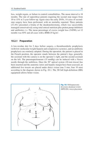
![96 G. Silecchia et al.
10.2.2 Sleeve Gastrectomy (SG)
Our personal technique is described here. Skeletonization of the greater gastric
curvature, starting at 6 cm from the pylorus and proceeding upward until the
angle of His, is performed using ultrasound dissection (Harmonic Scalpel,
Ethicon Endo-Surgery Inc, Cincinnati, OH, USA) or integration of both bipolar
and ultrasonic energies (Thunderbeat, Olympus, Japan). Complete exposure of
the left crus and the gastroesophageal junction is mandatory. Any hiatal defect,
if present, is routinely repaired. The sleeve is created using a linear stapler
(Echelon Flex Endopath, Ethicon Endo-Surgery Inc). The stapler is applied next
to a 42-Fr bougie placed next to the lesser curve. The stomach is then transected,
and a gastric pouch of 80–100 mL is created. Black (4.3 mm) and green (4.1 mm)
cartridges are used to transect the antrum, followed by two or three sequential
yellow cartridges (3.8 mm) for the gastric body and fundus. In case of revisional
SG, only black/green cartridges are used. The staple line is routinely reinforced
with Seamguard (W.L. Gore & Associates Inc, Newark, DE, USA). The last
cartridge is fired at least 1 cm lateral from the esophagogastric junction in order
to preserve vascularization of this critical area. During the vertical dissection, it
is important that the assistant carefully retract the stomach laterally to facilitate
the proper stapler line without torsion.
10.2.3 Duodenal Sinking
Using an ultrasound dissector or hook coagulator, the pylorus and the first
duodenal portion (3–5 cm) are meticulously prepared avoiding vascular or biliary
lesions. The duodenum is then divided ~2 cm from the pylorus using a linear
stapler with a blue cartridge (3.5 mm) reinforced with buttress material. It might
be necessary to complete mobilization of the upper margin of the duodenum as
far as the pylorus to facilitate the end-to-side duodenoileal anastomosis.
10.2.4 Ileoileal Anastomosis
The operating table is rotated ~15° to the left. The operator and assistant stand on
the patient’s left side. Once displayed, the caecum and the distal ileal limb, using
an atraumatic grasper marked at 10 cm, the common limb (100 cm), and the
alimentary limb (150 cm) are measured. At the latter level, we proceed with the
division of the small bowel using a 60-mm linear stapler with white cartridges
(2.5 mm). The distal part of the small intestine is anastomosed with the duodenal
stump (end-to-side duodenoileal anastomosis), while the proximal part will
make up the biliary limb to be anastomosed at the preset distance (100 cm from
the ileocecal valve). The side-to-side ileoileal anastomosis defines the common
intestinal limb. Placement of approaching stitches [polydioxanone (PDS) 2-0] on](https://image.slidesharecdn.com/bariatricandmetabolicsurgery-180301214153/85/Bariatric-and-metabolic-surgery-106-320.jpg)
![9710 Duodenal Switch
the antimesenteric side of the limb facilitates subsequent placement of the linear
stapler. After creating two small enterotomies, the jaws of the linear stapler with
60-mm vascular load (2.5 mm) are inserted to accomplish a large anastomosis.
The enterotomy is closed with 2-0 PDS barbed sutures.
10.2.5 Duodenoileal Anastomosis
Different methods for creating the duodenoileal anastomosis have been proposed
(Fig. 10.2):
• Hand-Sewen. Manual suturing, as described by Baltasar [18], is the
alternative to the mechanical stapler. End-to-side duodenoileal anastomosis
is performed in this case using a continuous manual 2-0 polypropylene
suture line in a double layer
• Side-to-Side. Side-to-side duodenoileal anastomosis is performed using a
linear stapler (30-mm blue cartridge inserted through small enterotomies
closed with PDS)
• End-to-Side. The inventor of the laparoscopic DS (Gagner) described an end-
to-side anastomosis created using a 21-mm circular stapler [10]. We adopted
this technique in 2002 [17].
Fig. 10.2 Duodenoileal anastomosis: end-to-side with circular stapler (A); hand sewn end-to-side
(B); side-to-side with linear stapler (C)](https://image.slidesharecdn.com/bariatricandmetabolicsurgery-180301214153/85/Bariatric-and-metabolic-surgery-107-320.jpg)
![9910 Duodenal Switch
10.3 Results
10.3.1 Mortality
Thirty-day mortality is low in different experiences. Hess et al. (open BPD-DS),
Bolckmans and Himpens (LBPD-DS) and Marceau et al. (open and LBPD-DS)
reported a perioperative mortality rate of 0.57%, 0%, 0.76%, and 0.1% respectively
[17, 19–22]. The most frequent causes of 30-day mortality following BPD-DS are
respiratory complications, especially in patients with obstructive sleep apnea or
pulmonary embolism. In the Blockmans and Himpens series, there was a mortality
rate of 1.9% (3/153) within 6 months after surgery [20]. After 20 years, Marceau et
al. reported a surgery-related mortality rate of 0.57% [21]. There was no mortality
in the two-stages LBPD-DS approach used by Silecchia et al. [17].
10.3.2 Weight Loss
With >1400 cases treated since 1988, Hess et al. reported a %EWL of 74% in
148 patients with a 10-year follow-up [19]. Recently, Blockmans and Himpens
reported a %EWL of 93% in 113 patients with a 10-year follow-up [20].
Marceau et al. analyzed the influence of common limb (CL) length on weight
loss; there was no statistically significant difference between a CL of 75 cm
[percentage of excess BMI loss (%EBMIL) 94.4%] and CL of 100 cm (%EBMIL
93.5%). Moreover, there was no statistically significant difference in terms of
weight regain between a CL of 75 and 100 cm (28.3% vs. 22.4%; p=0.49). When
analyzing the outcome in terms of weight loss and regain, superobese participants
(BMI>50 kg/m2
) had a significantly better result than the morbidly (not super-)
obese (%EBMIL 84 vs. 99%; weight regain 6 vs. 30%).
After a mean long-term follow-up of 9.8 years (2118 patients with >5-year
follow-up, 915 with >10-year follow-up) from open BPD-DS, Marceau et al.
reported a %EWL of 70.9%, and weight loss was maintained for the whole 20
years [21]. These data provide additional confirmation of the effectiveness of
the procedure, which gives long-term results comparable with BPD as regards
weight loss.
10.3.3 Revision and Other Operations
In large series with long-term follow-up [20, 21], reoperation for insufficient
weight loss ranged from 1.6% to 3.5%; re-sleeve, conversion to distal gastric
bypass, and shortening of the common channel are the indicated revision
procedures. Denutrition ranged from 1.4% to 10.6%, for which revisional
procedures are CL lengthening, conversion to normal anatomy, or feeding](https://image.slidesharecdn.com/bariatricandmetabolicsurgery-180301214153/85/Bariatric-and-metabolic-surgery-109-320.jpg)
![100 G. Silecchia et al.
jejunostomy. No differences were registered in terms of correlations with CL
length (75 vs. 100 cm) and denutrition. Other surgical procedures related to a
previous BPD-DS are cholecystectomy, internal hernia repair, incisional hernia
repair, surgery for invalidating reflux (hiatoplasty), and adhesiolysis.
10.3.4 Evolution of Comorbidities
Marceau et al. registered a total diabetes remission rate of 93.4%, with resolution
of metabolic syndrome in 89% of cases [21]. Blockmans and Himpens reported a
rate of complete diabetes remission of 87.5% without de novo or recurring diabetes
in a long-term follow-up (>10 years) [20]. In the same series, LDL cholesterol
dyslipidemia remission rate ranged from 37% to 95%, hypertriglyceridemia
remission rate from 35% to 89.7%, and arterial hypertension remission rate from
60% to 80.9%. However, they also registered an increase of gastroesophageal
reflux disease (GERD) from 15% preoperatively to 46.9% postoperatively
(43.8% being de novo) [20].
10.3.5 Bowel Habits
In BPD vs. DS analysis, Marceau et al. reported fewer daily bowel movements
and less diarrhea, vomiting, and bone pain after DS [21]. Blockmans and Himpens
reported 2.3 stools per day and 25.7% of patients with diarrhea (11.5% used
medications to control symptoms). When focusing on CL length in connection with
bowel habits, no significant differences were reported by the authors. Abdominal
bloating was mentioned by 46.9% and disturbing stool odors by 54% [20].
10.4 Complications (According to Clavien-Dindo Classification)
Hess et al. reported a reoperation rate of 3.7% for complications after open
BPD-DS [19]. Recently Blockmans and Himpens and Marceau et al. reported
an early (<30 days) surgical re-exploration of 10.5% and 3.5% respectively [20,
21]. Moreover, Biertho et al., after 1000 BPD-DS (228 laparoscopic, 772 open)
found no differences in terms of total major complications and reoperation rates
between laparoscopic and open approaches [22].
On the basis of the published clinical series of BPD-DS, complications can
be classified as perioperative (up to 30 days after operation), postoperative (up
to 6 months), and late (after 6 months). Based on severity, complications are
classified as major or minor (according to Clavien-Dindo grades).](https://image.slidesharecdn.com/bariatricandmetabolicsurgery-180301214153/85/Bariatric-and-metabolic-surgery-110-320.jpg)
![10110 Duodenal Switch
10.4.1 Perioperative and Postoperative Complications
10.4.1.1 Fistula (Grades IIIa–V)
Fistula can involve the gastric suture line after SG, the anastomosis, and the
duodenal stump. The incidence of suture-line leak after SG ranges between
0% and 4.6%. Biertho et al. reported no gastric leak after 228 LBPD-DS but
registered a 1.9% gastric leak rate after 772 open BPD-DS [22]. Blockmans and
Himpens. registered a gastric leak rate of 2.6% for primary procedures and 16.7%
for secondary procedures [20]. The critical areas for leak are the proximal third
part of the staple line and the transition points between sequential cartridges.
To prevent leak, many authors suggest reinforcing the suture line with buttress
material, running suture or biological fibrin glue. Anastomotic leak seems to
have the same incidence rate in LBPD-DS as in open series (2.5%).
Clinical presentation of the leak involves tachycardia (heart rate >120 beats/
minute), hypotension, fever, and abdominal pain. A spiral computed tomography
(CT) scan is the most accurate diagnostic tool, especially in cases of low-output
fistulas and when there are no signs of hemodynamic instability. The suture
line and anastomotic fistulas can be managed successfully by percutaneous
drainage plus total parenteral nutrition, gastric acid secretion inhibitors,
antibiotics (associated in selected cases with endoscopic endoprosthesis or
endoscopic internal drainage) (grade IIIa). In case of large dehiscence with
signs of sepsis (grade IV) or failure of conservative management (grade IIIb),
the surgical approach may be laparoscopic or open depending on patient status
and surgical-team experience. Operative management includes suturing gastric
or anastomotic dehiscence, a wide drain with or without creating a jejunostomy
in the biliopancreatic limb for decompression, enteral nutrition, and aggressive
supportive care.
10.4.1.2 Small-Bowel Obstruction (Grades IIIb, IV)
In their large series (2615 patients), Marceau et al. registered an obstruction rate
of 6% after open BPD-DS [21]. Internal hernias are the main cause of intestinal
obstruction and are secondary to limb incarceration in mesenteric defect.
Blockmans and Himpens reported an incidence of internal and incisional hernia
of 8% and 3.5%, respectively [20]. Small-bowel obstruction required surgery in
2.8% vs. the 1.3% reported after open and laparoscopic surgery in Biertho et al.’s
experience (p=0.2) [22].
Signs and symptoms can be misleading and unspecific (abdominal cramps,
nausea, vomiting). Plain abdomen X-rays, upper gastrointestinal (GI) series
or spiral CT scan can be diagnostically useful. Surgical management involves
adhesiolysis, reduction of herniated loops, and closure of mesenteric defects.
Careful closure of the mesenteric defect at the time of operation is the crucial
step in preventing this intestinal obstruction.](https://image.slidesharecdn.com/bariatricandmetabolicsurgery-180301214153/85/Bariatric-and-metabolic-surgery-111-320.jpg)
![102 G. Silecchia et al.
10.4.1.3 Bleeding (Grades II–IV)
Postoperative bleeding ranges from 1.7% to 10% and seems to be greater
following the laparoscopic procedure. However, larger laparoscopic series
report a postoperative haemorrhage rate of <3% [21, 22]. Bleeding can be
intraluminal (duodenoileal anastomosis, ileoileal anastomosis, SG). Clinically,
patients could present with anemia, hypotension, tachycardia, hematemesis,
and/or melena. Sometimes, site recognition and relative management represent
a challenge. In all cases, management includes serial blood count evaluation
and spiral angio-CT scan. Management differs according to bleeding timing.
If it occurs in the first postoperative day and is associated with hemodynamic
instability, reoperation is recommended. Endoscopy (with adrenaline injection,
electrocautery, or endoclips) can be useful. If bleeding occurs after – and
furthermore, in the presence of – hemodynamic stability, a conservative approach
(fluid administration, blood transfusion when needed) can be adopted. Hand-
sewing the duodenoileal anastomosis, using a six-rows vascular load stapler for
ileoileal anastomosis, and reinforcement materials over the stapler line have
proved to reduce the risk of bleeding.
10.4.1.4 Pulmonary Embolism (Grades II–V)
Pulmonary embolism (PE) is one of the main causes of death in bariatric surgery.
The incidence following BPD-DS ranges between 1 and 3% for open procedures
and 0.2 and 1% for laparoscopic procedures. In case of PE, the patient presents
dyspnea, tachycardia, hypoxemia, hypercapnia, and high D-dimer values. The
recommended diagnostic tests are color Doppler ultrasound of the lower limbs
and high-resolution pulmonary spiral CT.
10.4.1.5 Anastomotic Ulcer/Stenosis (Grades II, IIIa)
Anastomotic ulcer following BPD is reported in the literature at a percentage
between 3 and 10%. Little data are available on incidence rates of duodenal-
jejunal anastomotic ulcer and stricture after BPD-DS. In a series of 1000 patients,
Biertho et al. reported a bleeding ulcer rate of 0.2% and a stenosis rate of 1%
[22]. The laparoscopic approach (especially using circular mechanical stapler)
seems to be more frequently associated with these complications. Probably, the
routine use of postoperative proton pump inhibitors has considerably contributed
to the reduced incidence rate. Clinically, the patient can present with nausea,
vomiting, and fullness sensation. Rehospitalization for i.v. therapy and/or
endoscopic dilation is advisable.
10.4.2 Late Complications
10.4.2.1 Nutritional Deficiency
Following BPD-DS, duodenal bypass leads to iron-deficiency anemia. In
clinical practice, iron and vitamin B12
need to be supplemented in order to reduce](https://image.slidesharecdn.com/bariatricandmetabolicsurgery-180301214153/85/Bariatric-and-metabolic-surgery-112-320.jpg)
![10310 Duodenal Switch
the risk of anemia. Ileum bypass leads to lipid-soluble nutrient malabsorption
(vitamins A, D, E, K). In morbidly obese patients, osteomalacia, secondary
to vitamin D dislocation in peripheral fat, ranges between 8 and 30%. After
BPD-DS, osteomalacia increases up to 73% and can be symptomatic in 16% of
cases despite regular multivitamin supplement consumption. The pathogenesis
is caused by malabsorption of calcium and vitamin D. Supplementation with
specific preparations is advised. With supplementation, Marceau et al. reported
that after 20 years, vitamin B12
, folic acid, vitamin D, iron, ferritin, and albumin
levels were improved or unchanged from before BPD-DS [21]. Prevalence of
deficiencies for all nutritional markers remained below 2%, with no increases
over the last 5 years of follow-up. The only measured significant and persistent
nutritional marker was an increase, in 22% of patients, of parathyroid hormone
(PTH) values, which was correlated with lower calcium levels but not the
level of vitamin D. Only in a few cases was supplementation inadequate, due
to the lack of adaptation of the small bowel, and reoperation was necessary.
Reversing bypass, lengthening the common channel, or feeding jejunostomy
is mandatory. Recently, following 1000 BPD-DS procedures, Marceau et al.
reported a revisional rate for denutrition of 1% (6.7% when using the Scopinaro
BPD) [21].
10.4.2.2 Cholelithiasis (Grades II–IIIb)
Recently Blockmans and Himpens reported a cholecystectomy rate of 9.7% after
BPD-DS [20]. For this reason, some authors performed a routine cholecystectomy,
even during laparoscopic BPD-DS. However, in 2004, Barbaro et al. recorded a
lower incidence of gallstones and cholecystitis in obese patients treated with
ursodeoxycholic acid for 6 months after LBPD-DS [23]. Administering 600 mg/
day of ursodeoxycholic acid for at least 6 months following surgery is suggested
following one- and two-stage procedures. When cholelithiasis is present, it is
advisable to perform a cholecystectomy during LBPD-DS [24].
10.5 Conclusions
The excellent long-term weight loss and amelioration of obesity-related diseases
after BPD-DS have never been challenged. Buchwald et al. reported that long-
term results after BPD-DS are superior to the Roux-en-Y gastric bypass and
similar to BPD [11, 14].
BPD-DS is a complex procedure, particularly when performed
laparoscopically in superobese patients. This can explain the higher incidence
of complications following the laparoscopic compared with the open procedure.
However, it has been demonstrated that overall morbidity and mortality rates
in high-volume bariatric centers, using postlaparoscopic BPD-DS is similar
to open procedure. In high-risk superobese patients, the two stage LBPD-DS](https://image.slidesharecdn.com/bariatricandmetabolicsurgery-180301214153/85/Bariatric-and-metabolic-surgery-113-320.jpg)
![104 G. Silecchia et al.
seems to be best alternative for reducing mortality, reoperation, and major
complication rates.
References
1. Scopinaro N, Gianetta E, Civalleri D et al (1979) Bilio-pancreatic bypass for obesity: II.
Initial experience in man. Br J Surg 66:618–620
2. Scopinaro N, Gianetta E, Adami GF et al (1996) Biliopancreatic diversion for obesity at
eighteen years. Surgery 119:261–268
3. Scopinaro N, Adami GF, Marinari GM et al (1998) Biliopancreatic diversion. World J Surg
22:933–946
4. Scopinaro N, Marinari GM, Camerini GB et al (2005) Specific effects of biliopancreatic
diversion on the major components of metabolic syndrome: a long-term follow-up study.
Diabetes Care 28:2406–2411
5. Adami G, Murelli F, Carlini F et al (2005) Long-term effect of biliopancreatic diversion on
blood pressure in hypertensive obese patients. Am J Hypertens 18:780–784
6. Hess DS, Hess DW (1998) Biliopancreatic diversion with a duodenal switch. Obes Surg
8:267–282
7. Marceau P, Biron S, Bourque RA et al (1993) Biliopancreatic diversion with a new type of
gastrectomy. Obes Surg 3:29–35
8. Marceau P, Hould FS, Simard S et al (1998) Biliopancreatic diversion with duodenal switch.
World J Surg 22:947–954
9. DeMeester TR, Fuchs K, Ball C et al (1987) Experimental and clinical results with
proximal end-to-end duodenojejunostomy for pathologic duodenogastric reflux. Ann Surg
206:414–426
10. Ren CJ, Patterson E, Gagner M (2000) Laparoscopic biliopancreatic diversion with duodenal
switch: a case series of 40 consecutive patients. Obes Surg 10:514–523
11. Buchwald H, Oien D (2009) Metabolic/bariatric surgery worldwide 2008. Obes Surg
19:1605–1611
12. Angrisani L, Santonicola A, Iovino P et al (2015) Bariatric surgery worldwide 2013. Obes
Surg 25:1822–1832
13. SICOB - Italian Society of Bariatric and Metabolic Surgery (2016) Indagine conoscitiva:
Anno 2015. www.sicob.org/area_04_medici/00_indagine.aspx
14. Buchwald H, Avidor Y, Braunwald E et al (2004) Bariatric surgery: a systematic review and
meta-analysis. JAMA 292:1724–1728
15. Topart P, Becouarn G, Ritz P (2012) Comparative early outcomes of three laparoscopic
bariatric procedures: sleeve gastrectomy, Roux-en-Y gastric bypass, and biliopancreatic
diversion with duodenal switch. Surg Obes Relat Dis 8:250–254
16. Gagner M, Matteotti M (2005) Laparoscopic biliopancreatic diversion with duodenal switch.
Surg Clin North Am 85:141-149
17. Silecchia G, Boru C, PecchiaAet al (2006) Effectiveness of laparoscopic sleeve gastrectomy
(first stage of biliopancreatic diversion with duodenal switch) on co-morbidities in super-
obese high-risk patients. Obes Surg 16:1138–1144
18. Baltasar A (2007) Hand-sewn laparoscopic duodenal switch. Surg Obes Relat Dis 3:94-96
19. Hess DS, Hess DW, Oakley RS (2005) The biliopancreatic diversion with the duodenal
switch: results beyond 10 years. Obes Surg 15:408–416
20. Bolckmans R, Himpens J (2016) Long-term (>10 Yrs) outcome of the laparoscopic
biliopancreatic diversion with duodenal switch. Ann Surg [Epub ahead of print] doi:10.1097/
SLA.0000000000001622](https://image.slidesharecdn.com/bariatricandmetabolicsurgery-180301214153/85/Bariatric-and-metabolic-surgery-114-320.jpg)

![107
11
L. Angrisani (Ed), Bariatric and Metabolic Surgery,
Updates in Surgery
DOI: 10.1007/ 978-88-470-3944-5_11, © Springer-Verlag Italia 2017
A. Hasani (*)
Department of Clinical Medicine and Surgery, University of Naples Federico II
Naples, Italy
e-mail: ariolahasani@libero.it
Single Anastomosis Duodenoileal Bypass
with Sleeve Gastrectomy
Luigi Angrisani, Ariola Hasani, Antonio Vitiello,
Giampaolo Formisano, Antonella Santonicola,
and Michele Lorenzo
11.1 Introduction
The continuous research in the bariatric surgical field aims to offer more effective,
less invasive, simpler, and safer procedures in order to face the global challenge
of the obesity epidemic. As already occurred with the mini-gastric bypass or
one anastomosis gastric bypass, single anastomosis duodenoileal bypass with
sleeve gastrectomy is basically a one anastomosis biliopancreatic diversion
(BPD)-duodenal switch (DS) procedure and represents another alternative in the
armamentarium of the bariatric surgeon.
As often occurs with novel procedures, the nomenclature is not yet
standardized: it is defined according to different authors as single anastomosis
ileal bypass, loop duodenal switch, mini-DS, duodenoenteral omega switch,
stomach intestinal pylorus-sparing (SIPS) surgery. With some differences in
gastric tube size and intestinal bypass length, the principle is the same: it is less
demanding and eliminate the Roux limb and can potentially reduce postoperative
complications without losing metabolic effectiveness of the BPD-DS [1–4].
The BPD was introduced by Scopinaro et al. in 1976 and consisted of a distal
gastrectomywithalongRoux-en-Yreconstructioninwhichtheenteroenterostomy
is placed at a distal ileal level, namely, 50 cm from the ileocecal valve [5]. The
Scopinaro procedure was modified in 1988 by Hess et al. into the BPD-DS.
They proposed a vertical gastrectomy along the greater curvature to preserve the
integrity of the antropyloric region, as well as a modification of the length of the
common channel to 100 cm [6].
The major obstacles of BPD-DS are its technical complexity, complication
rates, and possibility of long-term nutritional issues. Nevertheless, BPD and](https://image.slidesharecdn.com/bariatricandmetabolicsurgery-180301214153/85/Bariatric-and-metabolic-surgery-116-320.jpg)
![108 L. Angrisani et al.
BPD-DS are actually considered among the best options for selected morbidly
obese patients with associated diseases. In an attempt to simplify such an effective
procedure and preserve its principles, the single anastomosis duodenoileal bypass
with sleeve gastrectomy (SADI-S) was first described in 2007 by Sánchez-
Pernaute and Torres [7]. SADI-S compared with DS eliminates the Roux-en-Y
gastric bypass by creating an omega loop, and because of pylorus preservation,
bile diversion is unnecessary as the natural barrier remains in place. Preservation
of the pylorus provides control of solid stool emptying, reducing the chances of
dumping syndrome and assisting in the maintenance of a physiologically based
rate of gastric emptying [4]. The immediate benefit is of the need for only one
anastomosis, thus saving operative time and potentially reducing postoperative
complications. Moreover, since it does not require a mesenteric opening, there is
a reduced risk of internal hernia.
SADI-S is a solution to determining a decrease in the postoperative and
nutritional effects of BPD-DS while maintaining weight and metabolic benefits
linked to fundus resection, pylorus preservation, duodenal exclusion, ileal
brake effect, and fat malabsorption [8, 9]. It also represents a valid option
for revisional surgery after failed sleeve gastrectomy (SG). With SADI, the
malabsorptive component is added to an essentially restrictive procedure, as
the DS is the natural complement to SG for improving weight loss outcomes
and sustainability. There is no consensus as to the best revisional procedure
after failed SG for poor weight loss. When there is evidence of dilation of the
sleeve construction, SADI may also be performed with a concomitant resleeve
gastrectomy [10–13]. Moreover, SADIS-S is a versatile procedure. According to
patient characteristics and surgeon preference, it can be performed with a narrow
gastric pouch and a long common channel (300 or 350 cm) or simply remain a
malabsorptive procedure with a short common channel (200 or 250 cm) and a
wider gastric pouch. However, to date, few data are available in the literature;
long-term results (>10 years) and randomized trials are expected.
11.2 Surgical Technique
The patient begins a semiliquid diet the day before the operation. Low-
molecular-weight heparin is administered according to body weight in single
daily subcutaneous doses, beginning the day before the operation and continuing
for at least 15 days. After induction of general anesthesia, the patient is placed
supine in a modified lithotomy position with legs abducted and positioned in
adjustable stirrups. Preparation is concluded by insertion of a Foley catheter and
a gastric tube. A body warmer and an intermittent compression device for deep
venous thrombosis prophylaxis are applied. Antibiotics are injected according to
the guidelines.](https://image.slidesharecdn.com/bariatricandmetabolicsurgery-180301214153/85/Bariatric-and-metabolic-surgery-117-320.jpg)
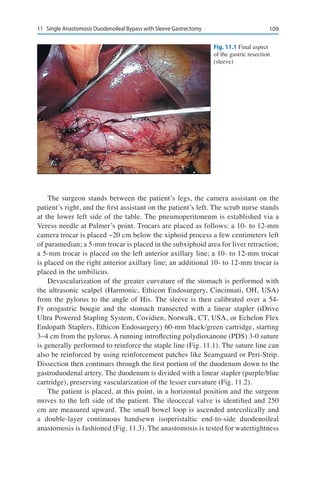
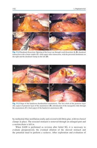
![11111 Single Anastomosis Duodenoileal Bypass with Sleeve Gastrectomy
the sleeved stomach, the procedure starts with the duodenal dissection and
preparation, followed by duodenal transection and duodenoileal anastomosis,
either performed mechanically or manually.
11.3 Outcomes
When analyzing outcomes of a relatively new procedure, it must first be
stressed that few studies are available, no long-term results can be discussed,
nomenclature is not yet standardized as well as the common channel length [200
cm vs. 250 cm vs. 300 cm vs. 350 cm, according to different studies and patient’s
preoperative body mass index (BMI)].
Most of the available studies have been published by Sánchez-Pernaute et al.
from Spain. Weight loss has been excellent in these first 3 years, approximating
100% of patients after the first postoperative year. Initial weight loss is even
greater than after a classic BPD-DS, which is probably correlated with a potent
ileal brake mediated by an enhanced secretion of peptide tyrosine-tyrosine
(PYY) and glucagon-like peptide-1 (GLP-1), which stimulate early satiety [9].
An updated report was published by the same group in 2013 on the first 100
patients, who had a mean percentage of excess weight loss (%EWL) of 95%,
which was maintained during the follow-up period. However, 3-year results were
available only for 19 of the 20 eligible patients.
Moreover, Sánchez-Pernaute et al. recently demonstrated the efficacy of
SADI-S in a subgroup of obese diabetic patients and in revisional surgery for
patients who experienced weight regain after SG as a stand-alone procedure [10].
Control of the disease, with glycolated hemoglobin (HbA1c) <6%, was obtained
in 70–84% in the long term, depending on the initial antidiabetic therapy, with
most patients being able to abandon antidiabetic therapy after the operation. An
8% recurrence rate in the first 5 years was registered among the 25 of 32 patients
eligible for long-term follow-up [14]. As far as revisional surgery after failed SG
is concerned, the authors reported outcomes on 16 patients with an initial BMI of
56.4 kg/m2
and a mean %EWL of 39.5% after a SG who underwent SADI with
a 250-cm common channel. Their results demonstrated good definitive weight
loss after the second procedure, increasing from an initial %EWL of 39.5% to a
final 72% after the duodenal bypass, with all patients losing >50% of their initial
excess weight. The authors concluded that the loop switch can be considered as
a suitable and simplified alternative to BPD-DS after failed SG.
Unpublished data from Mitzman et al. in their series of SIPS – which differs
from SADI-S in the size of the orogastric bougie (42 Fr vs. 54 Fr) and in the
length of the common channel (300 cm vs. 250 cm) – reported good weight loss
outcomes, reaching a mean %EWL of 72.3% after 1 year. The authors believe
that preserving 300 cm of intestine along with the ileocecal valve may reduce
the risk of malnutrition and diarrhea, and pylorus preservation may assist in](https://image.slidesharecdn.com/bariatricandmetabolicsurgery-180301214153/85/Bariatric-and-metabolic-surgery-120-320.jpg)
![112 L. Angrisani et al.
maintaining a physiologically based rate of gastric emptying, thus providing an
efficacious procedure that offers improved quality of life, reduction in hunger,
and increase in satiety while minimizing diarrhea and bowel movements [4].
Recently, an 18-month matched cohort analysis of single anastomosis loop
duodenal switch vs. Roux-en-Y gastric bypass was published by Cottam et al. in
which 54 patients who underwent RYGB were matched for sex and BMI with 54
patients who underwent SADI-S with a 300-cm-long common channel. SADI-S
produced similar but longer-sustained weight loss compared with RYGB, with
a reduced variability among patients, as demonstrated by the smaller confidence
intervals in their series. It also resulted in fewer complaints of nausea or other
upper-gastrointestinal (GI) complaints, with no anastomotic ulcers, resulting in
a lower rate of postoperative endoscopies when compared with the RYGB [15].
11.4 Complications (According to Clavien-Dindo Classification)
All bariatric procedures have inherent complications, but while mortality is an
easily quantifiable outcome parameter, overall morbidity and its severity are
poorly defined. The Clavien-Dindo Classification allows objective quantitation
of procedure safety [16]. Short-term postoperative complications include the
whole spectrum of events that may occur after a general or bariatric surgery
procedures, including leakage/fistula, bleeding, stenosis, and abdominal wall
complications.
11.4.1 Leakage (Grade II if Treated Conservatively, Grade IIIb if Treated
Endoscopically or Surgically)
Leakage (0.5–2%) can involve the gastric suture line after SG, the anastomosis,
and the duodenal stump. Clinical presentation is tachycardia, fever, abdominal
pain, and hypotension. An upper-GI X-ray with gastrografin can be useful
for diagnosis, but spiral angio-CT scan is the most accurate diagnostic tool.
Management can be conservative in case of low-output fistulas (nasogastric
tube, percutaneous drainage of fluid collections, total parenteral nutrition). If
conservative treatment fails or there is hemodynamic instability and signs of
peritonitis, a laparoscopic or laparotomic reoperation is indicated.
11.4.2 Bleeding (Grade II if Treated Conservatively, Grade IIIb if Treated
Endoscopically or Surgically)
Bleeding (2%) can be intraluminal (SG, duodenoileal anastomosis) or extralu-
minal. Clinical presentation is generally clear and characterized by hypotension,](https://image.slidesharecdn.com/bariatricandmetabolicsurgery-180301214153/85/Bariatric-and-metabolic-surgery-121-320.jpg)
![114 L. Angrisani et al.
entire afferent limb underneath the anastomosis and over to the right side of the
abdominal cavity, creating a partial bowel obstruction [17, 18].
11.5.2 Protein Malnutrition (Grade II if Treated Conservatively, Grade
IIIb if Treated Endoscopically or Surgically)
Protein malnutrition (0.5-3%) is the most serious complication of SADI-S, as
with any BPD. A sustained and rapid weight loss is invariably accompanied by
nutritional changes, which reflect the metabolic transformation of the organism.
Nutritional changes are characterized by hypoalbuminemia, anemia, asthenia,
edema, and alopecia. The pathogenesis of protein malnutrition is multifactorial,
being conditioned by some variables linked to the procedure (e.g., gastric volume,
intestinal lengths, individual digestion, proteic absorption capacity, amount of
lost endogenous nitrogen) and others linked to the patient, such as eating habits
and socioeconomic status. Protein malnutrition is generally limited to a single
episode during the first postoperative year and is often determined by inadequate
eating habits. In case of recurrent hypoalbuminemia, revisional surgery may be
necessary. As reported by Sánchez-Pernaute, in these cases, SADI-S is converted
to Roux-en-Y duodenal switch with a 300-cm alimentary limb and a 200- to
250-cm common channel by dividing the bowel in the efferent loop just distal to
the duodenoileostomy and bringing this end to the biliopancreatic limb 100 cm
proximal to the duodenoileostomy [9]. Lifelong follow-up of nutritional status
and supplementation are mandatory.
11.6 Conclusions
SADI-S is one of the most recent innovations in bariatric surgery. It represents
the continuous research and evolution in this field in an attempt to find more
effective and safer solutions when treating the obesity epidemic and metabolic-
related diseases. Though no single operation has been demonstrated to date to
be the unique solution for all patients with morbid obesity and severe metabolic
disease, preliminary results have shown that SADI-S, which is based on the
solid physiopathologic principles of BPD-DS, could potentially be a suitable
alternative for supermorbidly obese patients.
Standardization of nomenclature and technique, together with further studies
with long-term outcomes, are needed to confirm the safety and efficacy of
SADI-S in the routine surgical management of the obese population.](https://image.slidesharecdn.com/bariatricandmetabolicsurgery-180301214153/85/Bariatric-and-metabolic-surgery-123-320.jpg)
![11511 Single Anastomosis Duodenoileal Bypass with Sleeve Gastrectomy
References
1. Karcz WK, Kuesters S, Marjanovic G, Grueneberger JM (2013) Duodeno–enteral omega
switches—more physiological techniques in metabolic surgery. Wideochir Inne Tech
Maloinwazyjne 8:273–279
2. Lee WJ1, Lee KT, Kasama K et al (2014) Laparoscopic single-anastomosis duodenal-jejunal
bypass with sleeve gastrectomy (SADJB-SG): short-term result and comparison with gastric
bypass. Obes Surg 24:109–113
3. Zaveri H, Surve A, Cottam D et al (2015) Stomach intestinal pylorus sparing surgery (SIPS)
with laparoscopic fundoplication (LF): a new approach to gastroesophageal reflux disease
(GERD) in the setting of morbid obesity. Springerplus 4:596
4. Mitzman B, Cottam D, Goriparthi R et al (2016) Stomach intestinal pylorus sparing (SIPS)
surgery for morbid obesity: retrospective analyses of our preliminary experience. Obes Surg
[Epub ahead of print] doi:10.1186/s40064
5. Scopinaro N, Gianetta E, Civalleri D et al (1979) Bilio-pancreatic bypass for obesity: II.
Initial experience in man. Br J Surg 66:618–620
6. Hess DS, Hess DW (1998) Biliopancreatic diversion with a duodenal switch. Obes Surg
8:267–282
7. Sánchez-PernauteA, Rubio Herrera MA, Pérez-Aguirre E et al (2007) Proximal duodenal-ile-
al end-to-side bypass with sleeve gastrectomy: proposed technique. Obes Surg 17:1614–1618
8. Sánchez-Pernaute A, Rubio MÁ, Pérez Aguirre E et al (2013) Single-anastomosis duode-
noileal bypass with sleeve gastrectomy: metabolic improvement and weight loss in first 100
patients. Surg Obes Relat Dis 9:731–735
9. Sánchez-Pernaute A, Herrera MA, Pérez-Aguirre ME et al (2010) Single anastomosis duo-
denoileal bypass with sleeve gastrectomy (SADI-S). One- to three-year follow-up. Obes
Surg 20:1720–1726
10. Sánchez-Pernaute A, Rubio MÁ, Conde M et al (2015) Single-anastomosis duodenoileal
bypass as a second step after sleeve gastrectomy. Surg Obes Relat Dis 11:351–355
11. Gagner M, Rogula T (2003) Laparoscopic reoperative sleeve gastrectomy for poor weight
loss after biliopancreatic diversion with duodenal switch. Obes Surg 13:649–654
12. Baltasar A, Serra C, Pérez N et al (2006) Re-sleeve gastrectomy. Obes Surg 16:1535–1538
13. Dapri G, Cadière GB, Himpens J (2011) Laparoscopic repeat sleeve gastrectomy versus
duodenal switch after isolated sleeve gastrectomy for obesity. Surg Obes Relat Dis 7:38–44
14. Sánchez-Pernaute A, Rubio MÁ, Cabrerizo L et al (2015) Single-anastomosis duodenoileal
bypass with sleeve gastrectomy (SADI-S) for obese diabetic patients. Surg Obes Relat Dis
11:1092–1098
15. Cottam A, Cottam D, Medlin W et al (2015) A matched cohort analysis of single anastomosis
loop duodenal switch versus Roux-en-Y gastric bypass with 18-month follow-up. Surg
Endosc [Epub ahead of print] doi:10.1007/s00464-015-4707-7
16. Goitein D, Raziel A, Szold A, Sakran N (2016) Assessment of perioperative complications
following primary bariatric surgery according to the Clavien-Dindo classification:
comparison of sleeve gastrectomy and Roux-Y gastric bypass. Surg Endosc 30:273–278
17. Surve A, Zaveri H, Cottam D (2016) Retrograde filling of the afferent limb as a cause of
chronic nausea after single anastomosis loop duodenal switch. Surg Obes Relat Dis [Epub
ahead of print] doi:10.1016/j.soard.2016.01.018
18. Summerhays C, Cottam D, Cottam A (2016) Internal hernia after revisional laparoscopic
loop duodenal switch surgery. Surg Obes Relat Dis 12:e13–e15](https://image.slidesharecdn.com/bariatricandmetabolicsurgery-180301214153/85/Bariatric-and-metabolic-surgery-124-320.jpg)
![117
12
L. Angrisani (Ed), Bariatric and Metabolic Surgery,
Updates in Surgery
DOI: 10.1007/ 978-88-470-3944-5_12, © Springer-Verlag Italia 2017
D. Foschi (*)
Department of Biomedical Sciences Luigi Sacco, University of Milan
Milan, Italy
e-mail: diego.foschi@unimi.it
Ileal Interposition
Diego Foschi, Andrea Rizzi, and Igor Tubazio
12.1 Introduction
In the early 1980s, Koopmans and colleagues [1] first proposed the ileal
interposition (II) between the duodenum and jejunum as an experimental surgical
procedure to induce reduction of body weight in rodents and dogs. In 1999, Mason
[2] considered the increase in enteroglucagon (glucagon-like peptide-1) to be the
mechanism of action of II and recommended clinical controlled trials to assess
its ability to treat type 2 diabetes mellitus (T2DM) and obesity in humans. In
2006, De Paula et al. [3] described a short series of pathologically obese patients
treated by II in association with sleeve gastrectomy (SG) that showed very good
results for weight loss and resolution of obesity complications. Several clinical
trials investigated the effects of SG with ileal interposition to treat obese patients
affected by T2DM or metabolic syndrome (MS). The operation can be performed
with diversion of the duodenum (SG-DD-II) or not. In our experience, SG-DD-
II is very effective as a bariatric and metabolic operation, although it is highly
demanding and needs further evaluation before it will be considered for routine
clinical use.
12.2 Experimental Background of Ileal Interposition
The gastrointestinal tract has many intrinsic feedback mechanisms to control
secretory, motor, and hormonal functions. As a rule, the distal tract of the
intestine is able to regulate activity of the proximal tract by means of hormonal
changes [4–6]. Gastric secretion is regulated by the balance between gastrin and
somatostatin in the antral mucosa, but several hormones produced by duodenal
mucosa [gastric inhibitory peptide (GIP), cholecystokinin (CCK), and intestinal](https://image.slidesharecdn.com/bariatricandmetabolicsurgery-180301214153/85/Bariatric-and-metabolic-surgery-125-320.jpg)
![118 D. Foschi et al.
mucosa [glucagon-like peptide 1 and 2 (GLP-1 and -2)] are able to inhibit it.
GIP, CCK, and GLP are released by nutrient challenge from endocrine cells.
GIP is also known as a glucose-dependent insulinotropic peptide and stimulates
insulin secretion; the same effect is exerted by GLP-1 and peptide tyrosine-
tyrosine (PYY). These hormones, as well as CCK, pancreatic peptide (PP), and
oxyntomodulin also exert anorectic effects and help control food intake and body
weight. GIP, GLP-1 and -2, and PYY inhibit gastric emptying and contribute to
the regulation of gastrointestinal motility [6]. In the past, short-bowel syndrome
was treated by ileal interposition, which makes transit time longer. This effect
is mediated by GLP-1 secretion [7]. Furthermore, II reduces food intake and
decreases body weight [1] by the same mechanism. Since GLP-1 has a strong
incretin effect, II was found to improve glucose and lipid metabolism, delay
diabetes onset, and even cure it in experimental animals [7, 8]. From these results,
II was considered to be more effective than other operations for curing T2DM in
obese patients. However, De Paula (personal communication) found that II alone
was not really effective and proposed an association between SG and II [3].
12.3 Indications to Ileal Interposition Surgery
SG-II includes the restrictive effects of SG and the functional effects of ileal
interposition, which causes changes in lipid absorption and enterokine secretion.
After II, the interposed ileum is unable to absorb fats (because bile salts are
released distally in the jejunum) and is rapidly stimulated by chyme to release
GLP-1 and -2, fibroblast growth factor-19 (FGF-19), and other hormones that
reduce appetite and stimulate insulin secretion. SG-II is indicated mainly for
morbidly obese patients with obesity complications [9]. When the duodenum
is diverted (SG-DD-II), the operation is highly indicated for treating morbidly
obese or obese patients affected by T2DM or MS [10–12]. Moreover, SG with a
large remnant or a fundectomy [13] can be associated with II for T2DM patients
with a low (<30 kg/m2
) body mass index (BMI) to avoid excessive reduction of
body weight [14, 15].
12.4 Surgical Technique
The operation can be performed in two different ways: with or without duodenal
diversion [14]. Excluding the duodenum and upper part of the jejunum makes it
easier to control glucose homeostasis and allows better results in obese patients
with T2DM. We describe SG-DD-II as the standard procedure (Fig. 12.1).
The operation is performed under general anesthesia as a laparoscopic
procedure [3]. Preoperative preparation includes overnight fasting, preoperative](https://image.slidesharecdn.com/bariatricandmetabolicsurgery-180301214153/85/Bariatric-and-metabolic-surgery-126-320.jpg)
![11912 Ileal Interposition
bowel cleansing, perioperative antibiotics, and low-molecular-weight heparin
prophylaxis. A gastric tube and a urinary catheter are inserted. Seven trocars
are used. At the beginning of the operation, the first surgeon is situated between
the legs of the patient, who is in an extreme anti-Trendelenburg position. The
operation starts with SG after devascularization of the greater curvature of the
stomach 6-8 cm from the pylorus, which is achieved using an ultrasonic scalpel.
A 36-Fr orogastric calibration tube is placed along the lesser curvature of the
stomach toward the pylorus. Gastric resection is performed starting from the
antrum and continuing up to the angle of His using an Echelon 60-mm stapler,
with gold cartridges in the antrum and blue cartridges in the body of the stomach.
Bleeding from the suture line is stopped by bipolar coagulation, but a 3-0
polypropylene running invaginating suture is used in selected cases. After that,
dissection of the antrum and duodenum is done with ligature of the right gastric
vessels. Duodenum sectioning is performed 4 cm from the pylorus using a linear
blue or white cartridge staple. Opening a window in the mesocolon just above the
Treitz ligament allows transposition of the stomach and duodenum in the lower
abdomen. The stomach is sutured to the mesocolon to avoid internal hernia.
The second step is submesocolic and is performed from the left size of the
patient. Measurement of the total intestinal length is performed with traction,
along the anti-mesenteric border, using a 5-cm marked atraumatic grasper. In
the technique described by De Paula et al. [9], the first transection is done 30
cm from the ileocecal valve; we prefer to maintain 50 cm of distal ileum to
reduce the risk of biliary malabsorption. Then, an ileal tract 150- to 170-cm
long (50 cm in the first description of De Paula [3]) is prepared using a linear
stapler (white cartridge). The interposed ileum is anastomized to the duodenum.
We prefer a gastroileal anastomosis, especially when the duodenum is poorly
Fig. 12.1 Ileal interposition
associated to sleeve
gastrectomy and duodenal
diversion (reproduced with
permission from [16])](https://image.slidesharecdn.com/bariatricandmetabolicsurgery-180301214153/85/Bariatric-and-metabolic-surgery-127-320.jpg)
![120 D. Foschi et al.
perfused (Fig. 12.1). A distal anastomosis is done with the jejunum. Finally, the
intestinal circuit is restored by suturing the distal jejunum to the distal ileum. In
our technique, total intestinal tract length is 400 cm, with a 150-cm transposed
ileum (alimentary loop), 250-cm common tract, and a variable pancreatic-biliary
tract. The gastroileal anastomosis is performed using a 45-mm linear (blue
cartridge) stapler, intestinal anastomoses are performed functionally using 45-
mm linear (white cartridge) staplers. The enterotomy defects are closed with a
2-0 absorbable running suture. Mesenteric defects are closed using a continuous
3-0 polypropylene suture line. An abdominal tube is placed in the proximity of
the inferior tract of the gastric-line suture.
12.5 Postoperative Course and Complications (According to
Clavien-Dindo Classification)
De Paula et al. [9] reported a mean operation time (OT) of 188 min (range
125–330 min) and a median hospital stay (HS) of 3.3. days (range 3-63 days).
Higher values [OT 3.9 hours (3.1–8.2), HS 6.9 days (4–429] were reported by
Celik et al. [13]. Our mean OT was 290 min (180–480 min), with a mean HS
of 8 days (5–60 days). Mortality after SG-DD-II was low (from 0.27 to 0.4%);
in all cases, it was caused by septic complications associated with anastomotic
leakage. One patient died from myocardial infarction after suture-line leakage
of the SG (Clavien-Dindo [17] grade V). The overall rate of major postoperative
complications was low: from 4% in the series of De Paula et al. [9] to 6.1% in the
series of Celik et al. [13]. We had three cases out of 64 patients: two with leakage
and one with bleeding, all of whom required reoperation (Clavien-Dindo grade
IIIb). Minor complications were rare: there were three cases of wound infection
and wound hematoma (Clavien-Dindo grade I). Constipation and anorexia were
relatively frequent after discharge and may be considered typically related to
the operation. Furthermore, we had two late intestinal occlusions caused by
defects of the mesenteric folds; in both cases, surgical closure of the defect was
done (one patient also had jejunal resection for intestinal necrosis). Celik et al.
[13] described seven cases of diarrhea (2%) and one case of hypoglycemia. In
our experience, diarrhea was rare (two cases out of 60 patients) and reactive
hypoglycemia absent. The reoperation rate was between 1.3 and 7.22%, primarily
caused by gallstone disease. We had two more reoperations: one abdominoplasty
and one hernioplasty. The nonsurgical complication rate reported by Celik et al.
was very high, with 11 patients affected by neurological complications (3.05%).
However, it must be considered that all these series of SG-DD-II consisted
of T2DM patients who had a long-standing clinical course and a high rate of
diabetes-related complications.](https://image.slidesharecdn.com/bariatricandmetabolicsurgery-180301214153/85/Bariatric-and-metabolic-surgery-128-320.jpg)
![12112 Ileal Interposition
12.6 Clinical Results (Table 12.1)
In our series, SG-DD-II was a highly effective bariatric surgical procedure:
body weight loss was 40 ± 8 standard deviation (SD) kg at 1 year, with BMI
reduction of 12 ± 4 kg/m2
. Percentage of excess weight loss (%EWL) was 79.3
± 6.5%. Similar results were reported in all published experiences [13–15, 18],
and SG-DD-II must be considered one of the most powerful operations available
to reduce body weight in obese patients. Also, the complications of obesity
are strongly affected by SG-DD-II: MS, T2DM, hypertension, and obstructive
sleeping apnea syndrome resolve in the large majority of patients. If criteria
for T2DM evaluation proposed by the American Diabetes Association [19] are
considered, more than 90% of our patients have glycemia <100 mg/dL and
glycolated hemoglobin (HbA1c) <6 % without antidiabetic medication 1 year
after the operation. Similar results were reported by De Paula et al. [9, 11],
Kumar et al. [10], Tinoco et al. [12], and Celik et al. [13]. Our results after 5
years, in a case-control prospective study, showed persistent T2DM remission
in more than 90% of cases, with a significant difference (p<0.01) in comparison
with the medical therapy. Only one patient was resistant to SG-DD-II, and one
Table 12.1 Clinical, biochemical, and insulin changes after DDSG-II in T2DM obese patients
(reproduced with permission from [16])
Parameter Before DDSG-II After DDSG-II
Body weight (kg) 102.7 ± 4,2 69.5 ± 2.9**
BMI (kg/m2
) 38.6 ± 2.2 26 ± 1.3**
Fasting plasma glucose (mg dL) 168.8 ± 10.3 80 ± 2.1**
HbA1c (%) 7.6 ± 0.4 5.2 ± 0.1**
Insulin (μIU/mL) 16.1 ± 3.8 2.6 ± 0.4**
Vitamin B12
(ng/L)* 311.1 ± 33.3 332.2 ± 38.4
Folic acid (μg/L)* 6.3 ± 1.1 10.2 ± 1.8
Vitamin D (ng/mL) * 19.1 ± 2.6 15.13 ± 2.3
Total proteins (g/L) 68.1 ± 1.3 68.4 ± 0.7
Albumin (g/L) 4 0.2 ± 0.9 41.3 ± 0.7
Hemoglobin (g/dL) 13 ± 0.4 12.8 ± 0.2
DDSG-II duodenal diverted sleeve gastrectomy with ileal interposition, T2DM type 2 diabetes
mellitus, BMI body mass index, HbA1c glycated hemoglobin
* Dietary supplementation given postoperatively
** p<0.001, Mann-Whitney U test](https://image.slidesharecdn.com/bariatricandmetabolicsurgery-180301214153/85/Bariatric-and-metabolic-surgery-129-320.jpg)
![122 D. Foschi et al.
other patient relapsed 3 years after operation. Both patients had a long-lasting
T2DM clinical course, with autonomic neuropathy, demonstrated by a Tilt test.
We believe that T2DM patients affected by autonomic neuropathy should be
excluded from this operation. Furthermore, SG-DD-II is highly effective in
patients with a BMI <35 or < 30 kg/m2
(14, 15, 17). In such patients, a large SG
or fundectomy only is performed to reduce the risk of excessive weight loss, but
the metabolic effects of the operation seem to be very useful for these otherwise
unfavorable cases. Treatment side effects were mild; although the operation
cannot be classified as malabsorptive (only a few patients had diarrhea, and none
experienced protein malnutrition), since the intermediate cholesterol metabolites
(like 7α-hydroxy-4-cholesten-3-one. or 7αC4) were low [16]. It is important that
after SG-DD-II all patients assume a regime of daily vitamin and trace element
supplementation to avoid any long-term nutritional complications.
12.7 Biochemical and Hormonal Results (Table 12.1)
After SG-DD-II, glycemic and lipidic metabolic patterns improved impressively:
glycemia, low-density-lipoprotein (LDL) and total cholesterol and triglycerides
decreased to normal or subnormal values [11, 20]. The restrictive effect exerted
by SG and the inability of the interposed ileum to absorb lipids in the absence
of the biliary salts limit caloric intake and reduce body weight. The decrease in
adipose tissue induces very important changes in adipokines: leptin and resistin
decrease, whereas adiponectin increases [21], restoring the best metabolic
conditions to reduce lipotoxicity on the β-cell mass. Further changes induced by
SG-DD-II on gastrointestinal hormones play an important role in determining
the anorectic effects of the operation: the reduction of ghrelin secretion caused
by SG [22] or fundectomy (in low BMI patients) and the increase in GLP-1 -2
(Fig. 12.2), PYY, and GIP [20].
These hormonal changes are also effective in regulating glucose homeostasis
in T2DM patients. GLP-1 and -2, GIP, and PYY are proincretinic of insulin,
whereas ghrelin is able to increase insulin resistance [23]. After SG-DD-II,
fasting insulin secretion decreases and oral-glucose-stimulated insulin secretion
increases (Fig. 12.3). These effects are related to the decrease of insulin resistance
and the increase of β-cell sensitivity and secretion. Evaluation of β-cell function
using the euglycemic hyperinsulinemic clamp with both the intravenous [24] and
the oral glucose tolerance test [25] demonstrated that 1 year after the operation,
SG-DD-II doubled both insulin secretion and β-cell glucose sensitivity.
SG-DD-II also changes exposure of the ileal mucosa to the biliary salts,
with inhibition of 7αC4and increase of enterokine FGF-19 [19]. This hormone
stimulates hepatic buildup of glycogen and reduces glucose and triglycerides in
plasma, contributing to the overall metabolic effects of the operation [26].](https://image.slidesharecdn.com/bariatricandmetabolicsurgery-180301214153/85/Bariatric-and-metabolic-surgery-130-320.jpg)
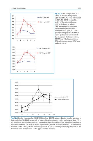
![124 D. Foschi et al.
12.8 Conclusions
Remission of T2DM after bariatric surgery is caused by reduction of insulin re-
sistance and increase of insulin secretion. Weight and adipose tissue reduction is
the most important factor mediating the improvement of glucose metabolism in
obese patients affected by T2DM; however, the role of hormonal changes induced
by surgery should also be considered. Suppression of ghrelin, the orexigenic and
lipogenetic hormone secreted by the stomach fundus; stimulation of GLP-1 and
-2 and PYY3-36 from endocrine enteric cells (hindgut mechanisms); and remov-
al of foregut (still unknown) mechanisms by duodenal exclusion are recognized
as very important in regulating glucose homeostasis. Ileal interposition is the
most powerful operation exerting hindgut effects (incretinic stimulation) but it
is unable to trigger T2DM remission. SG (which causes ghrelin suppression) in
association with II seems to be a good antidiabetic operation, but the best effects
occur when the duodenum is excluded from food stimulation. The technique
of SG-DD-II described by De Paula et al. [3] necessitates transposition of the
sleeved stomach into the submesocolic space, isoperistaltic transposition of the
terminal ileum (150 cm), and restoration of the intestinal circuit by a jejunoileal
anastomosis, entailing one gastric suture and three intestinal anastomoses. Op-
eration time is long, and SG-DD-II has a complication rate higher than SG alone.
However, the effects on glucose metabolism are very good, and more than 90%
of patients remain in T2DM remission after 5 years of follow-up. Weight reduc-
tion is very important in obese or morbidly obese patients but is quite limited in
patients with BMI <30 kg/m2
. For this reason, SG-DD-II should also be consid-
ered for nonobese patients affected by T2DM. Although diffusion of SG-DD-II
in the clinical setting is limited, if long-term results of T2DM remission > 90%
are confirmed, it would be fully considered in the surgical treatment for T2DM.
References
1. Koopmans HS, Sclafani A, Fichtner C et al (1982) The effects of ileal transposition on food
intake and body weight loss in VMH-obese rats. Am J Clin Nutr 35:284–293
2. Mason EE (1999) Ileal transposition and enteroglucagon/GLP-1 in obesity (and diabetic?)
surgery. Obes Surg 9:223–228
3. De Paula AL, Macedo ALV, Prudente AS et al (2006) Laparoscopic sleeve gastrectomy with
ileal interposition (“neuroendocrine brake”) – pilot study of a new operation. Surg Obes
Relat Dis 2:464–467
4. Schubert ML (2008) Hormonal regulation of gastric acid secretion. Curr Gastroenterol Rep
10:523–527
5. Wren AM, Bloom SR (2007) Gut hormones and appetite control. Gastroenterology
132:2116–2130
6. WU T, Rayner CK, Young RL et al (2013) Gut motility and enteroendocrine secretion. Curr
Opinion Pharmacol 13:928–934
7. StraderAD, Clausen TR, Goodin SZ et al (2009) Ileal interposition improves glucose tolerance
in low dose streptozotocin-treated diabetic and euglycemic rats. Obes Surg 19:96–104](https://image.slidesharecdn.com/bariatricandmetabolicsurgery-180301214153/85/Bariatric-and-metabolic-surgery-132-320.jpg)
![127
13
L. Angrisani (Ed), Bariatric and Metabolic Surgery,
Updates in Surgery
DOI: 10.1007/ 978-88-470-3944-5_13, © Springer-Verlag Italia 2017
R. Moroni (*)
Bariatric Surgery Unit, Department of Surgery, A.O. Brotzu
Cagliari, Italy
e-mail: rob.moron@tiscali.it
The Problem of Weight Regain
Roberto Moroni, Marco Antonio Zappa, Giovanni Fantola,
Maria Grazia Carbonelli, and Fausta Micanti
13.1 Introduction
The effectiveness of bariatric surgery has been demonstrated, and weight loss
can be simply obtained with a well-selected procedure. The real challenge of
bariatric surgery success is to obtain sustained weight loss. Long-term follow-up
after bariatric surgery (>5 years) is imperative to demonstrate surgical success
[1]. Weight regain may occur with all three of the most common procedures
(Roux-en-Y gastric bypass, sleeve gastrectomy and gastric banding), and it
depends on nutritional, psychological, and surgery-related factors. There is no
agreement in in the literature regarding the definition of weight regain, and it
is sometimes confused with a nonspecific definition of bariatric surgery failure
[2]. The percentage of excess weight loss (%EWL), percentage of weight regain,
regain after the nadir reached, persistent obesity, percentage of excess body mass
index loss (%EBMIL), and absolute body mass index (BMI) are definitions of
renewed weight gain. A gain of %EWL <50% and an absolute BMI >35 kg/m2
are the two most common definitions used in the literature. In order to obtain a
correct diagnosis of weight regain and to treat appropriately obesity recidivism,
nutritional, psychological, and surgical factors should be studied independently
and in an integrated approach [3].
13.2 Homeostatic Balances in Weight Regain
Changes in energy intake, and perhaps even in energy expenditure, seen after
bariatric surgery may be affected by alterations in gut and adipocyte hormones.
Although it has been suggested that gut hormones such as ghrelin, glucagon-](https://image.slidesharecdn.com/bariatricandmetabolicsurgery-180301214153/85/Bariatric-and-metabolic-surgery-134-320.jpg)
![128 R. Moroni et al.
like peptide-1 (GLP-1), and peptide tyrosine-tyrosine (PYY) may be involved
in postoperative weight homeostasis [4] due to observed decreases in ghrelin
concentrations and increases in GLP-1 and PYY after Roux-en-Y gastric bypass
(RYGB) and biliopancreatic diversion (BPD), other studies do not confirm a
clear relationship between these changes and weight reduction [5]. A reduction
in leptin and insulin serum concentrations may also play a role in weight regain
[6]. Moreover, the practice of regular physical activity influence weight-loss
maintenance [7]. In fact, patients who performed physical exercise on a regular
basis (three or four times per week, 30-min minimum each time) show a propensity
for the lowest weight regain [8]. As modifications of gastrointestinal function
after surgery increase, less dietary intervention is needed to induce weight loss.
In general, restrictive operations are more commonly associated with weight loss
failure than are other techniques with a malabsorptive component.
From the nutritional point of view, a low glycemic load and moderately high
protein content diet (or a diet based on the traditional Mediterranean dietary
pattern) combined with a physical activity program has been shown to effectively
treat weight regain in the short term [9]. Homeostatic eating control, regulated by
macronutrients that send satiety signals to the hypothalamus, may reduce high
glycemic index/low protein malnutrition, which modifies interaction between
gut and hypothalamus [10]. For this reason, homeostatic eating control may play
a critical role in weight regain treatment before considering redo surgery.
13.3 Nutritional Management of Weight Regain
Weight regain is most commonly related to noncompliance with dietary and
lifestyle instructions (inappropriate food choices and little physical activity) both
postoperatively and over the long-term follow-up [11]. Calorie intake is reduced
after bariatric surgery, but it increases 1–2 years after surgery, coinciding with
weight regain [12]. The problem of weight regain could be associated with
dumping syndrome, a common nutritional problem after gastric bypass. Patients
will often complain about lightheadedness and sweating after eating a high-
glucose meal or drinking fluids during the meal. This happens because high-
osmolarity foods – ice cream or pastries, for example – after bypassing much
of the stomach undigested, cause an osmotic overload upon entering the small
intestine. This osmotic overload brings fluid in the lumen of the small intestine,
causing a vagal reaction. During this uncomfortable feeling, patients prefer to eat
high-caloric-density foods to fight fatigue, thus causing insufficient weight loss
or weight regain.
Nutritional and lifestyle counseling may reduce the weight of patients with
previous weight regain, reducing body fat and improving the perspective of
weight maintenance over time [13]. In cases of severe or unremitting postopera-
tive weight gain, a multidisciplinary team (experienced surgeon, nutritionist, en-](https://image.slidesharecdn.com/bariatricandmetabolicsurgery-180301214153/85/Bariatric-and-metabolic-surgery-135-320.jpg)
![12913 The Problem of Weight Regain
docrinologist, psychologist, anesthetist, and specialized nurse) should consider
the possibility of redo surgery.
Although bariatric surgery is not a guarantee of success, post-surgery adher-
ence to lifestyle and nutritional recommendations – particularly to postoperative
management of diet progression – and a careful nutritional follow-up could reduce
the likelihood of weight regain and redo surgery in severely obese patients [14–16].
13.4 Psychologic and Psychiatric Factors of Weight Regain
There are very few studies investigating directly the clinical factors determining
weight regain. All we know about the topic comes from follow-up studies that
emphasize poor outcome after the first intervention, above all after banding and
gastric bypass [16].
Follow-up studies at 18–24 months and 5 years after bariatric surgery [17–29]
stress that the presence of a psychiatric disorder before bariatric surgery – such
as panic attack [20], depression [21], personality and addiction disorder [22,
23] – can reduce weight loss and determine poor outcome, above all when these
disorders were not recognized and treated at the first assessment.
Particular attention is now dedicated to the outcome of bariatric surgery for
patients suffering from maladaptive eating behaviors: grazing; sweet eating; and
eating disorders: binge-eating disorder (BED) and night-eating syndrome (NES)
at first bariatric surgery or that develops after it.
The bariatric outcome of BED patients is still controversial. Kalarchian et
al. [24] followed 96 RYGB patients for 2–7 years postsurgery and reported that
in those classified as binge eaters, body mass index (BMI) increased by 5.3 kg/m2
compared with a 2.4-kg/m2
increase in nonbinge eaters. Impulsive behavioral
traits were also a risk factor for weight regain following bariatric surgery at
2 years of follow-up [25]. On the other hand, Amianto et al. note that a BED
diagnosis, once considered a major contraindication to bariatric surgery, has
now been revised and that bariatric surgery interventions can now be suitable
for selected BED patients treated before and after surgery, even if the extent of
weight loss depends on the presence of binge episodes after the intervention [26].
Beck et al. emphasize that the weight loss obtained after bariatric surgery is
less than that of nonbingers and is strictly correlated with ineffectiveness and
high levels of impulsiveness that do not change in long-term follow-up studies
[27]. Maladaptive eating behaviors such as grazing or sweet eating can provoke
insufficient weight loss [28]. In particular, grazing is considered a high-risk
behavior due to its association with medium-level impulsiveness and high anxiety
levels [29]. Many studies emphasize that it can determine poor outcomes at first
intervention and after bariatric surgery [30]. Often, after the first intervention,
above all after sleeve gastrectomy, grazing appears as change in other eating
behaviors, such as sweet, binge, or nocturnal eating. The psychopathological](https://image.slidesharecdn.com/bariatricandmetabolicsurgery-180301214153/85/Bariatric-and-metabolic-surgery-136-320.jpg)
![130 R. Moroni et al.
trait of grazing clarifies why obese patients with the disorder achieve insufficient
weight loss and can also fail redo surgery without a psychiatric treatment before
it. Studies on sweet-eating show that the tendency to eat sweets needs to change
in the food reward system, and follow-up studies of bariatric surgery stress the
low incidence of sufficient weight loss in sweet eater. Behavioral therapy before
and after redo surgery seems to be effective for changing this behavior [31].
NES is characterized by a time-delayed pattern of eating relative to sleep,
where most food is consumed in the evening and night. A controlled study after
RYGB shows the persistence of nocturnal eating, with sufficient compliance to
diet during the day [32]. Nocturnal eating (often of sweets) determines insufficient
weight loss and persistence of comorbidities, such as diabetes.
Overall analysis of literature data indicates that the presence of eating disorder
and maladaptive behavior determine poor outcome after the primary surgery and
require a specific diagnosis and therapeutic program before redo surgery. It is
essential to rule out the possibility of the patient’s poor lifestyle habits being
responsible for weight recidivism. Patients with binge-eating and snack-eating
patterns or who consume high-caloric liquids may be at risk [33].
Bariatric surgery generally determines an improvement of psychological
factors, such as cognitive impairment, body image, better relationship with others,
and increased ability to cope with social pressure. However, not all patients
experience a real psychological benefit after bariatric surgery. Many continue to
struggle with feeling unable to comply with societal requests concerning body
image. Such condition can lead to poor compliance and insufficient weight loss,
thus intensifying the need for repeat surgery [34].
Redo bariatric surgery can now be considered the new challenge for surgeons,
psychiatrists, psychologists, and dieticians. The reason for failure of the first
intervention must be well examined, above all from a psychological perspective,
and weight regain can be influenced by failure of the primary psychological
assessment. Further studies are needed to highlight reasons for failure, and a more
careful multidisciplinary assessment is necessary in order to initiate effective
psychiatric/psychological therapies before and after redo surgery [35, 36].
13.5 Surgery-Related Factors of Weight Regain
Weight regain following bariatric surgery may be related to late complications or
procedural failures. We analyzed three of the most common bariatric procedures.
13.5.1 Laparoscopic Adjustable Gastric Banding
Success with Laparoscopic adjustable gastric banding (LAGB) is closely
related to appropriate follow-up, as adjustment is necessary to ensure adequate](https://image.slidesharecdn.com/bariatricandmetabolicsurgery-180301214153/85/Bariatric-and-metabolic-surgery-137-320.jpg)
![13113 The Problem of Weight Regain
restriction and weight loss. Gastric distension with smaller quantities of food
leads to an early satiation response and finally to weight loss. Since the gastric
muscle is physiologically expansible, the new gastric pouch could be enlarging
to accommodate food boluses. Therefore, over time, failure rates could be as
high as 40–50 % [37, 38]. Patient noncompliance is the commonest cause of
pouch dilatation and weight regain. In addition, surgical complications, such
as slippage (8%), reduces the compressive and restrictive effects [39–41].
Premature LAGB removal is another important etiology of weight regain,
as only 12% of such patients are able to maintain their reduced weight [41].
Band erosion (3.4–28%), band leak (0.6%), esophageal dilation (3.2%), port or
catheter leak (7.6%), port infection (1.2%), and patient noncompliance (e.g.,
perhaps due to esophageal reflux) are common causes of LAGB removal [39,
42]. Some authors argue that, if successful weight loss is achieved with LAGB,
band revision alone is a viable option in case of complications [39]. If the initial
LAGB achieved adequate weight loss and failure was related to band slippage or
pouch dilatation, laparoscopic SG (LSG) is a reasonable alternative [41]. In case
of severe gastroesophageal reflux disease, band intolerance/erosion, obstruction,
or insufficient weight loss, a gastric bypass or duodenal switch should be used
[40, 42–44].
13.5.2 Laparoscopic Sleeve Gastrectomy
LSG is a straightforward procedure with high success rates in terms of diabetes
resolution, comorbidity resolution, and weight loss. LSG has been considered a
stand-alone procedure since 2008, and low-evidence studies report data about
long-term follow-up weight regain [45–47]. Possible explanations for LSG
failure include dilatation of the residual stomach, calibration of the stomach with
an excessively large gastric bougie, and incomplete sectioning of the gastric
fundus. Himpens et al. reported a %EWL of 77.5 and 53.3%, respectively, at 3-
and 5-year follow-up, affirming that weight regain appears between the third and
sixth postoperative year [45].
The relation between weight regain and dilation of the gastric tube is a matter
of debate. Braghetto et al. [46] reported data on 15 LSG patients undergoing a
gastric volumetric computed tomography (CT) scan on postoperative day 3 and
between 24 and 36 months after surgery. No patient experienced weight regain
with a mean remnant gastric volume increased from 108 to 250 ml.
In 2006, Langer et al. reported a series of 23 obese patients with upper
gastrointestinal (GI) contrast studies at a mean follow-up of 20 months: weight
regain occurred in three patients but only one presented a gastric dilatation [47].
While LSG is considered a restrictive procedure, a metabolic/malabsorptive
procedure can be performed as redo surgery (i.e., gastric bypass or duodenal
switch); resleeving can be proposed if gastric dilatation occurs [45].](https://image.slidesharecdn.com/bariatricandmetabolicsurgery-180301214153/85/Bariatric-and-metabolic-surgery-138-320.jpg)
![132 R. Moroni et al.
13.5.3 Roux-en-Y Gastric Bypass
RYGB is the gold standard bariatric operation; however, a major concern in
late follow-up is weight regain. Despite its success, approximately 10–20 % of
patients undergoing RYGB experienced weight regain or failure to lose adequate
weight at 5-year follow-up [48]. Weight loss depends on restriction of intake
(gastric pouch) and metabolic/malabsorptive effects (gastrojejunal bypass). In
this setting, anatomical abnormalities are postulated to play a significant role
in weight regain [49, 50]. Loss of restriction caused by gastric pouch and/or
gastrojejunostomy (stoma) dilatation has been shown to be responsible for loss
of early satiety and weight regain in some patients [50]. Stomal dilatation is
one of the common causes of weight regain. Indeed, similar to gastric pouch
dilatation, stoma enlargement results in a great quantity of food being needed to
distend the gastric pouch in order to obtain patient satiation.
Yimcharoen et al. demonstrated that an enlarged gastric pouch was found
in almost one third of patients examined for weight regain [49]. Redo surgery
in such cases could be performed to reduce pouch size and create a smaller
stoma. Other options include injection of a sclerosant (sodium morrhuate) into
the stoma to scar it down, endoscopic plication of the gastric pouch and stoma
(StomaphyX and ROSE procedures), adding an adjustable or nonadjustable
gastric band. Gastrogastric fistula is another RYGB complication resulting in
weight regain. It occurs in 1.5 to 6% of patients [51]; weight regain could be
dependent on loss of both restrictive and metabolic/malabsorptive components of
RYGB. In gastrogastric fistula weight regain can occur over the long term. Food
can travel through the correct direction (gastrojejunal anastomosis) or through
the alternative direction (gastrogastric fistula); weight regain occurs when the
alternative direction is preferred than the correct one [52].
13.6 Conclusions
Bariatric surgery is the most effective weight loss intervention. Despite its proven
efficacy, weight regain is one of most considerable challenge after any bariatric
procedure. The etiology of weight regain is multifactorial and not clearly defined.
Nowadays, it is – and will become more so – an onerous public health issue,
taking into account the increasingly obese global population and a constantly
growing number of bariatric procedures around the world. According to the
American Society for Metabolic and Bariatric Surgery (ASMBS), as much as 50%
of patients regain 5% of body weight 2 years or more following their bariatric
procedures, and on long-term follow-up, approximately 20–30% of patients
regain most of the weight they lost [2, 53–55]. The causes leading to weight
regain are multifactorial and include anatomic and psychosocial factors, physical
inactivity, dietary habits, and endocrine/metabolic issues. Overall analysis of the](https://image.slidesharecdn.com/bariatricandmetabolicsurgery-180301214153/85/Bariatric-and-metabolic-surgery-139-320.jpg)
![137L. Angrisani (Ed), Bariatric and Metabolic Surgery,
Updates in Surgery
DOI: 10.1007/ 978-88-470-3944-5_14, © Springer-Verlag Italia 2017
V. Borrelli (*)
General and Bariatric Surgery Unit, Istituto di Cura Città di Pavia, Gruppo Ospedaliero
San Donato
Pavia, Italy
e-mail: vinborr@gmail.com
14Band Revision and Conversion to
Other Procedures
Vincenzo Borrelli and Giuliano Sarro
14.1 Introduction
Laparoscopic adjustable gastric banding (LAGB) is an effective technique that
guarantees satisfactory weight loss for morbidly obese patients [1]; however,
there is an increasing need for reoperation due to weight-loss failure or banding
complications. In case of band complications, there are different surgical options
to be considered, such as band removal or repositioning or conversion to a
different procedure.
Evidence shows that when the band is removed and no additional intervention
is performed, there is a very high percentage of weight regain. Rebanding has
often proven to be unsuccessful, and choosing this procedure is appropriate
only in cases of adequate weight loss and band leakage [2, 3]. Consequently,
when LAGB results in unsatisfactory weight loss and/or a complication occurs,
conversion to a different bariatric intervention should be performed. Although
the best surgical solution remains undetermined, laparoscopic Roux-en-Y
gastric bypass (LRYGB) and laparoscopic sleeve gastrectomy (LSG) are the
two more frequently adopted procedures [4–6]. Another option is conversion
to mini-gastric bypass/one anastomosis gastric bypass (MGB/OAGB) and other
malabsorptive procedures.
An important factor to consider is the timing of revision. Conversion from
LAGB to other procedures could be performed either in a single or a two-step
approach depending on the presence of certain complications (such as erosion)
that can make the stomach wall more complicated to be treated further [7–9].
In order to adopt the most successful strategy and avoid perioperative com-
plications, it is important to review patient’s overall condition and comorbidities](https://image.slidesharecdn.com/bariatricandmetabolicsurgery-180301214153/85/Bariatric-and-metabolic-surgery-143-320.jpg)
![138 V. Borrelli and G. Sarro
following a tailored multidisciplinary approach (surgeon, nutritionist, endosco-
pist, radiologist, and psychologist).
14.2 Technique
14.2.1 Selection and Timing of the Procedure
In order to identify the more suitable procedure for each patient, different variables
must be taken into account: preoperative studies, body mass index (BMI) pre- and
post-LAGB, and comorbidities, as well as patient personal preference. Patients
must undergo an esophagogastroduodenoscopy to identified gastroesophageal
reflux, gastric ulcers or band erosion, and an upper gastrointestinal X-ray with
barium swallow to evaluate band position, pouch size, and presence of esophageal
dilation. Moreover, the nutritionist’s clinical investigation (anthropometrics,
dietary intake, weight history) and psychologist’s research for eating disorders
are mandatory to identify the best procedure. Biochemical parameters, including
metabolic and nutritional parameters, must also be examined.
The following conditions are suggestive for LSG:
• no hiatal hernia
• no gastroesophageal reflux disease (GERD)
• no diabetes
• iron or vitamin deficiency
• history of extensive abdominal surgery
• superobese patient programmed for staged surgery
• patient’s preference.
On the other hand, the following circumstances and conditions are suggestive
for LRYGB:
• hiatal hernia
• GERD
• esophageal dilation
• diabetes
• slippage or gastric pouch dilatation.
The use of malabsorptive procedures must be limited to patients failing to
lose weight after gastric banding (nonresponders) without band complications.
For this group, preoperative supervision by a nutritionist is highly important.
Patients showing evidence of infection/slipping/erosion are more suitable for a
two-step procedure. LSG is commonly performed in two steps, while LRYGB is
a one-stage procedure.
14.2.2 Surgical Technique
Even if all procedures are performed like primary surgery, there are some important
details to be highlighted due the presence of anatomic alterations [10, 11]. To dissect](https://image.slidesharecdn.com/bariatricandmetabolicsurgery-180301214153/85/Bariatric-and-metabolic-surgery-144-320.jpg)
![140 V. Borrelli and G. Sarro
Table14.1Conversionfromlaparoscopicadjustablegastricbanding(LAGB)tolaparoscopicRoux-en-Ygastricbypass(LRYGB)
Author Numberof Median Mortality Overall Main Reoperation %EWLafter
patients follow-up rate morbidity morbidity LRYGB
Wornietal.[12] 301 NA 1/301(0.3%) 91/301(30.2%) NA 11/301(3.7%) NA
Perathoneretal.[13] 108 3.4+2.5years 0 49/108(45.3%) NA 9/108(8.3%) 37.7%
Emousetal[9] 257 30months 0 NA 21/257(8.7%) NA 67%
Weberetal.[14] 32 12months 0 NA 6/32(18.7%) NA NA
Jenningsetal.[15] 55 2years 0 NA NA NA 59.4%
Hiietal.[16] 82 1years 1/82(1.2%) 38/82(46.3%) 11/82(13.4%) NA 50%
vanWageningenetal[17] 47 5.5years 0 NA 8/47(17%) NA NA
%EWLpercentageofexcessweightloss,NAnotavailable
Table14.2Conversionfromlaparoscopicadjustablegastricbanding(LAGB)tolaparoscopicsleevegastrectomy(LSG)
Author Numberof Median Mortality Overall Main Reoperation %EWLafter
patients follow-up rate morbidity morbidity LSG
Yazbeketal.[18] 90 12months 0 NA 14.4% 6.6% 61.3%
Acholonuetal.[19] 15 12months 0 NA 13.3% 6.6% NA
Folettoetal.[20] 52 20months 1/52(1.9%) NA 5.7% NA 41.6%
Jacobsetal.[21] 32 26months 0 NA 3.1% NA 60%
Abu-GazalaandKeidar[22] 18 14months 0 NA 5.5% NA 69.7%
Daprietal.[23] 27 18.6months 0 NA 0 NA 34.8%
Silecchiaetal.[24] 76 24months 0 17.1% 0 0 78.5%
%EWLpercentageofexcessweightloss,NAnotavailable](https://image.slidesharecdn.com/bariatricandmetabolicsurgery-180301214153/85/Bariatric-and-metabolic-surgery-146-320.jpg)
![14114 Band Revision and Conversion to Other Procedures
14.5 Revisional Indications
The primary considerations in revisional surgery reported in the literature [9,
12–24] are shown in Tables 14.1 and 14.2.
References
1. O’Brien PE, MacDonald L, Anderson M et al (2013) Long-term outcomes after bariatric
surgery: fifteen-year follow-up of adjustable gastric banding and a systematic review of the
bariatric surgical literature. Ann Surg 257:87–94
2. Suter M (2001) Laparoscopic band repositioning for pouch dilatation/slippage after gastric
banding: disappointing results. Obes Surg 11:507–512
3. Müller MK, Attigah N, Wildi S et al (2008) High secondary failure rate of rebanding after
failed gastric banding. Surg Endosc 22:448–453
4. Gonzalez-Heredia R, Masrur M, Patton K et al (2015) Revisions after failed gastric band:
sleeve gastrectomy and Roux-en-Y gastric bypass. Surg Endosc 29:2533–2537
5. Marin-Perez P, Betancourt A, Lama A et al (2014) Outcomes after laparoscopic conversion
of failed adjustable gastric banding to sleeve gastrectomy or Roux-en-Y gastric bypass. BJS
101:254–260
6. Coblijn UK, Verveld CJ, van Wagensveld BA, Lagarde SM (2013) Laparoscopic Roux-en-Y
gastric bypass or laparoscopic sleeve gastrectomy as revisional procedure after adjustable
gastric band: a systematic review. Obes Surg 23:1899–1914
7. Stroh C, Weiner R, Wolff S et al (2015) One- versus two-step Roux-en-Y gastric bypass
after gastric banding: data analysis of the German Bariatric Surgery Registry. Obes Surg
25:755–762
8. Stroh C, Benedix D, Weiner R et al (2014) Is a one-step sleeve gastrectomy indicated as a
revision procedure after gastric banding? Data analysis from a quality assurance study of the
surgical treatment of obesity in Germany. Obes Surg 24:9–14
9. Emous M, Apers J, Hoff C et al (2015) Conversion of failed laparoscopic adjustable gastric
banding to Roux-en-Y gastric bypass is safe as a single-step procedure. Surg Endosc
29:2217–2223
10. Fronza JS, Prystowsky JB, Hungness ES et al (2010) Revisional bariatric surgery at a single
institution. Am J Surg 200:651–654
11. Hii MW, LakeAC, Kenneled C, Hopkins GH (2012) Laparoscopic conversion of failed gastric
banding to Roux-en-Y gastric bypass. Short-term follow-up and technical considerations.
Obes Surg 22:1022–1028
12. Worni M, Østbye T, ShahAet al (2013) High risks for adverse outcomes after gastric by-pass
surgery following failed gastric banding. Ann Surg 257:279–285
13. Perathoner A, Zitt M, Lanthaler M et al (2013) Long-term follow-up evaluation of revisional
gastric bypass after failed adjustable gastric banding. Surg Endosc 27:4305–4312
14. Weber M, Müller MK, Michel JM et al (2003) Laparoscopic Roux-en-Y gastric bypass, but
not rebanding, should be proposed as rescue procedure for patients with failed laparoscopic
gastric banding. Ann Surg 238:827–834
15. Jennings NA, Boyle M, Mahawar K et al (2013) Revisional laparoscopic RYGB following
failed laparoscopic adjustable gastric banding. Obes Surg 23:947–952
16. Hii MW, Lake AC, Kenfield C, Hopkins GH (2012) Laparoscopic conversion of failed
gastric banding to RYGB. Short term follow-up and technical consideration. Obes Surg
22:1022–1028
17. van Wageningen B, Berends FJ, Van Ramshorst B, Janssen IF (2006) Revision of failed
laparoscopic adjustable gastric banding to Roux-en-Y gastric bypass. Obes Surg 16:137–141](https://image.slidesharecdn.com/bariatricandmetabolicsurgery-180301214153/85/Bariatric-and-metabolic-surgery-147-320.jpg)
![143L. Angrisani (Ed), Bariatric and Metabolic Surgery,
Updates in Surgery
DOI: 10.1007/ 978-88-470-3944-5_15, © Springer-Verlag Italia 2017
M. Foletto (*)
Center for the Study and the Integrated Management of Obesity, Department of Medicine,
University Hospital of Padua
Padua, Italy
e-mail: mirto.foletto@unipd.it
15Sleeve Revision and Conversion to Other
Procedures
Mirto Foletto, Alice Albanese, Maria Laura Cossu,
and Paolo Bernante
15.1 Introduction
Laparoscopic sleeve gastrectomy (LSG) is one of the most common bariatric
procedures performed worldwide [1, 2]. Asignificant worldwide rise in prevalence
has been reported as 0–37% in the period 2003–2013, with LSG currently
being the most frequently performed procedure in the North America and Asia/
Pacific [2]. This steep increase is probably due to the common perception of
LSG as a safe and easy procedure, with no need for gastrointestinal anastomosis,
short operative time, and increasing evidence of successful outcomes. Other
advantages in comparison with more complex bariatric procedures have been
reported, such as the absence of side effects of bypass procedures (dumping
syndrome, marginal ulcers, malabsorption, small-bowel obstruction, and internal
hernia) and a better quality of life over gastric banding [3, 4].
LSG consists of gastric tubulization achieved by a partial resection of the
stomach on an orogastric bougie that varies in size from 32 to 40 Fr. Dissection
of the greater curvature is started 4 to 6 cm proximal from the pylorus to the
angle of His. The resection is completed using multiple applications of the linear
stapler. The staple line can be reinforced with suture, fibrin glue, or buttress
material in selected patients [5].
LSG was originally introduced as first step of a staged strategy for superobese
and high-risk patients [6, 7]. Over the last decade LSG gained popularity among
surgeons as a standalone primary or revisional bariatric procedure [8, 9] due to its
technical feasibility, low morbidity and mortality rates, resolutions of metabolic
comorbidities, excellent weight loss in the short- and mid-term, despite the](https://image.slidesharecdn.com/bariatricandmetabolicsurgery-180301214153/85/Bariatric-and-metabolic-surgery-149-320.jpg)
![144 M. Foletto et al.
paucity of long-term data. The percentage of excess weight loss (%EWL) after 1
year is comparable with that of laparoscopic Roux-en-Y gastric bypass (LRYGB)
and can exceed 70%. Comorbidity resolution is also comparable with LRYGB
[10–16]. While LSG is becoming one of the leading bariatric procedures, there
are still some quandaries regarding the complex management of leaks and some
issues related to insufficient weight loss/weight regain and gastroesophageal
reflux disease (GERD) in the long run.
Failure is usually multifactorial, involving poor patient adherence to
prescribed lifestyle modifications, procedural failure, and operative error.
Increasingly, as the prevalence of LSG increases, the need for robust options in
revisional therapy after failure also becomes progressively more important, as
occurred with adjustable gastric banding and vertical banded gastroplasty. To
date, however, evidence on revision after failed LSG is limited in the literature,
with the majority of available studies reporting on small patient cohorts with
short-term follow-up. Actually, revisional rates range from 5.5% to 11%
[17–19]. According to the Fifth International Consensus Summit for Sleeve
Gastrectomy, conversions after failed sleeve due to weight-loss failure was 4.8
%, whereas it was 2.9 % due to reflux, with an overall revisional rate of 7.7%
[20]. These outcomes are relevant not only for superobese patients for whom –
after successful initial weight loss – a second step is offered to achieve further
weight loss, but also for morbidly obese patients who underwent LSG as a stand-
alone procedure or those suffering from post-LSG complications.
15.2 Reasons for LSG Conversion
15.2.1 Weight Regain or Insufficient Weight Loss
Weight regain or insufficient weight loss after LSG is a major concern, as
described for other bariatric procedures. Unsatisfactory weight loss is commonly
considered when the percentage of excess weight loss (%EWL) is <50% at
1-year follow-up. Dilation of the gastric tube is a point of debate. As discussed
by Nedelcu et al. [21], studies on computed tomography (CT) volumetric scan
of LSG would be needed to assess whether gastric dilation is secondary to an
incomplete fundus resection. In their series of 61 patients, the authors describe
42 primary (gastric pouch) and 19 secondary (gastric tube) dilations following
barium swallow.
Persistence of gastric fundus is not only associated with less restriction but
also with persistence of high levels of ghrelin. Ghrelin is a hormone responsible of
appetite during fasting, with a peak level before consumption. Ghrelin reduction
is associated with less appetite and lowered calorie intake. Surgical technique is
a key point to avoid incomplete fundus resection. Some tips and tricks need to be
kept in mind, such as the importance of exposing the left crus of the diaphragm](https://image.slidesharecdn.com/bariatricandmetabolicsurgery-180301214153/85/Bariatric-and-metabolic-surgery-150-320.jpg)
![14515 Sleeve Revision and Conversion to Other Procedures
during the procedure, size of the orogastric bougie, and complete mobilization of
the posterior gastric wall [22, 23].
15.2.2 Gastroesophageal Reflux Disease
ItisstillcontroversialwhetherLSGpromotes,worsens,orimprovesrefluxdisease,
as available data in literature are not conclusive [24–27]. Gastroesophageal
reflux disease (GERD) is reported in 20–30% of sleeved patients in the long
term, although Petersen et al. found an increase in lower esophageal sphincter
pressure after surgery in 37 patients, which should have a protective effect
against reflux after LSG [28]. A narrow sleeve can lead to GERD and dysphagia
and be further worsened when a hiatal hernia is present. Stricture or angulation
of the stomach at the incisura or incompetence of the lower esophageal sphincter
can be important factors associated with reflux. Moreover, an undissected fundus
is a major determinant risk factor for GERD. In these cases, resizing the sleeve
in combination with hiatal hernia repair represents a valid treatment option, as
reported by Parikh and Gagner [29]. Nedelcu et al. reported four patients with
complete remission of GERD after a second gastric sleeving procedure [21].
LRYGB represents probably the most effective option to treat GERD after LSG.
van Laarhoven et al. showed that patients converted to LRYGB for dysphagia or
GERD were free of complaints at follow-up [30].
15.2.3 Late or Recurrent Leak/Stenosis
Late or recurrent leak usually requires surgical treatment. Surgical options may
vary from conversion to LRYGB or to Roux-en-Y fistulojejunostomy and total
gastrectomy, depending on fistula location and the condition of the stomach [31].
Obstruction/stricture at the incisura angularis can be successfully managed with
pneumatic balloon dilation. If the procedure fails, LRYGB represents the most
effective option, though successful stricturoplasty and seromyotomy have been
described [32].
15.3 Revisional Procedures
Data available on LSG conversion or revision are mostly from small clinical
series or short-term results. Resleeve gastrectomy (Re-LSG), one-anastomosis
gastric bypass (OAGB), LRYGB, and biliopancreatic diversion-duodenal switch
(BPD-DS) are all accepted options for conversion, although it is preferable to
use a tailored approach to the individual patient. Recently, a case of laparoscopic
diverted resleeve with ileal transposition was reported [33].](https://image.slidesharecdn.com/bariatricandmetabolicsurgery-180301214153/85/Bariatric-and-metabolic-surgery-151-320.jpg)
![146 M. Foletto et al.
15.3.1 BPD-DS
Weiner et al. [26] showed that BPD-DS is more effective in achieving and
maintaining weight loss than LRYGB, although the risk of complications and
malnutrition sequelae is higher [34, 35]. BPD-DS is a well-established technique
and part of a planned strategy in superobese patients [36, 37]. Iannelli reported
that results in terms of weight loss of single-stage versus staged BPD-DS were
similar, with an average %EWL of 73%, but in the staged approach, the risk of
postoperative complications was lower [36]. Moreover, 65% of their patients had
a satisfactory weight loss with primary LSG only.
15.3.2 LRYGB
Many studies show higher weight loss with conversion to BPD-DS than LRYGB.
LRYGB plays an important role in resolving functional problems, such as GERD
or dysphagia, after LSG. GERD symptoms mostly disappear after conversion to
LRYGB, with resection of the cardial part of the sleeve to fashion a gastric pouch
[13, 37]. When BPD-DS is performed without resizing the sleeve, it is unlikely
to successfully resolve GERD/dysphagia.
15.3.3 Re-LSG
Re-LSG has a reasonable role when the gastric remnant is dilated or fails to give
sufficient restriction [21, 29, 38]. It can also be effective in terms of weight loss
and GERD resolution when some portion of the gastric fundus was left over
during the primary sleeve procedure, although available data are from a small
number of patients. Nedelcu et al. reported a series of 38 LSG patients converted
to Re-LSG after a mean of 37.4 months, with a mean %EWL of 67% at 19.9
month follow-up [21]. Recently, Alsabah et al. [39] reported that outcomes of
Re-LSG were comparable with conversion to LRYGB in terms of %EWL at 1
year (57% vs. 61.3%, p NS) in a retrospective analysis of 36 patients.
15.3.4 OAGB
OAGB was initially proposed as a primary bariatric procedure by Musella et al.
in 1997 [40] and is gaining increasing acceptance among the surgical community
as a primary treatment for morbid obesity, though initial concerns related to the
omega reconstruction, biliary reflux, and a potential increased risk for cancer.
Recently, long-term results were published by Chevallier et al. [41] on revisional
OAGB after failed restrictive procedures, with a mean %EBMIL of 66% at 5
years. Though promising, it is worthwhile considering that the cohort was limited
to 30 patients and that only four of them had primary LSG.](https://image.slidesharecdn.com/bariatricandmetabolicsurgery-180301214153/85/Bariatric-and-metabolic-surgery-152-320.jpg)
![14715 Sleeve Revision and Conversion to Other Procedures
15.4 Conclusions
Morbid obesity is a chronic disease requiring lifelong therapy, and revisional
surgery rates will probably continue to increase due to the higher number of
bariatric procedures performed worldwide. As for other chronic comorbid
diseases, some patients will respond well to initial therapy, whereas others will
experience a partial response, and there will be a subset of nonresponders or who
experience recurrent or persistent disease or complications.
Reoperative bariatric surgery is considered more challenging than primary
procedures and is seemingly associated with a higher complication rate. Risk and
complication rates are acceptable when procedures are performed by experienced
surgeons. Revision and conversion procedures should be taken into consideration
after failure of primary LSG—as with any other bariatric procedure—although
literature data are limited.
It seems reasonable to resleeve if a dilated pouch is found on upper-
gastrointestinal contrast series and good results reported in terms of weight
loss. While LRYGB offers the most satisfactory results in case of refractory/
de novo GERD symptoms, esophagitis, and narrowing of the incisura angularis
[42], BPD-DS represents the most effective procedure as revision or the second
step of a staged approach in terms of weight loss and comorbidity resolution.
However, the risk of complications and malnutrition sequelae is higher. Few data
are available on OAGB, although initial results are promising.
Currently, most decisions to pursue revisional bariatric surgery are based on
the preference of individual surgeons and centers rather than on clear evidence.
Ultimately, before these revisional options following LSG failure attain
widespread acceptance as effective options to control weight regain, dedicated
randomized controlled trials are required to compare long-term weight loss and
complication rates, which are both significant factors in the surgeon’s decision-
making process. Such information with definitely provide results for an evidence-
based tailored approach to redo surgery.
References
1. Ponce J, Nguyen NT, Hutter M et al (2015) American Society for Metabolic and Bariatric
Surgery estimation of bariatric surgery procedures in the United States, 2011–2014. Surg
Obes Relat Dis 11:1199–1200
2. Angrisani L, Santonicola A, Iovino P et al (2015) Bariatric surgery worldwide 2013. Obes
Surg 25:1822–1832
3. Freeman RA, Overs SE, Zarshenas N et al (2014) Food tolerance and diet quality following
adjustable gastric banding, sleeve gastrectomy and Roux-en-Y gastric bypass. Obes Res Clin
Pract 8:e115–e200
4. Strain GW, Kolotkin RL, Dakin GF et al (2014) The effects of weight loss after bariatric
surgery on health-related quality of life and depression. Nutr Diabetes 4:e132
5. Gagner M, Brown M (2015) Update on sleeve gastrectomy leak rate with the use of
reinforcement. Obes Surg 26:146–150](https://image.slidesharecdn.com/bariatricandmetabolicsurgery-180301214153/85/Bariatric-and-metabolic-surgery-153-320.jpg)
![14915 Sleeve Revision and Conversion to Other Procedures
29. Parikh M, Gagner M (2008) Laparoscopic hiatal hernia repair and repeat sleeve gastrectomy
for gastroesophageal reflux disease after duodenal switch. Surg Obes Relat Dis 4:73–75
30. van Laarhoven KJHM, Janssen IMC, Berends FJ (2015) Secondary surgery after sleeve
gastrectomy: Roux-en-Y gastric bypass or biliopancreatic diversion with duodenal switch.
Surg Obes Relat Dis 11:771–778
31. Brethauer SA, Kothari S, Sudan R et al (2014) Systematic review on reoperative bariatric
surgery. Surg Obes Relat Dis 10:952–972
32. Dapri G, Cadière GB, Himpens J (2009) Laparoscopic seromyotomy for long stenosis after
sleeve gastrectomy with or without duodenal switch. Obes Surg 19:495–409
33. Çelik A, Ugale S, Ofluoglu H (2015) Laparoscopic diverted resleeve with ileal transposition
for failed laparoscopic sleeve gastrectomy: a case report. Surg Obes Relat Dis 11:e5–7
34. Sovik TT, Aasheim ET, Taha O et al (2011) Weight loss, cardiovascular risk factors, and
quality of life after gastric bypass and duodenal switch: a randomized trial. Ann Inter Med
155:281–291
35. Botella Romero F, Milla Tobarra M, Alfaro Martinez JJ et al (2011) Bariatric surgery
in duodenal switch procedure: weight changes and associated nutritional deficiencies.
Endocrinol Nutr 58:214–218 [Article in Spanish]
36. Iannelli A, Schneck AS, Topart P et al (2013) Laparoscopic sleeve gastrectomy followed by
duodenal switch in selected patients versus single-stage duodenal switch for superobesity:
case-control study. Surg Obes Relat Dis 9:531–538
37. Dapri G, Cadiere GB, Himpens J (2011) Superobese and super-superobese patients: 2-step
laparoscopic duodenal switch. Surg Obes Relat Dis 7:703–708
38. Dapri G, Cadiere GB, Himpens J (2011) Laparoscopic repeat sleeve gastrectomy versus
duodenal switch after isolated sleeve gastrectomy for obesity. Surg Obes Relat Dis 7:38–43
39. AlSabah S, Alsharqawi N, Almulla A et al (2016) Approach to poor weight loss after
laparoscopic sleeve gastrectomy: re-sleeve vs. gastric bypass. Obes Surg [Epub ahead of
print] doi:10.1007/s11695-016-2119-y
40. Musella M, Milone M, Deitel M et al (2016) What a mini/one anastomosis gastric bypass
(MGB/OAGB) is. Obes Surg 26:1322–1323
41. Chevallier JM, Arman GA, Guenzi M et al (2015) One thousand single anastomosis (omega
loop) gastric bypasses to treat morbid obesity in a 7-year period: outcomes show few
complications and good efficacy. Obes Surg 25:951–958
42. De Groot NL, Burgerhart JS, Van De Meeberg PC et al (2009) Systematic review: the effects
of conservative and surgical treatment for obesity on gastro-oesophageal reflux disease.
Aliment Pharmacol Ther 30:1091–1102](https://image.slidesharecdn.com/bariatricandmetabolicsurgery-180301214153/85/Bariatric-and-metabolic-surgery-155-320.jpg)
![151L. Angrisani (Ed), Bariatric and Metabolic Surgery,
Updates in Surgery
DOI: 10.1007/ 978-88-470-3944-5_16, © Springer-Verlag Italia 2017
D. Tassinari (*)
Bariatric and Metabolic Surgery Unit, Azienda Ospedaliera-Universitaria Pisana
Pisa, Italy
e-mail: tassinaridaniele@alice.it
16RYGB Revision and Conversion to
Other Procedures
Daniele Tassinari, Rudj Mancini, Rosario Bellini, Rossana Berta,
Carlo Moretto, Abdul Aziz Sawilah, and Marco Anselmino
16.1 Introduction
Obesity is a pathology that is rapidly increasing; the World Health Organization
has, in fact, calculated that up to 10% of the world population is obese, with
an estimated 2.3 billion people being overweight and 700 million being obese
[1]. Bariatric surgery is universally recognized as the most efficient treatment
for obesity and its related comorbidities. There is a continuous rise in the
number of bariatric procedures performed worldwide, and it is estimated that
25% of patients who undergo bariatric surgery need another surgical procedure
to improve quality of life and prevent reoccurrence of obesity [2, 3]. Although
sleeve gastrectomy (SG) has become the most popular operation carried out in
the USA, Canada, and Asia Pacific – and is increasing in popularity – Roux-
en-Y gastric by-pass (RYGB), as described in a recent review by Angrisani
et al. [4], despite a fall from 65.1% in 2003 to 45% in 2013, is still the most
widely used surgical procedure in the world, with 95% of procedures carried out
laparoscopically [4]. RYGB allows patients to achieve excellent results as far
as weight loss and overall improvement are concerned, as well as resolution of
related comorbidities, such as hypertension, dyslipidemia, sleep apnea, and type
2 diabetes mellitus (T2DM). However, despite the renowned efficiency of this
surgical procedure, 15–35% of patients do not obtain the desired results [5–7].
For some of these patients, following a detailed multidisciplinary evaluation
(psychiatrist, psychologist, endocrinologist, radiologist, bariatric surgeon),
which analyzes the causes of RYGB failure, it may be opportune to propose
corrective procedures. To this day, various therapeutic strategies have been](https://image.slidesharecdn.com/bariatricandmetabolicsurgery-180301214153/85/Bariatric-and-metabolic-surgery-156-320.jpg)
![152 D. Tassinari et al.
described, both endoscopic and surgical, some with the aim of incrementing
the restrictive component of RYGB and others with the purpose of increasing
malabsorption. The decision to propose another surgical operation to a patient
who failed RYGB must be evaluated with care. In revision surgery, morbidity
and mortality are higher with respect to primary bariatric procedures, and weight
loss results are lower. The right choice of candidate, scrupulous preoperative
evaluation, surgeon experience, and above all choice of the most appropriate
revisional surgery, reduce risks and increase chances of success [8–10].
16.2 Cause of Unsatisfactory Weight Loss or Weight
Regain After RYGB
Failure of RYGB can be characterized by insufficient weight loss after the
operation or by acceptable weight loss followed by weight regain, which can
sometimes increase back to preoperation weight and in some cases even higher
than before the operation. The most widely used definition of unsatisfactory
weight loss in the literature consists of percentage of excess weight loss (%EWL)
<50% with a body max index (BMI) >35 kg/m2
at 18 months postoperation [11].
Before initiating a therapeutic pathway with a patient whose RYGB has failed,
it is essential to understand the causes of failure. It is particularly important to
understand if technical errors have been made during the primary procedure, if
there are anatomic causes that arose after surgery, or if the cause is due to the
patient’s behavior sabotaging the operation. In fact, according to Dykstra et al.,
causes of failure can be behavioral, psychological, and/or anatomical [12].
16.2.1 Behavioral Causes
By not following a diet and frequently consuming food with a high calorie
count, such as fatty foods, sweets, and snacks, associated with a lack of physical
exercise makes it difficult to reach the desired results and maintain them in
the long term, even for patients whose operation was carried out successfully
and did not present anatomic alterations. Patients need a strict follow-up with
a dietician (four to five visits a year) associated with encouragement to keep
a food diary (food records) and to control their weight regularly. By doing so,
patients will be able to attain acceptable weight loss and maintain it over the
long term [13]. Treating patients who do not show anatomic abnormality, such
as gastrojejunostomy and/or gastric-pouch dilation, or a gastrogastric fistula, the
reoperation for weight recidivism is destined to fail if the maladaptive behavioral
problem is not treated.](https://image.slidesharecdn.com/bariatricandmetabolicsurgery-180301214153/85/Bariatric-and-metabolic-surgery-157-320.jpg)
![15316 RYGB Revision and Conversion to Other Procedures
16.2.2 Psychological Causes
Rutledge et al. demonstrated that obese patients with two or more related
psychiatric pathologies who undergo RYGB have a 6.4-times greater probability
of failure than patients with nonpsychiatric pathologies when it comes to losing
weight or regaining weight 1 year after the operation [14]. Eating disorders
– such as grazing, sweet eating, binge eating, or night eating – are frequent
negative predictive factors of an individual’s capacity to maintain weight loss;
other disorders are depression, alcohol and drug abuse, and personality disorders
[13, 15]. Psychiatric disorders and eating behavior should be investigated,
diagnosed, and treated before primary bariatric surgery in order to predict failure.
If diagnosed following weight regain, post-RYGB patients would be advised
to undergo behavioral therapy. Leahey et al. sustain that behavioral therapy is
not only fundamental for such patients but also gives them a greater chance to
complete the behavioral treatment followed after rather than before RYGB [16].
16.2.3 Anatomical Causes
Anatomical causes are generally morphological alterations that lead to loss of the
restrictive component of RYGB. The most important among these alterations are
enlargement of the gastric pouch and gastrojejunostomy and the development of a
gastrogastric fistula. Rawlins et al. and Heneghan et al. reported that enlargement
of the gastric pouch and/or gastrojejunostomy was present in 70% of patients
with weight recidivism following RYGB [17, 18]. A gastrogastric fistula is a
rarer complication but is not to be neglected. It varies from 1.3% in patients
whose stomach is divided at pouch formation [19] to 49% after nondivided pouch
RYGB [20]. The presence of a gastrogastric fistula, which reduces the sense of
early satiety, leads to weight regain and is also associated with symptoms of
refractory gastroesophageal reflux disease (GERD) and marginal ulcers. Patients
whose RYGB has failed due to anatomical causes are those who would benefit
most from corrective surgery.
16.3 Preoperative Assessment
Evaluation of a patient who has experienced weight loss failure or weight regain
after RYGB is very complex and requires collaboration of different medical
professionals. A multidisciplinary evaluation is indispensable to understand the
causes that led to RYGB failure and to guide the patient toward the most suitable](https://image.slidesharecdn.com/bariatricandmetabolicsurgery-180301214153/85/Bariatric-and-metabolic-surgery-158-320.jpg)
![154 D. Tassinari et al.
therapeutic strategy. The main figures involved in the decision-making process
are psychiatrist/psychologist, dietitian, endocrinologist, radiologist, endoscopist,
and bariatric surgeon.
16.3.1 Psychological Evaluation
The psychological/psychiatric evaluation is fundamental. It is necessary to
understand whether there are psychiatric diseases and/or eating disorders at
the root of failure that stopped the patient from following dietary and lifestyle
indications, which are indispensable to attaining and maintaining positive results.
A psychologist also has the important task of understanding whether the patient’s
expectations in terms of weight loss after RYGB are realistic, if the patient is
psychologically ready to face new surgery, is aware of the importance of follow-
up, and is well prepared to follow it.
16.3.2 Dietary and Endocrinological Evaluation
The dietitian evaluates how the patient’s dietary habits have changed after under-
going RYGB and if strategies to circumvent the limiting mechanisms of calorie
introduction created by the operation have been applied. Patients with a restric-
tive mechanism that is still efficient (small gastric pouch and gastrojejunostomy)
can sidestep RYGB by frequently eating small amounts of food and by eating and
drinking contemporarily, which speed up progression of the alimentary bolus and
consequently obtain rapid emptying of the gastric pouch (polyphagia, grazing).
When proposing a new operation to these patients, it must be considered that
an additional restriction would probably not be of great help. We believe that
increasing the malabsorption quota through distalization of the RYGB (DRYGB)
or reconversion to normal anatomy followed by SG with duodenal switch (DS),
as a single or two-step procedure, is more appropriate for these patients. Other
patients can, in time, force and beat the restrictions of RYGB dilating the gastric
pouch and gastrojejunostomy, managing to progressively assume greater quan-
tities of food (hyperphagia). In this case, an additional restriction is indicated.
Procedures that could be proposed in this case are refashioning of the “anasto-
motic complex” (dilated pouch, enlarged gastrojejunostomy, and candy cane)
by resecting it or using a plication suture and/or adding a band (adjustable or
not). Furthermore, dumping syndrome, which is a powerful dissuader of sugar
consumption in patients who suffer from it, is to be considered one mechanism
by which RYGB leads to persistent loss of weight [21]. Some years after surgery,
patients often no longer notice the dumping phenomenon, which leads some of
them to consume an evergrowing quantity of sweet foods, which undermines the
function of RYGB, even when the restrictive component is preserved. Patients
diagnosed as sweet/sugar eaters, even after RYGB, should not undergo a new op-](https://image.slidesharecdn.com/bariatricandmetabolicsurgery-180301214153/85/Bariatric-and-metabolic-surgery-159-320.jpg)
![15516 RYGB Revision and Conversion to Other Procedures
eration, as it is highly likely to result in one more failure. These patients should
be guided toward appropriate dietary counseling.
Endocrinological evaluation is another fundamental component in the choice
of appropriate surgery, especially in relation to comorbidity treatment that has
demonstrated itself to be resistant to RYGB. If T2DM, dyslipidemia, or hyper-
tension remain after RYGB, endocrinologists are likely to suggest operations
with a greater component of malabsorption, such as DRYGB or biliopancreatic
diversion with duodenal switch (BPD-DS) once they are sure that patients are
well prepared to accept a strict follow-up and are motivated to diligently follow
the nutritionist’s advice to avoid the rise of serious nutritional deficiencies.
16.3.3 Upper Endoscopy and Upper Gastrointestinal
Contrast Study
Detailed study of the upper gastrointestinal (GI) tract is of great value to
understanding the causes of RYGB failure and to best program eventual reoperation.
An upper endoscopy and a contrast upper GI study are indispensable. Endoscopy
allows diagnosing the presence of a gastrogastric fistula, anastomotic ulcers, stoma
enlargement, acid or bile reflux, and other signs of flaws and anatomical alterations
[9]. A large gastric pouch/gastrojejunostomy has been found endoscopically in
~70% of patients with recurrent weight gain after RYGB [22].
A contrast upper GI study is also essential because it provides a dynamic
image, helping define the anatomy as constructed in the primary operation,
to estimate transit speed and relative gastric emptying, and to allow assess to
pouch size and gastrojejunostomy diameter. In the literature, a large gastric
pouch has been defined as a pouch >6 cm in length or 5 cm in width, and a
wide gastrojejunostomy has been defined as an anastomosis with a diameter >2
cm. During such study, measurements must be obtained when the gastric pouch
and gastrojejunostomy are maximally distended. As reported in a recent study
by Wang et al., patients with images of a dilated pouch/ gastrojejunostomy are
significantly more likely to benefit from a surgical reduction of pouch size or
gastrojejunostomy width than from medical weight-loss programs (90% vs. 21%)
[23]. Contrast upper GI study also aids in gastrogastric fistula diagnosis. Thus,
according to Brethauer et al., we strongly believe that endoscopy and upper-GI
contrast studies are essential and complementary in evaluating weight regain
after bariatric surgery [24].
16.3.4 Surgical Evaluation
Preoperative surgical evaluation is a complex procedure given that the surgeon
must to take into account multidisciplinary indications as well as the patient’s
needs. However, the effective technical feasibility of the operation based on local](https://image.slidesharecdn.com/bariatricandmetabolicsurgery-180301214153/85/Bariatric-and-metabolic-surgery-160-320.jpg)
![156 D. Tassinari et al.
anatomical conditions after RYGB must be evaluated. Once the most appropriate
operation for the patient has been identified, the surgeon must be able to
consider the risks and benefits of a second operation. For technical or anatomical
reasons, such as the presence of tenacious adhesions and acute or chronic tissue
inflammation, the choice of redo procedure at times needs to be changed, even
during surgery. When planning the surgical approach, instruments that provide
the most information and that guide the technical decisions of the surgeon are
review of operative notes from the primary bariatric operation, upper endoscopy,
and contrast upper-GI study. A decision the surgeon must make is whether to
use an open or a laparoscopic technique. In accordance with other experienced
authors, such as Gagner et al. and Cohen et al., we believe that laparoscopy is
feasible and safe in most revisional procedures involving the upper-GI tract,
even if the primary operation was performed using the open technique [2, 25].
Tissue visualization is magnified and permits better identification of anatomical
boundaries. It also reduces postoperative pain, wound complications, and
length of hospital stay. At times, the presence of multiple adhesions can make
revisions involving the small bowel more difficult. Extended adhesiolysis can
lead to complications such as bowel perforation, ureteral injury, and bleeding.
However, we believe that in expert hands, practically all cases can be dealt
with laparoscopically. The choice of surgical technique must be evaluated by
the surgeon according to each individual case and the surgeon’s own skill and
experience.
16.4 Surgical and Endoscopic Options After Failed RYGB
16.4.1 Banded or Fobi Ring Procedure and Pouch Resizing
RYGB is still the most frequent bariatric surgical procedure in the world, with
a percentage of 45% of bariatric surgery procedures [4, 26]. However, in some
cases, RYGB failure can come about due to insufficient body weight loss or
weight regain over the years. Causes of weight gain can be the increase of intake
volume determined by dilation of the gastric pouch, dilation of the anastomotic
gastrojejunal complex, dilation of gastric pouch or presence of a gastrogastric
fistula. However, changes in eating behavior – above all, hyperphagia – seem
to be at the root of most weight gain after RYGB. Chin et al., in a preoperative
radiological study [27] found no gastric pouch dilation in patients with weight
regain, thus indicating salvage banding when a patient shows hyperphagic eating
behavior. Salvage banding can be carried out by placing an adjustable gastric
band or a nonadjustable silicone ring via laparoscopy at the gastric pouch level
to restore restriction. This mechanism should be fixed with two nonabsorbable
suturing stitches to the gastric pouch to avoid slippage; all studies show increased
weight loss with this procedure after RYGB failure. Gobble et al. [28] reported](https://image.slidesharecdn.com/bariatricandmetabolicsurgery-180301214153/85/Bariatric-and-metabolic-surgery-161-320.jpg)
![15716 RYGB Revision and Conversion to Other Procedures
11 patients with an adjustable band had a mean %EWL of 20.8 ± 16.9% after
an average of 13.2 ± 10.3 months; Bessler et al. [29], in 22 patients, reported
percentages at 1, 2, 3, 4 and 5 years, respectively. of 29, 43.5, 51, 33, and 34%.
It may be possible to reduce the number of operations for revisional surgery
after gastric bypass by placing a band – adjustable or not – at the gastric pouch
level during primary RYGB, especially with patients with a high BMI. Fobi [30]
reported a %EWL of 69.8% at 10 years in patients who had a nonadjustable
silicone band during primary RYGB. Placing a band around the gastric pouch
in RYGB is also indicated for patients with reactive hypoglycemia, as it slows
down gastric-pouch emptying, hence improving or resolving symptomatology.
According to the literature, long-term complications can occur in ~18% of cases
[26] and are represented mainly by device erosion or slippage and dysphagia,
which all require removal.
Pouch and/or anastomotic gastrojejunal complex resizing are suggested in
cases of RYGB failure related to anatomic dilation of the pouch, gastrojejunal
anastomosis, and alimentary limb/candy cane documented on upper-GI series
and after having excluded eating disorders, as mentioned above. Dilation of the
gastric pouch can be divided into primary (wide construction during primary
RYGB) or secondary (dilated over time). The gastric pouch can be considered
dilated if – during an upper gastrointestinal endoscopy – it is possible to carry
out an internal retroversion or if the horizontal diameter of the pouch, evaluated
during upper-GI contrast, is 1.5 times the height of the vertebral body (2.5 cm).
Gastric pouch dilation can also be evaluated on computed tomography (CT) scan
with oral contrast and 3D reconstruction.
After adequate freeing of the gastric pouch and anastomotic gastrojejunal
complex with left pillar exposition, the surgical technique involves a vertical
gastrectomy using a linear stapler along a 36-Fr bougie. The procedure can be
associated with simultaneous silicone-ring placement (Fig. 16.1). Remaking the
gastrojejunal anastomosis is necessary in cases of marked anastomosis dilation,
documented via gastrointestinal endoscopy, and in the presence of Roux limb
dilation, documented during contrast upper-GI study. Anastomotic reconstruction
associated with a fundectomy of the gastric remnant is essential in the presence
of a gastrogastric fistula secondary to a perforating ulcer of the gastric pouch.
Iannelli et al. [31] demonstrated how pouch resizing was possible via
laparoscopy in 90% of cases and that %EWL was, respectively, 75, 72.2, and
68.6% at 6, 12, and 18 months of follow-up, with a complication rate of 30%.
However, Flanagan [32] proved that gastric-pouch enlargement is a gradual
phenomenon present in all patients with RYGB and is not related to weight
gain. O’Connor and Carlin [33] also showed how variation within 10–20 cm3
in pouch dimensions is not related to weight gain. Furthermore, according to
the literature, pouch resizing during secondary dilations attains lower weight
loss and hence failure probably, for the reason that in these patients, an essential
element at the root of RYGB failure is not anatomical dilation of the pouch but an
underlying eating disorder. Therefore, placement of an adjustable gastric band,](https://image.slidesharecdn.com/bariatricandmetabolicsurgery-180301214153/85/Bariatric-and-metabolic-surgery-162-320.jpg)
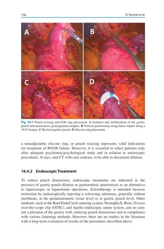
![15916 RYGB Revision and Conversion to Other Procedures
16.4.3 Conversion to Distal RYGB
An increase in the quota of malabsorption may be indicated, with the aim of
optimizing weight loss, for patients who have an intact restrictive RYGB
mechanism (gastric pouch volume <30 ml and gastrojejunostomy <1.5 cm) and
who, during preoperative evaluation, have been diagnosed with eating disorders
such as polyphagia/grazing. This can be done surgically, increasing the length of
the alimentary limb, the biliopancreatic limb, or both. Distalization of the bypass
consists of recreating a common limb that has a mean length of ~100–150 cm
(from 75 to 300 cm depending on the author). A common channel <75 cm is
not acceptable, as it carries life-threatening protein calorie malnutrition (PCM)
[34]. Sugerman et al. [35] were the first to report the results of 27 conversions
from RYGB to DRYGB. The alimentary limb was lengthened from 40 cm to
145 cm and, without measuring the biliopancreatic limb, anastomosed to the
ileum 50–150 cm from the ileocecal valve. The first five patients had a common
channel that measured 50 cm, and results in nutritional terms were catastrophic.
Two patients died of hepatic failure. In the other patients, construction of a
common limb of 150 cm was sufficient to avoid fatal complications. However,
18% of patients required total parenteral nutrition, and 9% eventually required
a new operation to lengthen the common channel. Results in terms of weight
loss were satisfactory, with a mean %EWL of 69% and complete resolution of
comorbidities at 5 years after adding malabsorption. Fobi and colleagues [36]
reported 65 patients who, after failed RYGB and Fobi pouch operation as first
revisional procedure, were converted to DRYGB, moving the Roux limb distally
halfway down the common channel (common limb: 200–300 cm). However,
23% of patients developed PCM to such an extent that it required total parenteral
nutrition and/or percutaneous gastrostomy feedings, and 9% required re-revision
to short-limb gastric bypass. Brolin and Cody [37] reported a series of 30 patients
with unsatisfactory weight loss after RYGB and who were converted to DRYGB
by detaching the biliopancreatic limb from the Roux limb, reanastomosing it to
the ileum 75–100 cm proximal to the ileocecal valve, and revising or creating
a new pouch/gastrojejunal anastomosis: 47.9% of patients obtained a %EWL of
at least 50% at 1 year postoperatively, 7.4% developed PCM, and 3.7% had to
undergo another operation to lengthen the common channel by reducing the Roux
limb length to 150 cm. Rawlins et al. [17] reported a series of 29 patients who
underwent conversion from a 150-cm proximal Roux limb to a DRYGB (common
channel 100 cm), creating a long biliopancreatic limb. In that study, 31% of
patients developed PCM, 21% required total parenteral nutrition, 27% developed
nonclinical secondary hyperparathyroidism because of vitamin D deficiency, and
82 and 61% experienced vitamin D and vitamin A deficiency, respectively. One
patient (3.4%) required re-revision to provide an additional 100-cm length to the
common channel; mean %EWL was 68.8% at 5-year follow-up. A study recently
published by Caruana et al. [38] demonstrated that conversion from RYGB to
DRYGB can be carried out safely and efficiently, with an acceptable compromise](https://image.slidesharecdn.com/bariatricandmetabolicsurgery-180301214153/85/Bariatric-and-metabolic-surgery-164-320.jpg)
![160 D. Tassinari et al.
between weight loss and PCM, if the percentage of bypassed intestine during
revisional operation is <70%. In their study, the Roux limb was detached at the
previous jejunojejunostomy, brought distally, and anastomosed to the ileum at
a distance from the cecum ranging between 120 and 300 cm. A threshold of
70% of small-bowel bypassed from the food stream was identified. Patients in
whom this threshold was exceeded experienced a higher percentage of PCM and
diarrhea compared with those below the threshold. Patients with ≥70% of the
small bowel bypassed had a mean %EWL of 47%, whereas those with <70% had
a mean %EWL of 26% at 2-year follow-up. Although in terms of weight loss
results were more modest, taking into account the pros and cons, exceeding the
threshold of 70% is not recommended.
Revision of short-limb gastric bypass to DRYGB is a good option to enhance
weight loss but is complicated by an increased incidence of PCM. We believe
that the proposal by Caruana et al. [38] not to use fixed measurements when
constructing the DRYGB but to personalize the operation case by case without
exceeding the threshold of 70% of bypassed intestine is the safest method.
However, we strongly recommend DRYGB only for selected patients who show
they are able to follow the nutritional indications given and adhere to a strict
follow-up program.
16.4.4 Conversion of RYGB to Biliopancreatic Diversion with
Duodenal Switch
Literature shows that BPD-DS is the best operation in bariatric surgery for long-
term sustained weight loss. Many studies have demonstrated that BPD-DS allows
maintenance of a %EWL of 70–80%, even at 5 and 10 years from operation
[39]. BPD-DS can also be suggested as revisional surgery after RYGB with the
aim of optimizing weight loss by increasing the malabsorption component. This
conversion should provide an additional %EWL of 15–20%, even in patients
who obtained good weight loss after RYGB. Technically, conversion to BPD-
DS is the most complex procedure of all possible revisions and can be done
in one or two stages either open or laparoscopically. Laparoscopic conversion
requires extensive experience in both advanced laparoscopy and bariatric
surgery. The operation consists of two different procedures: the first one is
reversing the operation back to the normal gastrointestinal anatomy followed by
a sleeve gastrectomy; the second is the classic BPD-DS. The surgeon performs
four anastomoses: gastrogastrostomy, duodenoileostomy, ileoileostomy, and
jejunojejunostomy to reconnect the old Roux limb. It is necessary to take
down the gastrojejunostomy anastomosis and to reconstruct gastric continuity
with a gastrogastrostomy that can be done with a circular or linear stapler or
can be hand sewn. At this point, a sleeve gastrectomy is performed along a
bougie. There are differing opinions regarding appropriate bougie size [39–41].
Gagner et al. [42] suggest a 60-Fr bougie to permit adequate protein intake and](https://image.slidesharecdn.com/bariatricandmetabolicsurgery-180301214153/85/Bariatric-and-metabolic-surgery-165-320.jpg)
![16116 RYGB Revision and Conversion to Other Procedures
digestion. Then, duodenoileostomy and ileoileostomy are performed to create
a long Roux-en-Y with a 100-cm common channel and a 150-cm alimentary
limb, as described by Rabkin et al. [43] or basing the Roux-en-Y construction
on the percentage of total bowel length (common limb 8–12%; alimentary limb
35–45%), as suggested by Hess and Hess [44]. Finally, jejunojejunostomy of
the previous RYGB is resected, and continuity is re-established by anastomosis
of the biliopancreatic limb and proximal end of the old Roux limb. Given the
technical complexity of the operation, even in expert hands, conversion from
RYGB to BPD-DS is encumbered by a percentage of complications that cannot be
neglected. Keshishian et al. [40] reported a series of 31 patients who underwent
laparotomic conversion from RYGB to BPD-DS. In their study, the postoperative
leak rate was 15%, and one half of the leaks required surgical intervention.
Another large series of laparotomic conversions from RYGB to BPD-DS was
reported by Greenbaum et al. [39]; 30% of major complications were described,
including a leak rate of 22%, 2% small-bowel obstruction, 2% stenosis of the
vertical sleeve, 2% duodenoenterostomy stenosis requiring endoscopic dilation,
and 2% pulmonary embolism. Reoperation rate was 7%. To our knowledge, the
only large series of laparoscopic conversions from RYGB to BPD-DS is the
one reported by Gagner et al. [42] (12 patients). Four patients (33%) developed
stricture at the gastrogastrostomy: three were performed using a circular stapler,
and one was hand sewn; no leaks were reported. According to the literature,
after conversion to BPD-DS, the mean %EWL at the 1- and 2-year follow-up is
59–63% and 69–77%, respectively [39, 40, 42].
We conclude that although conversion to BPD-DS for failed RYGB is highly
effective, it is encumbered by a high percentage of complications that cannot be
ignored. A nutritional follow-up is fundamental for these patients, together with
vitamin supplements that must be taken throughout their lives, just as in primary
BPD-DS.
16.5 Conclusions
RYGB is one of the most effective procedures in bariatric surgery, but treatment
of failed RYGB is challenging. The first and essential step in the clinical
management of these patients is to determine the reasons behind its failure.
It is crucial to diagnose psychological issues, psychiatric diseases, and eating
disorders that need to be treated by counseling. Equally important is to identify
morphological alterations or anatomical flaws that could lead to weight regain
or failure to lose weight by using upper endoscopy and contrast upper-GI study.
Following an accurate preoperative evaluation, the decision to refer a patient
to revisional surgery must be multidisciplinary. To this day, various surgical
strategies have been described, some with the aim of adding restriction to the
primary RYGB and others with the purpose of increasing malabsorption. We](https://image.slidesharecdn.com/bariatricandmetabolicsurgery-180301214153/85/Bariatric-and-metabolic-surgery-166-320.jpg)
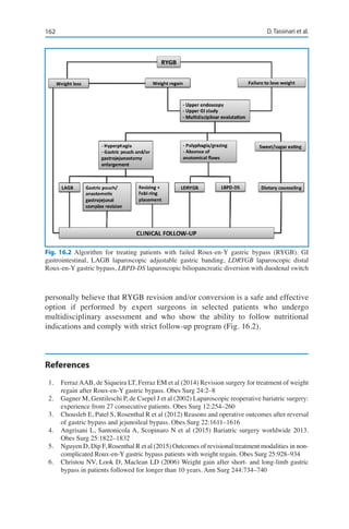
![165L. Angrisani (Ed), Bariatric and Metabolic Surgery,
Updates in Surgery
DOI: 10.1007/ 978-88-470-3944-5_17, © Springer-Verlag Italia 2017
A. Santonicola (*)
Gastrointestinal Unit, Department of Medicine and Surgery, University of Salerno
Salerno, Italy
e-mail: antonellasantonicola83@gmail.com
17The Problem of Gastroesophageal Reflux
Disease and Hiatal Hernia
Paola Iovino, Antonella Santonicola, and Luigi Angrisani
17.1 Introduction
The most widely accepted definition of gastroesophageal reflux disease (GERD)
is a condition that develops when stomach contents cause troublesome symptoms
and/or complications [1]. GERD is a common condition with multifactorial
pathogenesis affecting about 10–20% of the Western population [2]. Some
contributing factors play a role in the provocation of GERD, specifically,
dysfunction of the esophagogastric junction (EGJ). The EGJ consists of three
components: the lower esophageal sphincter (LES), the crural diaphragm, and
the anatomical flap valve. It is structurally and functionally designed to act as an
antireflux barrier in which the tonically contracted smooth muscle of the LES is
surrounded by oblique gastric fibers that are attached to the striated muscle of
the crural diaphragm by the phrenoesophageal ligament [3]. The right crus of
the diaphragm forms a sling that surrounds the distal esophagus and acts as an
extrinsic sphincter by augmenting the high-pressure zone of the LES. When a
proximal displacement of EGJ occurs, which is likely caused by the weakening or
rupture of the phrenoesophageal ligament [4], a hiatal hernia is present because a
spatial dissociation of the antireflux barrier at the EGJ into the intrinsic sphincter
and extrinsic sphincter crural diaphragm exists [5]. The most comprehensive
classification scheme [6] recognizes four types of hiatal hernia (Fig. 17.1):
• Type I or sliding hernia (>95% of cases) is characterized by a widening
of the muscular hiatus and circumferential laxity of the phrenoesophageal
membrane, allowing a portion of the gastric cardia to herniate upward.
• Type II is characterized by a localized defect in the phrenoesophageal
membrane, while the gastroesophageal junction remains fixed to the preaortic
fascia and the median arcuate ligament.](https://image.slidesharecdn.com/bariatricandmetabolicsurgery-180301214153/85/Bariatric-and-metabolic-surgery-170-320.jpg)
![166 P. Iovino et al.
• Type III has elements of both types I and II, with progressive enlargement
of the hiatus that allows increasing amounts of fundus and LES to migrate
through the hiatus.
• Type IV is associated with a massive defect in the phrenoesophageal
membrane, allowing not only the LES and gastric fundus to herniate, but also
other abdominal organs, such as the pancreas, spleen, omentum, and/or small
and/or large intestine.
The presence of hiatal hernia is considered an independent risk factor for
GERD [7]. It has been reported that ~75% of individuals with esophagitis and
90% of patients with Barrett’s esophagus (BE) have a hiatal hernia [8, 9] Among
the different types of hiatal hernia, type I (sliding) is closely associated with
GERD [10]. Although sensitivity and specificity are not ideal, heartburn and
regurgitation are considered typical symptoms sufficient to make a presumptive
diagnosis of GERD [1], taking into account patient age and in the absence of
other concerning symptoms or signs (the so-called alarm signs), which include
dysphagia, odynophagia, weight loss, gastrointestinal (GI) bleeding, vomiting,
family history of cancer, and epigastric mass. In such situations, upper-GI
Esophagus
Esophagogastric
junction
Diaphragm
Stomach
Herniated
Stomach
Type I
Type II
Type III
Type IV
Diaphragm
Diaphragm Diaphragm
Herniated
Stomach
Herniated
Stomach
Stomach
Stomach
Esophago-
gastric
junction
Esophago-
gastric
junction
Esophago-
gastric
junction
Esophagus
Esophagus
Esophagus
Herniated
Stomach
Herniated
transverse
colon
Transverse colon Omentum
Fig. 17.1 Schematic representation of different types of hiatal hernia](https://image.slidesharecdn.com/bariatricandmetabolicsurgery-180301214153/85/Bariatric-and-metabolic-surgery-171-320.jpg)
![16717 The Problem of Gastroesophageal Reflux Disease and Hiatal Hernia
endoscopy should be considered. However, the most sensitive and objective
means for assessing reflux is ambulatory 24-h pH impedance monitoring.
Classically, the diagnosis of hiatal hernia relies on its presence during endoscopy
or barium swallow study [11, 12]. The diagnosis of hiatal hernia with these
techniques has several limitations. One limitation is that these are snapshot
techniques, and the presence of a hiatal hernia is consequently considered an all-
or-nothing phenomenon. Furthermore, diagnosis of a small hiatal hernia could be
challenging [13]. A recent study suggests that high-resolution manometry (HRM)
has a high sensitivity and specificity (92 and 93%, respectively) for detecting a
hiatal hernia [13], allowing a dynamic evaluation of EGJ with a more accurate
analysis. The combination of endoscopy and HRM could reach a sensitivity
of 98% in the diagnosis of hiatal hernia, making it redundant to perform an
additional barium esophagogram for the preoperative diagnosis of hiatal hernia.
Nowadays, intraoperative findings of hiatal hernia are considered the reference
standard for its diagnosis and classification [14].
17.2 GERD and Hiatal Hernia in the Obese Population
Obesity is considered an independent risk factor for GERD. Obese patients tend
to have more severe erosive esophagitis and have a greater risk of developing
BE and adenocarcinoma of the esophagus compared with individuals of normal
weight [15, 16]. The exact pathophysiological link between obesity and GERD
has not been completely determined as yet. Among the proposed mechanisms
are the greater frequency of transient low esophageal sphincter relaxation
(TLESR), increased prevalence of esophageal motor disorders, diminished LES
pressure, as well as presence of hiatal hernia [16, 17]. Obese patients, in fact, are
more than three times as likely to have hiatal hernia compared with nonobese
individuals [18]. A recent study on 142 obese patients who were candidate to
primary bariatric surgery [19] reveals the presence of hiatal hernia in about 23%
of cases, which were, for the most part, asymptomatic. Although ~50–70% of
patients undergoing bariatric surgery have asymptomatic hiatal hernia [20],
obese patients show higher symptoms score, abnormal acid exposure at 24-h
ambulatory pH-metry, and lower LES pressure compared with controls [21].
Other authors confirmed these data, showing that 73% of morbidly obese patients
had some abnormal 24-h pH monitoring findings and 51.7% had an elevated
DeMeester score [22].
In conclusion, GERD with or without hiatal hernia can manifest in a variety
of forms: from no visible esophageal injury at endoscopy, also called nonerosive
reflux disease (NERD), to esophageal injury or erosive reflux disease (ERD);
from metaplasia of the squamous esophageal mucosa to a columnar phenotype or
BE. It is not clear whether these manifestations are part of a continuous spectrum
or distinct phenotypes of GERD [23, 24]. However, this wide range of clinical](https://image.slidesharecdn.com/bariatricandmetabolicsurgery-180301214153/85/Bariatric-and-metabolic-surgery-172-320.jpg)
![168 P. Iovino et al.
conditions increases the need of more preoperative investigations and influences
the choice of procedure.
17.3 GERD and Bariatric Surgery
17.3.1 Gastric Banding
Literature data shows that ~80% of patients with gastric banding (GB) report
resolution of GERD symptoms at short-term follow-up [25, 26]. GERD remission
after GB was also evaluated using 24-h pH and manometry recordings at a mean
follow-up of 19 months, revealing a significant decrease in total number of
reflux episodes, total reflux time, and DeMeester score [27]. In patients with
preoperative hiatal hernia, GB was performed with concomitant hiatal hernia
repair with good results [28]. It has been hypothesized that the unfilled Lap-
Band, when placed more proximally at the EGJ, could be an effective antireflux
device, probably because it creates a longer intra-abdominal pressure zone or
pulls the stomach more into the abdomen in the presence of a hiatal hernia [29].
Three years after GB, some authors described the resolution of GERD symptoms
[30], whereas others reported new-onset GERD in 20.5% of patients [31]. Pouch
formation is a crucial event in the occurrence of GERD after GB. Uncorrected
band placement is considered the primary cause of early pouch dilation, whereas
late pouch dilation is attributed to the inclusion of fundus above the band
[29]. Another possible cause of GERD after GB is esophageal dilation. It is
hypothesized that the inflated band reduces trans-stomal flow, causing reduced
esophageal clearance, stasis of ingested food, and subsequently its reflux [32].
Given data on increased adverse outcomes, including GERD. in the long term
with laparoscopic adjustable gastric banding (LAGB), as well as the high rate of
reoperation or conversion to a more definitive bariatric surgery, the procedure is
expected to be used less frequently going forward [33].
17.3.2 Sleeve Gastrectomy
Current data about the effect of sleeve gastrectomy (SG) on GERD are still
controversial [34]. At short follow-up after SG, some authors reveal a discrete
percentage of GERD remission [35, 36] and others a worsening of GERD,
demonstrating a high prevalence of de novo erosive esophagitis [37] or a
significant decrease of LES pressure with an increase of DeMeester score [38].
Data on long-term effects of SG are still scarce: Himpens et al [39] showed a
biphasic pattern of GERD prevalence after sleeve gastrectomy, with 21.8% de
novo proton pump inhibitor (PPI) use 1 year after SG, which improved to 3.1%](https://image.slidesharecdn.com/bariatricandmetabolicsurgery-180301214153/85/Bariatric-and-metabolic-surgery-173-320.jpg)
![16917 The Problem of Gastroesophageal Reflux Disease and Hiatal Hernia
at 3 years but increased again to 23% at 6 years. In our recently study [40],
we described at 5-year follow-up the resolution of GERD symptoms in 65%
of patients with preoperative body mass index (BMI) ≤50 kg/m2
and 44% in
those with BMI >50 kg/m2
; we also reported new-onset GERD in 15 and 8%,
respectively. Other studies reported a similar percentage of GERD resolution,
from 53 to 60% [41, 42], with new-onset GERD seen in 11% of patients [41]. The
reduction of weight and visceral adiposity and/or an accelerated gastric emptying
might explain the positive effect of SG on GERD [34, 43]. Conversely, multiple
factors may explain GERD worsening after SG [34]. One proposed mechanism
is alteration of the angle of His, which normally acts as a valve to prevent reflux
of stomach contents into the esophagus. Himpens et al. [39] showed that after
3 years, the rate of GERD after SG decreased, potentially due to restoration
of the angle of His. Another possible factor is LES dysfunction. Specifically,
after transection near the angle of His during gastrectomy, the sling fibers at the
fundus are divided, which can subsequently decrease LES pressure. A significant
decrease in LES pressure was, in fact, reported 3 months after SG [44]. The role
of concomitant hiatal hernia repair during SG is still debated. At short term,
some authors found an improvement of GERD symptoms [45, 46]. However,
our recent study demonstrated no significant improvement in frequency-intensity
of typical GERD symptoms in obese patients with GERD and hiatal hernia
who underwent SG with concomitant hiatal hernia repair 16 ± 8 months earlier
[36]. Recently, Samakar et al. [47] confirmed our results, demonstrating that
at mean 2-year follow-up period, two thirds of symptomatic patients remained
symptomatic after LSG with concomitant hiatal hernia repair and that 15.6 % of
previously asymptomatic patients developed de novo reflux symptoms. We agree
with the hypothesis of authors who suggest the routine repair of small hiatal
hernias may contribute to LES dysfunction by disrupting the normal anatomical
barriers to reflux in order to perform the repair. Potential repair breakdown may
also lead to worsening of the hiatal hernia, given the dissection that takes place
in order to perform the cruroplasty.
17.3.3 Roux-en-Y Gastric Bypass
Roux-en-Y gastric bypass (RYGB) is associated with a good outcome in
regard to GERD. Symptom resolution or improvement has been described in
several studies [48, 49]. Madalosso et al. [49] reported a significant reduction
of GERD symptoms, reflux esophagitis, and DeMeester scores after 39 ± 7
months. The positive effect of RYGB on GERD may be explained by multiple
factors. Creation of a small gastric pouch and separation of most of the stomach
drastically reduce the acid that could promote regurgitation. Importantly, bile
reflux is also eliminated due to biliary diversion. In fact, in patients undergoing
surgery for morbid obesity, RYGB is the procedure of choice for patients with
concomitant severe GERD.](https://image.slidesharecdn.com/bariatricandmetabolicsurgery-180301214153/85/Bariatric-and-metabolic-surgery-174-320.jpg)
![170 P. Iovino et al.
A recent variation on RYGB is the omega-loop gastric bypass, also known
as the mini-gastric bypass or one-anastomosis gastric bypass. There have been
concerns about the proximity of the biliary flow to the gastric tube in this procedure
compared with RYGB and the subsequent potential for both biliary reflux and
esophagitis. However, a recent study using HRM and 24-h pH impedance
monitoring performed both before and 1 year after omega-loop gastric bypass
demonstrated that this procedure did not cause de novo gastroesophageal reflux
or esophagitis [50]. Other data on the risk of developing de novo GERD after
this particular procedure is needed, especially as there is a lack of studies of this
procedure in obese patients with GERD.
In our opinion, in patients with a moderate to large hiatal hernia and/or severe
esophagitis and/or BE, RYGB with or without hiatal hernia repair should be
preferred.
References
1. Vakil N, van Zanten SV, Kahrilas P et al (2006) The Montreal definition and classification
of gastroesophageal reflux disease: a global evidence-based consensus. Am J Gastroenterol
101:1900–1920
2. Katz PO, Gerson LB, Vela MF (2013) Guidelines for the diagnosis and management of
gastroesophageal reflux disease. Am J Gastroenterol 108:308–328
3. van Herwaarden MA, Samsom M, Smout AJ (2004) The role of hiatus hernia in gastro-
oesophageal reflux disease. Eur J Gastroenterol Hepatol 16:831–835
4. Curci JA, Melman LM, Thompson RW et al (2008) Elastic fiber depletion in the supporting
ligaments of the gastroesophageal junction: a structural basis for the development of hiatal
hernia. J Am Coll Surg 207:191–196
5. Sloan S, Rademaker AW, Kahrilas PJ (1992) Determinants of gastroesophageal junction
incompetence: hiatal hernia, lower esophageal sphincter, or both? Ann Intern Med
117:977–982
6. Kahrilas PJ, Kim HC, Pandolfino JE (2008) Approaches to the diagnosis and grading of
hiatal hernia. Best Pract Res Clin Gastroenterol 22:601–616
7. Boeckxstaens GE (2007) Review article: the pathophysiology of gastro-oesophageal reflux
disease. Aliment Pharmacol Ther 26:149–160
8. Zagari RM, Fuccio L, Wallander MA et al (2008) Gastro-oesophageal reflux symptoms,
oesophagitis and Barrett’s oesophagus in the general population: the Loiano-Monghidoro
study. Gut 57:1354–1359
9. Cameron AJ (1999) Barrett’s esophagus: prevalence and size of hiatal hernia. Am J
Gastroenterol 94:2054–2059
10. Hyun JJ, Bak YT (2011) Clinical significance of hiatal hernia. Gut Liver 5:267–277
11. Ott DJ, Gelfand DW, Chen YM et al (1985) Predictive relationship of hiatal hernia to reflux
esophagitis. Gastrointest Radiol 10:317–320
12. Patti MG, Goldberg HI, Arcerito M et al (1996) Hiatal hernia size affects lower esophageal
sphincter function, esophageal acid exposure, and the degree of mucosal injury. Am J Surg
171:182–186
13. Weijenborg PW, van Hoeij FB, Smout AJ, Bredenoord AJ (2015) Accuracy of hiatal
hernia detection with esophageal high-resolution manometry. Neurogastroenterol Motil
27:293–299](https://image.slidesharecdn.com/bariatricandmetabolicsurgery-180301214153/85/Bariatric-and-metabolic-surgery-175-320.jpg)
![172 P. Iovino et al.
37. Tai CM, Huang CK, Lee YC et al (2013) Increase in gastroesophageal reflux disease
symptoms and erosive esophagitis 1 year after laparoscopic sleeve gastrectomy among obese
adults. Surg Endosc 27:1260–1266
38. Gorodner V, Buxhoeveden R, Clemente G et al (2015) Does laparoscopic sleeve gastrectomy
have any influence on gastroesophageal reflux disease? Preliminary results. Surg Endosc
29:1760–1768
39. Himpens J, Dobbeleir J, Peeters G (2010) Long-term results of laparoscopic sleeve gastrec-
tomy for obesity. Ann Surg 252:319–324
40. Angrisani L, Santonicola A, Hasani A et al (2015) Five-year results of laparoscopic sleeve
gastrectomy: effects on gastroesophageal reflux disease symptoms and co-morbidities. Surg
Obes Relat Dis pii:S1550-7289(15)00855-2
41. Rawlins MP, Brown CC, Schumacher DL (2013) Sleeve gastrectomy: 5-year outcomes of a
single institution. Surg Obes Relat Dis 9:21–25
42. Hirth DA, Jones EL, Rothchild KB et al (2015) Laparoscopic sleeve gastrectomy: long-term
weight loss outcomes. Surg Obes Relat Dis pii: S1550-7289(15)00049-0
43. Melissas J, Leventi A, Klinaki I et al (2013) Alterations of global gastrointestinal motility
after sleeve gastrectomy: a prospective study. Ann Surg 258:976–982
44. Burgerhart JS, Schotborgh CA, Schoon EJ et al (2014) Effect of sleeve gastrectomy on
gastroesophageal reflux. Obes Surg 24:1436–1441
45. Soricelli E, Iossa A, Casella G et al (2013) Sleeve gastrectomy and crural repair in obese
patients with gastroesophageal reflux disease and/or hiatal hernia. Surg Obes Relat Dis
9:356–361
46. Ruscio S, Abdelgawad M, Badiali D et al (2015) Simple versus reinforced cruroplasty in
patients submitted to concomitant laparoscopic sleeve gastrectomy: prospective evaluation
in a bariatric center of excellence. Surg Endosc [Epub ahead of print] doi:10.1007/s00464-
015-4487-0
47. Samakar K, McKenzie TJ, Tavakkoli A et al (2016) The effect of laparoscopic sleeve gas-
trectomy with concomitant hiatal hernia repair on gastroesophageal reflux disease in the
morbidly obese. Obes Surg 26:61–66
48. Mejía-Rivas MA, Herrera-López A, Hernández-Calleros J et al (2008) Gastroesophageal
reflux disease in morbid obesity: the effect of Roux-en-Y gastric bypass. Obes Surg
18:1217–1224
49. Madalosso CA, Gurski RR, Callegari-Jacques SM et al (2016) The impact of gastric bypass
on gastroesophageal reflux disease in morbidly obese patients. Ann Surg 263:110–116
50. Tolone S, Cristiano S, Savarino E et al (2016) Effects of omega-loop bypass on esophago-
gastric junction function. Surg Obes Relat Dis 12:62–69
51. Jeur AS (2010) Review of literatures on laparoscopic prosthetic repair of giant hiatal hernia
than pure anatomical repair of crura. World J Lap Surg 3:85–90](https://image.slidesharecdn.com/bariatricandmetabolicsurgery-180301214153/85/Bariatric-and-metabolic-surgery-177-320.jpg)
![173L. Angrisani (Ed), Bariatric and Metabolic Surgery,
Updates in Surgery
DOI: 10.1007/ 978-88-470-3944-5_18, © Springer-Verlag Italia 2017
P. Gentileschi (*)
Bariatric Surgery Unit, University of Rome Tor Vergata
Rome, Italy
e-mail: gentileschi.paolo@gmail.com
18Diabetes Surgery: Current Indications
and Techniques
Paolo Gentileschi, Stefano D’Ugo, and Francesco Rubino
18.1 Introduction
Being pathologically overweight is an epidemic problem, with increasing
numbers; global obesity rates have doubled in the last two decades, reaching 500
million in 2008. The current prevalence of 7–10% in children and adolescents
is predicted to double by 2025, leading to a significant increase in obesity-
related diseases [1, 2]. A large number of those patients will have multiple
comorbidities, which will create a drastic negative impact on society, health-
care systems, and life expectancy. Nonsurgical approaches to treating obesity,
including a combination of lifestyle modifications, diets, and drugs, have shown
limited long-term success. This concept is particularly true for obese people
with type 2 diabetes mellitus (T2DM). The estimated worldwide prevalence of
T2DM among adults was 285 million in 2010, and this is projected to increase
to 439 million by 2030. Managing diabetes has historically been medical, based
on lifestyle interventions combined with pharmacotherapy. However, although
the pharmacological armamentarium to treat T2DM has expanded considerably,
few patients are able to achieve and maintain optimal glycemic targets in the
long term [3, 4].
In contrast to nonsurgical treatments, bariatric surgery has been consistently
shown to induce greater and more sustained weight loss; a systematic review
reported an overall percentage of excess weight loss (%EWL) of >60% with
bariatric surgery and ~70 with gastric bypass. Moreover, with the widespread
adoption of minimally invasive laparoscopic techniques, bariatric surgery
has become safer. Current mortality risk rates are in the range of 0.2–0.5%,
similar to those of other commonly performed operations, such as laparoscopic
cholecystectomy [5]. Long-term results are as equally encouraging. The Swedish](https://image.slidesharecdn.com/bariatricandmetabolicsurgery-180301214153/85/Bariatric-and-metabolic-surgery-178-320.jpg)
![174 P. Gentileschi et al.
Obese Subjects Research Program, a nonrandomized but controlled longitudinal
study, documented sustained weight loss at 10, 15 and 20 years (17, 16, and
18% respectively) in operated patients compared with minimal or no results in
matched controls undergoing conventional weight loss treatment [6].
Several gastrointestinal operations have been described over recent years
aimed primarily at producing significant and durable weight loss in morbidly
obese patients. These procedures have also been shown in multiple trials to
induce remission or dramatic improvement of T2DM and other obesity-related
comorbidities. While improvement of diabetes and other metabolic disorders is an
expected outcome of weight loss by any means, evidence from both experimental
animal studies and clinical investigations suggests that these effects are partly
independent of weight loss. This knowledge provided a rational to the idea of
a “diabetes surgery” specifically aimed at treating T2DM [7]. Following this
new option for diabetic patients, the concept of “metabolic surgery” has rapidly
emerged in the scientific community to more broadly indicate a surgical approach
aimed at controlling metabolic illnesses, not just excess weight.
Efficacy and safety of bariatric surgery to treat T2DM in obese patients have
been demonstrated in many papers published in recent years. The scientific
community has recommended the use of bariatric surgery in patients with
diabetes and body mass index (BMI) >35 kg/m2
and as an alternative treatment
option in patients with BMI 30–35 kg/m2
inadequately controlled with optimal
medical regimens [8].
18.2 Metabolic Surgery: Mechanisms of Glycemic Control
Morbidly obese individuals with T2DM undergoing bariatric surgery reach
sustained weight loss and substantial improvement in glucose metabolism. In
many cases, good glycemic control is maintained without insulin injections or
even medications [16, 17]. The first explanation for this effect is major weight loss;
indeed, surgery is a primary trigger for increased insulin sensitivity, accompanied
by cellular and tissue changes at multiple levels in the metabolism of glucose.
However, growing evidence indicates that the antidiabetic mechanisms of some
of these operations cannot be explained by changes in caloric intake and body
weight alone. In fact, rearrangement of the gastrointestinal anatomy seems to
play an important role, independently of weight loss, as shown at different levels:
• Using the duodenojejunal bypass (DJB) model in rodents, excluding the
proximal small intestine (duodenum and proximal jejunum) from the passage
of food contributes to the resolution (or improvement) of diabetes after other
diversionary procedures [Roux-en-Y gastric bypass (RYGB), biliopancreatic
diversion (BPD)] independently of weight loss.
• Diabetic patients after surgery, mainly RYGB and BPD, show a rapid im-
provement or resolution of T2DM long before substantial weight loss occurs.](https://image.slidesharecdn.com/bariatricandmetabolicsurgery-180301214153/85/Bariatric-and-metabolic-surgery-179-320.jpg)
![17518 Diabetes Surgery: Current Indications and Techniques
• Different bariatric procedures can achieve similar weight loss but different
glucose homeostasis. For example: RYGB achieves greater improvement
of glucose tolerance and beta-cell function than an equivalent magnitude
of weight loss achieved by purely gastric-restrictive bariatric surgery, such
as laparoscopic adjustable gastric banding (LAGB) or following calorie
restrictions.
The exact molecular mechanisms underlying improved glycemic control after
gastrointestinal surgery remain unclear. In spite of anatomical and functional
differences between procedures, glucose homeostasis is improved after all
these types of operations, probably as an expression of partial overlap in the
mechanisms of action. However, given the specific physiological role of the
stomach and various intestinal segments in regulating glucose homeostasis, it is
also plausible that different gastrointestinal surgeries may have distinct effects
and mechanisms of action.
Several studies showed the involvement of many key peptides believed to
have a role in regulating insulin secretion, including incretin peptides, especially
glucagon-like peptide-1 (GLP-1) and peptide tyrosine-tyrosine (PYY) [18].
These factors are produced by intestinal enteroendocrine cells in response to
ingestion of carbohydrates or fats, which in turn cause the release of insulin
from the pancreas and induce satiety/reduced appetite; their changes after
bariatric surgery could potentially explain the effects on obesity and diabetes.
The surgical trigger for these changes is not clear; recent studies suggest the
importance of modifications of gastric anatomy and physiology (emptying) more
than the intestinal bypass per se [19].
One antidiabetic effect of bariatric surgery is weight independent; a number
of experimental investigations have postulated changes in intestinal nutrient-
sensing, regulating insulin sensitivity; disruption of vagal afferent and efferent
innervations;perturbationsofbileacidmetabolism;tastealterations;enhancement
of intestinal glucose uptake in the alimentary limb after diversionary procedures;
and downregulation of one or more anti-incretin factors [7, 20–23]. However,
despite much research, the exact pathway to diabetes improvement remains
unidentified.
18.3 Bariatric/Metabolic Surgery in Patients with Type 2
Diabetes
18.3.1 Patients with BMI >35 kg/m2
Several papers in recent years have clearly documented results of metabolic
surgery on T2DM and associated comorbidities in patients with a BMI >35 kg/m2
.
Buchwald et al. in a meta-analysis of 22,094 diabetic patients, reported an overall
77% remission rate. The mean procedure-specific resolution of T2DM was 48%](https://image.slidesharecdn.com/bariatricandmetabolicsurgery-180301214153/85/Bariatric-and-metabolic-surgery-180-320.jpg)
![176 P. Gentileschi et al.
for LAGB, 68% for vertical banded gastroplasty (VBG), 84% for RYGB, and
98% for BPD. However, these results were mainly from retrospective studies
with short follow-up [5].
The multicentre Swedish Obese Subjects (SOS) study, a large prospective
observational study, compared bariatric surgery LAGB (n = 156), VBG (n =
451), and RYGB (n = 34) versus conservative medical management in a group
of well-matched obese patients. At 2 years, 72% of patients in the surgical group
achieved remission of T2DM compared with 21% in the medically treated arm.
At 10 years, the relative risk (RR) of incident T2DM was three times lower and
the rates of recovery from T2DM three times greater for patients who underwent
surgery compared with individuals in the control group. The proportion of
individuals in whom remission was sustained at 10 years declined to 36% in the
surgical group and 13% in the medical group [24].
More recently, sleeve gastrectomy (SG) has gained popularity because
of its low morbidity, reasonably quick operative time, no anastomosis, and
its effectiveness in obtaining effective weight loss and controlling metabolic
disease. A systematic review by Gill et al. of 27 studies involving 673 patients
(mean follow-up of 13.1 months) reported a T2DM resolution rate of 66.2%
in obese individuals and improved glycemic control in 26.9%. This result has
been confirmed by many other papers, showing that SG also represents a very
effective procedure both in terms of weight reduction and control of T2DM [25].
Efficacy of bariatric/metabolic surgery has been showed also in several
randomized controlled trials (RCT) comparing medical versus surgical
management of T2DM. The STAMPEDE (Surgical Treatment and Medications
Potentially Eradicate Diabetes Efficiently) trial compared intensive medical
therapy alone versus medical therapy plus RYGB or SG in 150 obese patients
(BMI 27–43 kg/m2
) with uncontrolled T2DM. The proportion of patients who
reached the primary end point [glycolated hemoglobin (HbA1c) of 6.0% at 1
year) was 12% in the medical group, 42% in the RYGB group, and 37% in the
SG group. No patient in the RYGB group required diabetes medications, unlike
patients in the SG group (although the study was not powered to demonstrate
differences between surgical procedures, these findings suggest a potentially
greater antidiabetic effect of RYGB). Surgery showed a better outcome also for
secondary end points, including BMI, body weight, waist circumference, and the
homeostasis model assessment of insulin resistance (HOMA-IR). Although the
study presented promising results, follow-up was limited to 12 months, with no
reports about long-term control and risk of disease relapse [26].
In the Diabetes Surgery Study (DSS) trial, 120 patients were randomized to
receive intensive lifestyle and medical therapy with or without RYGB. The pri-
mary endpoint was a composite outcome including achievement of HbA1c <7.0%,
low-density lipoprotein (LDL) cholesterol <100 mg/dL, and systolic blood pres-
sure <130 mmHg. After 12 months, 28 participants [49%; 95% confidence interval
(CI) 36–63%) in the gastric bypass group and 11 (19%; 95% CI 10–32%) in the
lifestyle-medical management group achieved the primary end points [27].](https://image.slidesharecdn.com/bariatricandmetabolicsurgery-180301214153/85/Bariatric-and-metabolic-surgery-181-320.jpg)
![17718 Diabetes Surgery: Current Indications and Techniques
In 2012, a randomized clinical trial by Mingrone et al. reported results at 2
years of 60 patients (BMI ≥35 kg/m2
) with T2DM of at least 5-year duration and
an HbAlc level of ≥7.0% comparing conventional medical therapy to RYGB
and BPD. Diabetes remission (fasting glucose level <100 mg/dL and an HbA1c
level of <6.5% in the absence of pharmacological therapy) occurred in no
patient in the medical therapy group versus 75% in the RYGB group and 95%
in the BPD group. All patients in the surgical group were able to discontinue
pharmacotherapy (oral hypoglycemic agents and insulin) within 15 days after the
operation. At 2 years, surgical patients had the greatest degree of improvement
in HbA1c levels [28].
Published literature data about long-term remission of T2DM after metabolic
surgery are limited. The SOS study in 2014 presented results of the prospective
matched-cohort study on diabetes control and micro-macrovascular diabetes
complication comparing 270 controls to 343 surgical patients. Surgery was in
the form of gastric banding (GB) (61), vertical banded gastroplasty (VBG) (227),
or RYGB (55). For diabetes assessment, the median follow-up time was 10 years
in both groups. For diabetes complications, the median follow-up time was 17.6
and 18.1 years in control and surgery groups, respectively. Diabetes remission
(defined as blood glucose <110 mg/dL and no medication) at 15 years was 6.5%
for control patients and 30.4% for bariatric surgery patients (p<0.001). With
long-term follow-up, the cumulative incidence of microvascular complications
was 41.8:1000 person-years for controls and 20.6:1000 person-years in the
surgery group (p<0.001). Macrovascular complications were observed in
44.2:1000 person-years in controls and 31.7:1000 person-years for the surgical
groups(p=0.001). This long-term data, although not randomized, shows how
metabolic surgery is associated with more frequent diabetes remission and fewer
complications than standard medical treatments [29].
The first longer-term results from a randomized trial were published by
Mingrone et al. in 2015 [30]. The authors reported data of 5-year outcome from
a trial specifically designed to assess management of T2DM comparing surgery
versus medical treatments. From the group of 60 patients recruited between April
and October 2009, they analyzed glycemic and metabolic control, cardiovascular
risk, medication use, quality of life, and long-term complications. Overall,
19 (50%) of the 38 surgical patients [seven (37%) of 19 in the gastric bypass
group and 12 (63%) of 19 in the BPD group] maintained diabetes remission at
5 years compared with none of the 15 patients in the medically treated group
(p=0.0007). Hyperglycemia relapse occurred in eight (53%) of the 15 patients
who achieved 2-year remission in the gastric bypass group and seven (37%) of
the 19 patients who achieved 2-year remission in the BPD group. Eight (42%)
patients who underwent gastric bypass and 13 (68%) who underwent BPD had an
HbA1c concentration of ≤6.5% (≤47.5 mmol/mol) with or without medication,
compared with four (27%) medically treated patients (p=0.0457). Surgical
patients lost more weight than medically treated patients, but weight changes did
not predict diabetes remission or relapse after surgery. Both surgical procedures](https://image.slidesharecdn.com/bariatricandmetabolicsurgery-180301214153/85/Bariatric-and-metabolic-surgery-182-320.jpg)
![178 P. Gentileschi et al.
were associated with significantly lower plasma lipids, cardiovascular risk,
and medication use. Five major complications of diabetes (including one fatal
myocardial infarction) arose in four (27%) patients in the medical group compared
with only one complication in the gastric bypass group and no complications in
the BPD group [30].
These findings show that bariatric surgery is more effective than medical
treatment for the long-term control of obese patients with T2DM. Compared
with medical treatments, surgery results in sustained remission of diabetes in a
significant number of patients and in a greater reduction of cardiovascular risk,
diabetes-related complications, and medication use, including use of insulin and
cardiovascular drugs.
18.3.2 Patients with BMI <35 kg/m2
Observational studies and RCTs show with growing evidence that patients
with lower BMI may obtain benefits regarding T2DM and comorbidity control
following metabolic surgery. A prospective study of 66 diabetic patients with
BMI 30–35 kg/m2
who underwent RYGB showed remission of diabetes in
~90% and T2DM improvement in ~10% after a follow-up of 6 years. This was
also associated with a cessation of pharmacotherapy for T2DM in the majority
of patients and reduction of medication use (and withdrawal of insulin when
previously used) in the remaining [31].
An RCT by Dixon et al. in 2008 included individuals with BMI 30–35 kg/m2
comparing LAGB to medical treatment. They showed at 2-year follow-up remission
of T2DM in 73% in the surgical group and 13% in the conventional therapy
group; moreover, insulin sensitivity and levels of triglycerides and high-density
lipoprotein (HDL) cholesterol were improved after surgery [32]. Positive results
were also seen in the DSS trial including less obese patients with diagnosis
of T2DM and follow-up of 12 months. Surgery was associated with greater
improvement in HbA1c, LDL-cholesterol, and blood pressure that with medical
treatment [27].
These preliminary results highlight the possible role of metabolic surgery
even in treating T2DM in patients not classified as morbidly obese. However,
despite this data, to define the exact role of surgery in this setting, more RCTs
and longer follow-up results are required, along with assessments of the impact
of surgery on vascular complications of diabetes.
18.4 Diabetes Surgery: A New Point of View
Over the years, the term used for surgical procedures in morbidly obese patients
has been primarily “bariatric surgery.” The main endpoint of these procedures is](https://image.slidesharecdn.com/bariatricandmetabolicsurgery-180301214153/85/Bariatric-and-metabolic-surgery-183-320.jpg)
![17918 Diabetes Surgery: Current Indications and Techniques
reduction of body weight and BMI. However, it became clear early that benefits
and mechanisms of action of bariatric surgery extend beyond weight loss: T2DM
and associated comorbidities can dramatically improve after surgery. This has
led to the development of a new concept of surgery, called diabetes or metabolic
surgery. The primary intent of this surgical approach is to control metabolic
alterations/hyperglycemia, not only to reduce weight. This influences patient
selection, with important clinical care implications. A recent study demonstrated
that patients treated in metabolic surgery units have a higher incidence of T2DM
and comorbidities plus lower BMI than those undergoing surgery in a bariatric
unit [33]. Applying the concept of diabetes/metabolic surgery parameters
to evaluate outcomes of surgery need to be redefined. Success and failure of
this treatment should be assessed by remission or improvement of T2DM and
associated comorbidities of the metabolic syndrome, rather than simply checking
weight loss. In diabetes surgery, success parameters to consider are then no
longer BMI and weight but mainly HbA1c levels, C peptide, fasting glycemia,
insulin levels, lipid profile, and similar indexes.
Asinotherfieldsofmedicine,diabetesandmetabolicsurgerymeansintegration
of knowledge and multidisciplinary expertise to provide a combination of medical
and surgical treatments. These have to be seen not as alternative strategies but
as complementary options, with the same goal of optimizing disease control and
achieving cure when possible.
References
1. World Health Organization, Global Health Observatory (GHO) data (2008) Obesity, situation
and trends. http://www.who.int/gho/ncd/risk_factors/overweight/en
2. McPherson K, Marsh T, Brown M (2007) Tackling obesities: The future choices: mod-
elling future trends in obesity and their impact on health. UK Government Foresight
Programme, Government Office for Science, London. https://www.researchgate.net/
publication/242459208_Tackling_Obesities_The_Future_Choices-Modelling_Future_
Trends_in_Obesity_Their_Impact_on_Health
3. Atallah R, Filion KB, Wakil SM et al (2014) Long-term effects of 4 popular diets on weight
loss and cardiovascular risk factors: a systematic review of randomized controlled trials.
Circ Cardiovasc Qual Outcomes 7:815–827
4. Leblanc ES, O’Connor E, Whitlock EP et al (2011) Effectiveness of primary care-relevant
treatments for obesity in adults: a systematic evidence review for the US Preventive Services
Task Force. Ann Intern Med 155:434–447
5. Buchwald H, Avidor Y, Braunwald E et al (2004) Bariatric surgery: a systematic review and
meta-analysis. JAMA 292:1724–1737
6. Sjostrom L (2013) Review of the key results from the Swedish Obese Subjects (SOS) trial:
a prospective controlled intervention study of bariatric surgery. J Intern Med 273:219–234
7. Rubino F, Gagner M (2002) Potential of surgery for curing type 2 diabetes mellitus. Ann
Surg 236:554–559
8. Dixon JB, Zimmet P, Alberti KG et al (2011) Bariatric surgery for diabetes: the International
Diabetes Federation takes a position. J Diabetes 3:261–264](https://image.slidesharecdn.com/bariatricandmetabolicsurgery-180301214153/85/Bariatric-and-metabolic-surgery-184-320.jpg)
![183L. Angrisani (Ed), Bariatric and Metabolic Surgery,
Updates in Surgery
DOI: 10.1007/ 978-88-470-3944-5_19, © Springer-Verlag Italia 2017
G.D. De Palma (*)
Department of Clinical Medicine and Surgery, University of Naples Federico II
Naples, Italy
e-mail: giovanni.depalma@unina.it
19Endoluminal Procedures
Giovanni Domenico De Palma, Alfredo Genco,
Massimiliano Cipriano, Gaetano Luglio, and Roberta Ienca
19.1 Introduction
Obesity is a very well recognized health-care problem worldwide due to its
increasing prevalence (>35% in USA) [1], to the positive correlation between body
mass index (BMI) and obesity-related comorbidities and mortality, and to huge
costs related to its treatment overall (up to 21% of US health expenditures) [2].
Current treatment options include lifestyle intervention, weight-loss medications,
and bariatric surgery. Lifestyle interventions, including diet and exercises, have
demonstrated to be only modestly effective, with 5–10% of total body weight
loss at 1 year and a significant rate of weight regain later on [3, 4]. If combined
with lifestyle interventions, pharmacotherapy may lead to 5–11% of total body
weight loss [5], but – with the exception of orlistat – there are no data available
on weight-loss maintenance beyond 2-year follow-up due to the recent approval
of some of the novel drugs by the US Food and Drug Administration (FDA).
Bariatric surgery is demonstrated to be significantly effective: gastric bypass,
for example, may allow a percentage of excess weight loss (%EWL) of 62–74%
at 1 year, as demonstrated in a recent meta-analysis [6], and the same review
reports relatively low morbidity rates after bariatric surgery (10–17%) and a low
reoperation rate (6–7%). Nevertheless, even in the minimally invasive surgery
era, less than 1% of severely obese patients will ask for surgery, probably due to
costs related to procedures in some countries, the fear of lack of reversibility, and
the reluctance of primary care physicians to refer patients to a bariatric surgeon
[7]. In this scenario, endoscopic bariatric therapy (EBT) might be considered
as a further clinical tool to fill in the gap among the different treatment options.
EBTs have been shown superior to lifestyle interventions alone and obesity
medications; moreover, the low morbidity rate, the reversibility of some](https://image.slidesharecdn.com/bariatricandmetabolicsurgery-180301214153/85/Bariatric-and-metabolic-surgery-187-320.jpg)
![184 G.D. De Palma et al.
procedures, and the lower BMI threshold required in order to access EBTs, might
be appealing to some patients and referring physicians [6, 8]. As outlined in the
2013 guidelines by the American Heart Association (AHA)/American College
of Cardiology (ACC)/The Obesity Society (TOS), EBTs are appropriate for
inclusion in the obesity treatment algorithm – other than for individuals with
major comorbidities wo are not ideal candidates for surgery – when lifestyle
changes are not sufficient alone, when ideal BMI criteria are satisfied, and as a
bridge treatment to major bariatric procedures [9].
Mechanisms of weight loss of bariatric procedures are not related to me-
chanical restriction or malabsorption only, as initially believed, but trigger physi-
ological alterations in gastrointestinal (GI) motility, gut neuroendocrine system,
autonomic signalling, and gut microbiota, which are also related to diabetes
control. Thanks to a wide variety of novel technologies, EBTs can reproduce
some anatomic alterations and consequent physiological effects of major surgi-
cal procedures proven to be effective in treating severe obesity. EBTs also offers
the potential advantage of reversibility, repeatability, minimal invasiveness, and,
perhaps, cost savings.
19.2 Endoscopic Bariatric Therapies
EBTs can be divided into two main groups: (1) gastric, and (2) small-bowel
endoscopic interventions [10].
19.2.1 Gastric Interventions
Gastric interventions aim to reduce the gastric reservoir, thus inducing an early
satiety and modulating the action of gastric mechanical and chemical receptors,
orexigenic hormones, and gastric emptying.
19.2.1.1 Intragastric Balloon
Intragastric balloon (IGB) treatments are certainly the best known option, and
different devices are available. This approach will be described in detail in the
next section.
19.2.1.2 Aspiration Therapy
Aspiration therapy is a novel approach based on the use of a specifically
designed tube, the AspireAssist (A-tube) (Aspire Bariatrics Inc, King of Prussia,
PA, USA), to perform a percutaneous endoscopic gastrostomy, which is then
connected to the AspireAssist device, which allows aspiration of ~30% of the
meal 20 min after ingestion.](https://image.slidesharecdn.com/bariatricandmetabolicsurgery-180301214153/85/Bariatric-and-metabolic-surgery-188-320.jpg)
![18519 Endoluminal Procedures
19.2.1.3 Gastroplasty Techniques
Gastroplasty attempts to decrease gastric capacity not by occupying space but
by altering the gastric anatomy. Endoscopic sleeve gastroplasty (ESG) aims to
reduce gastric volume by placing a series of full-thickness, closely spaced sutures
through the gastric wall from the prepyloric antrum to the gastroesophageal
junction using an endoscopic stitching device (OverStitch; Apollo Endosurgery
Inc, Austin, TX, USA); thus, stomach volume is reduced along the greater
curvature, creating a sleeve effect. Good results in terms of efficacy have been
reported, with a %EWL between 30 and 40% at 6 months [11, 12]. Long-term
results are awaited.
19.2.2 Small-Bowel Interventions
The second group of endoscopic procedures is represented by small-bowel
interventions. The proximal small bowel plays an important role in nutrient
absorption, glucose homeostasis, and production of gut peptides with entero-
endocrine functions, thus regulating the sense of satiety and rate of insulin
production. The mechanism of action is why bypassing this tract may help in
weight loss and diabetes treatment. The EndoBarrier gastrointestinal liner
system (GI Dynamics Inc, Lexington, MA, USA) aims to create a functional
duodenojejunal bypass using a kind of impermeable tube-shaped Teflon liner,
extending from the duodenal bulb for 65 cm into the small bowel, creating a
mechanical barrier and preventing absorption of pancreaticobiliary secretions,
which then will mix with food more distally in the gastrointestinal tract. The
sleeve is removed endoscopically 12 months after insertion. Similarly, the
gastroduodenal bypass sleeve is a 120-cm-long fluoropolymer sleeve, which is
placed from the gastroesophageal junction using a combined endoscopic and
laparoscopic approach. The procedure should theoretically mimic the Roux-
en-Y gastric bypass, as the stomach, duodenum, and proximal jejunum are all
excluded from food transit.
19.2.3 Implementation
It should be noted that most of these procedures have not been implemented in
routine clinical practice or approved for bariatric indications, at least in USA.
Therefore, to better define the role of such techniques in clinical scenarios, a
joint task force of the American Society of Gastrointestinal Endoscopy (ASGE)
and the American Society for Metabolic and Bariatric Surgery, attempted to
define acceptable thresholds of safety and efficacy for EBTs. The threshold for
major adverse events was set to <5%, while the threshold for efficacy as primary
treatment was set at %EWL of 25% 1 year after a procedure [13].](https://image.slidesharecdn.com/bariatricandmetabolicsurgery-180301214153/85/Bariatric-and-metabolic-surgery-189-320.jpg)
![18719 Endoluminal Procedures
the complication of ulcers extremely rare. The IGB has shown to be minimally
invasive, being a removable device with a high safety and efficacy profile.
To date the BIB – and recently its evolution, the Orbera intragastric balloon,
sold by Apollo Endosurgery (Austin, TX, USA) – remains the most frequently
used and best known brand, although several types of balloons (Heliosphere
bag, Spatz adjustable intragastric balloon, ReShape Duo, Elipse, Obalon) are
available. However, most information in this section refers to the Orbera.
19.3.1 Indications and Contraindications
The Orbera and other IGB are indicated in association with a specific diet
treatment in patients with a history of obesity (at least 5 years) after numerous
failures of dietary treatment only or as bridge to surgery [14, 15].
19.3.1.1 Indications
• Grade 1 obese (BMI 30–34.9 kg/m2
) patients with obesity-related comorbidi-
ties whose improvement requires mandatory weight loss
• High-risk grade 2–3 obese (BMI 35-49.9 kg/m2
) or superobese (BMI >50 kg/m2
)
patients before any other type of surgery (bariatric, orthopedic, cardiac, etc.), as
they could benefit from presurgical weight loss to reduce operative risk and
incidence of postoperative complications [16].
• Patients who refuse bariatric surgical procedures to help them achieve
satisfactory weight loss or, at least, weight stability
• Presurgical tests to evaluate compliance with a pre-established diet program
by candidates for restrictive surgery and whose eating behavior is difficult to
pinpoint. At the moment, there are no specific limitations as regards age, so
the device can be used in children.
19.3.1.2 Contraindications
• Voluminous hiatal hernia (>5 cm);
• Esophagitis (>grade II)
• Duodenal ulcer
• Potential bleeding conditions of the upper gastrointestinal tract
• Outcomes of prior major surgical operations on the gastrointestinal tract and/
or certified adhesion syndrome
• Neoplasias
• Chronic treatment with gastric irritants or anticoagulants
• Major or noncollaborative psychiatric disorders
• Alcohol or drug dependence
• Certified pregnancy
• All inflammatory pathologies of the gastrointestinal tract (e.g., Crohn’s
disease)](https://image.slidesharecdn.com/bariatricandmetabolicsurgery-180301214153/85/Bariatric-and-metabolic-surgery-191-320.jpg)
![190 G.D. De Palma et al.
urine) or symptoms indicating a possible complication, an immediate clinical
evaluation, and possibly an esophagogastroduodenoscopy, is essential. Decubital
ulcers or gastrectasia indicate the need to remove the BIB.
Before discharge, the patient is made aware of the importance of optimum
hydration, as well as ongoing urine checks in order to diagnose in time premature
BIB rupture or a possible valve leak. All patients are informed of the increased
chance of balloon rupture if it remains in the gastric cavity for longer than the
prescribed 6 months.
At the end of the 6th month the BIB will be removed. Then, the following
alternatives are evaluated: (a) starting the patient of on a maintenance diet
program; (b) consensual placement of a second BIB (multiple treatment); (c)
performing a previously planned bariatric surgery.
19.3.7 Outcomes
19.3.7.1 Weight Loss and Impact on Comorbidities
To date, the largest experience with Orbera/BIB was published in 2005 [17] by
the Italian Lap-Band and BIB group (GILB). In that study of 2,515 patients with
a mean initial BMI of 44.4 kg/m2
and a mean excess weight of 59.5 kg, a mean
BMI loss of 4.9 kg/m2
and mean %EWL of 33.9% were achieved. Consistently,
6-month treatment demonstrated its effectiveness in resolving or improving
obesity-related comorbidities in 44.3 and 44.8% of patients, respectively, while
no changes were reported in 10.9%. These data are similar to those registered
in our own experience of 1596 placements in 1503 patients from 1998 to 2013;
at the end of the treatment, patients showed a mean BMI loss of 7.1 kg/m2
and
a mean %EWL of 34.4%. In our series, weight loss was greater than diet alone
[18] and drastically affected the progression of obesity-related diseases, with a
resolution or improvement of hypertension, visceral fat and liver volume [19],
dyslipidemia, sleep apnea, and joint disease in 90, 86, 80 and 61%, respectively.
Moreover, Orbera/BIB is a repeatable treatment. In fact, in a study published by
our group, patients who received two consecutive intragastric balloon treatments
achieved significantly better postintervention weight loss than those who followed
a 7-month diet program after balloon removal [20]. These data are supported by
Lopez-Nava et al., who showed that patients continue to lose weight following
the second balloon treatment. Data concerning long-term weight-loss outcomes
[21] are encouraging. Within the framework of our experience from 1998 to
2006, we retrospectively evaluated weight-loss outcomes in 45 patients with a
60-month follow-up after balloon removal. The rate of success, intended as an
%EWL>25%, was 69% at the time of intragastric balloon removal and 30% after
60 months. In addition, investigating the association between long-term weight-
loss outcome results and other factors such as initial BMI, age, and sex, factors](https://image.slidesharecdn.com/bariatricandmetabolicsurgery-180301214153/85/Bariatric-and-metabolic-surgery-194-320.jpg)
![19119 Endoluminal Procedures
predicting long-term success were female gender, age <35 years, and initial BMI
35–40 kg/m2
.
Data from GILB showed that diabetic patients undergoing IGB treatment
achieved disease resolution or improvement in 32.8 and 54.8% of patients,
respectively. Consistently, high rates of resolution or improvement of metabolic
syndrome, hypertension, hypertriglyceridemia, type 2 diabetes mellitus, and
hypercholesterolemia have been registered in different studies after balloon
placement. The metabolic impact of intragastric balloon seems to be maintained
in patients whose weight loss percentage 1 year after balloon removal is >10% and
has been ascribed mainly to the weight-loss-related decrease in insulin resistance.
However, these changes seem to occur earlier in obese (BMI <40 kg/m2
) than in
morbidly obese patients (BMI >40 kg/m2
) despite a similar weight loss, as shown
by Mirošević et al. [22]. The authors speculated difference may be explained
by lower adipose tissue mass and consequent lower leptin and proinflammatory
cytokine levels.
19.3.8 Complications (According to Clavien-Dindo Classification)
IGB treatment has acceptable complication rates [23]. Data from the Italian
experience with BIB/Orbera, accounting for 3252 patients, showed that the
overall incidence of complications was 3.1% (103 patients) [24].
19.3.8.1 Major Complications (Grades III–IV)
The Italian study reported that 32 patients (0.9%) experienced major
complications:
– gastric obstruction (19; 0.58%) requiring balloon removal in 16 cases
– gastric ulceration (5; 0.15%)
– gastric perforation (5; 0.15%); four patients had previously undergone
surgery: three at the gastric level (Nissen fundoplication, vertical gastroplasty,
and gastric band removal because of intragastric migration) and one due to
prior thoracic-abdominal trauma. Three patients were managed surgically;
two patients (0.06%) died.
19.3.8.2 Minor Complications (Grades I–II)
The Italian study also reported the following minor complications in 71 patients:
– psychological intolerance (13; 0.9%) requiring early balloon removal
– esophagitis (39; 1.2%) diagnosed during balloon removal and probably due
to the discontinuation of proton pump inhibitors (PPI).
Although device breakage occurred in 19 patients (0.58%), it is important to
emphasize that 17 of these patients did not undergo balloon removal within the
6-months period, as advised by the manufacturer.](https://image.slidesharecdn.com/bariatricandmetabolicsurgery-180301214153/85/Bariatric-and-metabolic-surgery-195-320.jpg)
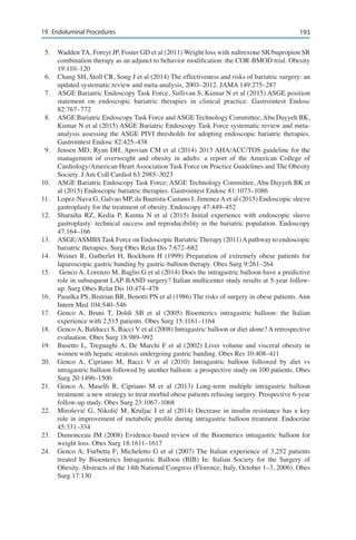
![195L. Angrisani (Ed), Bariatric and Metabolic Surgery,
Updates in Surgery
DOI: 10.1007/ 978-88-470-3944-5_20, © Springer-Verlag Italia 2017
M. Lucchese (*)
General, Metabolic and Emergency Unit, Department of Surgery, Santa Maria Nuova Hospital
Florence, Italy
e-mail: mlucch@iol.it
20Other Bariatric Procedures
Marcello Lucchese, Stefano Cariani, Enrico Amenta,
Ludovico Docimo, Salvatore Tolone, Francesco Furbetta,
Giovanni Lesti, and Marco Antonio Zappa
20.1 Introduction
Bariatric surgery can still be considered a “young” surgery in its continuous
evolution, even though the first surgical procedure was performed 70 years ago
[1]. Surgical pioneers developed innovative procedures that initially caused
malabsorption only, then restricted volume intake, and eventually both techniques
were combined [2]. Variations, alterations, and modifications of these original
procedures, combined with the concept of weight-loss surgery that eventually
became known as metabolic surgery, are the demonstration of this continuous
surgical improvement.
The intense efforts to follow and document outcomes after bariatric proce-
dures have led to the evolution of modern bariatric surgery. Many authors have
developed different technical solutions to solve some of the problems with what
is considered standard procedures.
One such procedure is the Roux-en-Y gastric bypass on vertical banded
gastroplasty (RYGB on VBG), a variation of the standard RYGB, with the goal
of developing a gastric bypass allowing exploration of the excluded stomach and
biliary tract [3, 4]. A similar procedure associated with resection of the gastric
fundus was also proposed [5].
Intragastric balloon (IGB) and adjustable gastric banding (AGB) have been
proposed as sequential systematic treatments to achieve a degree of weight loss
before a definitive RYGB procedure [6].
Even in the malabsorptive procedures, some alternative procedures have
been proposed. Hallberg proposed a modification of the jejunoileal bypass by
adding anastomosis of the excluded intestinal tract with the gallbladder, thus](https://image.slidesharecdn.com/bariatricandmetabolicsurgery-180301214153/85/Bariatric-and-metabolic-surgery-198-320.jpg)
![196 M. Lucchese et al.
avoiding postoperative diarrhea due to dramatic reduction in the absorption of
biliary juice [7–10].
In this chapter, we present some bariatric procedures that are still considered
alternative because, although they have been published in the literature, they
are not performed universally, but are regularly performed in some centers, and
follow-up results are reported for many patients. In the following paragraphs
these procedures are presented by the authors who first proposed the technique
or by surgeons regularly performing it.
20.2 Biliointestinal Bypass
The biliointestinal bypass (BIBP) is a malabsorptive procedure. Indications and
limits of this technique are those commonly adopted for this category of bariatric
procedures. The BIBP was introduced into clinical practice by Hallberg [7] and
Eriksson [11] in the late 1970s, as a modification of the jejunoileal bypass. The
aim was to reduce the side effects linked with the bypassed limb. The technical
peculiarity of BIBP involves an anastomosis between the gallbladder and the
proximal stump of the excluded jejunal limb (Fig. 20.1)
Fig. 20.1 Biliointestinal bypass (BIBP) with cholecystojejunal anastomosis (CJA) and jejunoileal
anastomosis (JIA) (reproduced with permission from [12])](https://image.slidesharecdn.com/bariatricandmetabolicsurgery-180301214153/85/Bariatric-and-metabolic-surgery-199-320.jpg)
![19720 Other Bariatric Procedures
20.2.1 Surgical Technique
The BIBP can be performed with the laparoscopic (usually four to six trocars,
according to surgeon’s preference) or open approach. The BIBP consists of
the following steps (Fig. 20.1): Identification of the duodenojejunal flexure
(D-J flexure, or Treitz ligament); measurement of the small bowel from a point
marking the first 30 cm of jejunum; measurement of the last 12–18 cm of ileum
starting from the ileocecal valve; section of the jejunum 30 cm from the D-J
flexure; side-to side jejunoileal anastomosis 12–18 cm from the ileocecal valve;
cholecystojejunal anastomosis (with possible removal of gallstones through the
surgical access to the gallbladder for anastomosis), using a linear mechanical
stapler (30 or 45). In our experience, we opt for a less malabsorptive BIBP,
sectioning the jejunum 40 cm distal to the Treitz ligament and anastomosing it to
40 cm proximal to the ileocecal valve. BIBP strength lies in its total reversibility
in case of failure or complications due to malabsorption. Moreover, this is the
only malabsorptive procedure in which the stomach is not resected; therefore,
a restrictive intervention can be considered as a revisional procedure in case of
failed weight loss. At the same time, the duodenum and main bile duct can be
explored with standard endoscopic procedures, if required, since the intestinal
bypass involves the small bowel distal to the papilla.
20.2.2 Rationale for the Procedure
BIBP creates nutrient malabsorption by reducing the absorbing surface
(considering an alimentary limb of 45–80 cm, plus the duodenum) and thus
accelerates transit. The cholecystojejunal anastomosis diverts a large portion of
the bile, thus reducing the absorption of lipids. Theoretically, bile washout in
the excluded limb can also prevent the formation of gallstones and bacterial
overgrowth while assuring enterohepatic bile-salt circulation. All these features
lead to a lower number of bowel movements per day and the dramatic decrease
of serious complications related to jejunoileal bypass. Furthermore, BIBP does
not expose the patient to the risk of anemia related to a deficiency of intrinsic
factors (since the stomach and last portion of the ileum are entirely preserved),
and a small residual flow of bile though the duodenum can guarantee satisfactory
absorption of vitamins A, D, E, and K.
20.2.3 Results
In a multicenter Italian series [13] consisting of 1030 patients, the average
weight loss after 1 year was reported to be 14–33% of initial weight, with body
mass index (BMI) loss of 18–35%. These results were confirmed by authors’](https://image.slidesharecdn.com/bariatricandmetabolicsurgery-180301214153/85/Bariatric-and-metabolic-surgery-200-320.jpg)
![198 M. Lucchese et al.
experience [10] with 347 obese patients. Weight loss reach a plateau within 18–
24 months after surgery, with a percentage of excess weight loss (%EWL) of
60–70%. Weight loss remained >60% at 5-year follow-up; 2.5–5% of patients
experience insufficient weight loss. In the first months after surgery, diarrhea
occurred approximately five to seven times a day. After weight stabilization,
bowel movements occurred two to three times day on average.
BIBPcauses a drastic reduction in serum levels of cholesterol and triglycerides,
which remains constant over the years. In the first postoperative months, there
is a significant increase in liver enzymes glutamic oxaloacetic transminase
(GOT) and glutamate pyruvic transaminase (GPT), indicating liver overload.
These values begin to normalize in the third month after surgery due to better
liver function, which entails a reduction of hepatic overload itself. All diseases
related to obesity and the metabolic syndrome show a dramatic improvement
or a complete remission [8]. In particular, glycemic control improves markedly
and therefore drastically reduces the need for drugs or insulin. Remission
was recently reported in 83% of cases with type 2 diabetes mellitus (T2DM)
preoperatively [10].
20.2.4 Complications
20.2.4.1 Early Complications
BIBP is burdened with complications within the first postoperative month by
the same rate of complications described for all abdominal surgery involving
intestinal anastomoses (i.e., bleeding, leaks, infections). The reported mortality
rate is ~0.5–1%. The most frequent complication is represented by inflammation
(~68% of cases). Severe diarrhea, due to dietary restrictions in most cases,
appears more rarely and is accompanied by electrolyte imbalances and requires
appropriate infusional nutritional support. If not treated, diarrhea can also cause
perianal abscesses and fistulas [9].
20.2.4.2 Late Complications
Cholelithiasis appears in ~4% of patients. It can be treated medically using
dissolving therapy with bile acid analogs), although in most cases, it is due
to cholecystojejunal anastomotic stenosis, which requires surgical revision
using a laparoscopic approach. Oxalic nephrolithiasis (5.3%) is caused by an
inappropriate diet. Abdominal colicky pain due to altered gut microbiota and
consequent gas formation and migrant polyarthralgias affecting distal joints is
episodic (4.3%). The recurrent abdominal pain is treated successfully in most
cases with a course of antibiotics orally. Protein malnutrition is rare but is usually
due to inadequate protein intake or absorption and electrolyte malabsorption.
These complications are serious and require intensive medical treatment and
sometimes intravenous nutritional support and revisional surgery [10].](https://image.slidesharecdn.com/bariatricandmetabolicsurgery-180301214153/85/Bariatric-and-metabolic-surgery-201-320.jpg)
![19920 Other Bariatric Procedures
20.3 Roux-en-Y Gastric Bypass on Vertical Banded
Gastroplasty
Roux-en-Y gastric bypass on vertical banded gastroplasty (RYGB-on-VBG), is
a technique derived from the standard RYGB with the goal of creating a gastric
bypass in which it is possible to perform a standard endoscopic and an oral contrast
study of the excluded stomach. The concept was conceived in 2002 after a pilot
study performed with a functional gastric bypass with the same aim [14, 15]. In the
midterm, the RYGB-on-VBG procedure reached similar outcomes as the standard
techniques of gastric bypass both in terms of weight loss and incidence of surgical
complications [3]. Moreover, it demonstrated similar good results when performed
in other bariatric centers, thus proving to be highly standardized and reproducible
while enabling diagnostic evaluation of the bypassed stomach and biliary tract
[4]. Progressively, outcomes of RYGB-on-VBG have been publicly presented and
discussed in international meetings [16–18]. Similar results were obtained when
used as a revisional procedure after failure of previous restrictive techniques [19].
Long-term results following RYGB-on-VBG are available as from a single-center
experience [16], that at present has a 12-year clinical follow-up.
While achieving weight loss outcomes as good as to those after the standard
procedure, RYGB-on-VBG enables traditional diagnostic evaluation of the
bypassed stomach [20].Although RYGB-on-VBG is technically more demanding
than standard RYGB, the low rate of early and late complications suggests
considering the procedure in selected patients with chronic conditions in the
gastric remnant, in whom it is advisable to avoid creating blind, unexplorable
sections of the digestive tract.
20.3.1 Surgical Technique
The definitive technique of RYGB-on-VBG has already been described [3]. In
brief, gastric pouch construction is fashioned as during the Mason/MacLean
VBG, with a gastrojejunostomy (2 cm in diameter) to a 150-cm Roux-en-Y
limb performed proximally to the pouch outlet (1 cm in diameter). The outlet is
encircled with a soft polytetrafluoroethylene (PTFE) band to prevent enlargement
of the passage between the pouch and the remnant (Fig. 20.2). The gastric outlet
inner diameter is standardized over a 38-Ch endogastric tube. Initially, the
RYGB-on-VBG was performed using only the open approach; in 2009, using the
laparoscopic approach also became an option [21].
20.3.2 Outcomes
According to the most recent data of a single-center experience [15] – presently
based on 456 patients who received a RYGB-on-VBG as primary operation from](https://image.slidesharecdn.com/bariatricandmetabolicsurgery-180301214153/85/Bariatric-and-metabolic-surgery-202-320.jpg)
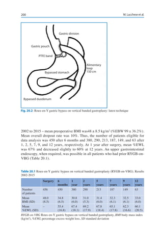
![20120 Other Bariatric Procedures
20.3.3 Complications
Six patients (1.3%) had surgical treatment for early complications (Clavien-
Dindo grade IIIb); mortality was 0.6% following two grade IVa and one grade
IVb medical complication. Late complications were seven (1.5%) band erosions
(four patients had surgical revision), five (1%) vertical staple-line disruption
(three patients had surgical revision), and four (0.8%) anastomotic ulcers [treated
with proton pump inhibitors (PPI)].
20.4 Roux-en-Y Gastric Bypass with Fundectomy and
Explorable Stomach
Most bariatric surgeons consider the standard laparoscopic Roux-en-Y gastric
bypass (LRYGB) the ideal procedure for treating severe obesity. The problem
with this procedure is that the bypassed stomach cannot be explored, and thus
there is no opportunity to diagnose and treat diseases of the stomach, duodenum,
and main bile duct. In March 2001, Lesti developed a model of gastric bypass,
making subsequent modifications up to the final version used since January
2007. The current model of LRYGB with fundectomy and explorable stomach
[LRYGB(FES)], in which the gastric fundus is removed, is based on the use of a
device that allows the stomach to be isolated from passage of the food bolus but
also to remain examinable by endoscopy, providing diagnostic and/or operative
possibilities. (Fig. 20.3). The reversible LRYGB(FES) has similar results to
the standard model. Removing the gastric fundus leads to a marked decrease
Fig. 20.3 Roux-en-Y gastric bypass
with fundectomy (Lesti’s technique)
Alimentary Limb
200 cm
Biliopancreatic
Limb
200 cm
Common Channel
> 350 cm](https://image.slidesharecdn.com/bariatricandmetabolicsurgery-180301214153/85/Bariatric-and-metabolic-surgery-204-320.jpg)
![202 M. Lucchese et al.
of ghrelin, a hormone that increases the sense of hunger, and to an increase in
peptide tyrosine-tyrosine (PYY) and glucagonlike peptide-1 (GLP-1), hormones
that block hunger and play an important role in controlling diabetes.
20.4.1 Surgical Technique
This laparoscopic procedure is done with four 10- to 12-mm trocars and one 5-mm
trocar. The gastrocolic ligament, opened at Van Goethem’s point, is sectioned
toward the angle of His: attention is paid to cutting the short vessels. A 36-Fr bougie
is introduced into the stomach, and the linear stapler–cutter device is fired beginning
at Van Goethem’s point at the great curvature toward the lesser curvature 7 cm from
the cardias. Three firings of the stapler parallel to the bougie are applied to make
the pouch as narrow as possible (20–30 mL). An expanded polytetrafluoroethylene
(ePTFE) band 1-mm thick, 5- to 7-cm long, and 1-cm wide is placed 7 cm from the
cardias to gently close the end of the pouch. The mean length of the ePTFE band
is 57 mm (range 53–65 mm). The jejunum, identified at the Treitz ligament and
followed distally for 200–220 cm, is pulled cephalad toward the gastric pouch in the
antecolic position after separation of the greater omentum.
The gastrojejunal anastomosis is made side to side, 2.5- to 3-cm wide in the
posterior wall of the pouch, starting 5 cm from the cardias, using an endocutter blue
cartridge (Echelon). The jejunumileum anastomosis is made 200–220 cm from the
pouch, side to side, 2.5- to 3-cm wide. Lesti’s technique includes a long biliary
limb: first, because the gastroanastomosis is made substantially with the last part
of the jejunum, that is very important for the theory of hindgut incretin secretion;
second, because in the case of failure, it is very easy to convert the procedure into
a long distal gastric bypass. The common channel was >350 cm in all cases.
Mean operative time was 152 (108–227) min, postoperative stay was 4.74 (3–
8) days, and operative mortality zero The only early postoperative complication
was one important intra-abdominal bleeding occurring on the second
postoperative day, which required open surgery; the point of bleeding was not
found. Two cases of internal hernia occurred after 3 and 25 months, respectively;
both revisions were performed laparoscopically without complications. No leak,
migration, ulcer, or stricture occurred in either the early or late follow-up, and
there were no minor complications during the 60 months of follow-up.
20.4.2 Outcomes
We report the results of 454 severely obese patients who underwent a LRYGB(FES)
with the ePTFE band between January 2007 and December 2014 [5]. All patients
were selected according to criteria for bariatric surgery proposed by the US
National Institutes of Health (NIH) Consensus of 1991 and replicated and updated
by the Italian Society of Obesity Surgery (SICOB) Patients eligible for bariatric](https://image.slidesharecdn.com/bariatricandmetabolicsurgery-180301214153/85/Bariatric-and-metabolic-surgery-205-320.jpg)
![20320 Other Bariatric Procedures
surgery (BMI >40 kg/m2
or BMI >35 kg/m2
with obesity-related comorbidities)
underwent evaluation by a multidisciplinary bariatric team at our center for obesity
disorders. The population comprised 288 women and 166 men with a mean age of
43.6 (range 27–68) years and a preoperative BMI of 48.2 (range 36.7–58.3) kg/
m2
, weight 136,48 (range 98.3–158.5) kg, and excess weight 58.2 (range 34.5–
7.2) kg. Existing comorbidities were T2DM 24.2%, arterial hypertension 31.3%,
metabolic syndrome 21.9%, obstructive sleep apnea syndrome 26,4%, depression
15.8%, and gastroesophageal reflux 36.4%. At 5-years follow-up, resolution was
seen in T2DM 68.5%, hypertension 67.4%, obstructive sleep apnea syndrome
60.5%, gastroesophageal reflux 84.8%; improvement was seen in T2DM 21.4%,
hypertension 26.1%, obstructive sleep apnea syndrome 39.5%, gastroesophageal
reflux 15.2%, and arthritis 100%. The % EWL at 1 year in 414 was 77.8%, at 3 years
in 278 patients 84.4%, and at 5 years in 166 patients 79.6%. Preoperatively, BMI
was 48.2 kg/m2
and at 5 years 30.7 kg/m2
, with a mean loss of 17.5 kg/m2
.
20.5 Functional Gastric Bypass
The functional gastric bypass (FGB) is consistent with laparoscopic adjustable
gastric banding (LAGB), which creates the pouch of a long-limb gastric bypass
when fully inflated. The aim of the procedure is to obtain complete diversion
of food into the alimentary limb, with the band fully inflated, while allowing
band deflation to balance efficacy and to allow access to the excluded stomach
with a standard endoscope if necessary (diagnostic-operative in case of suspicion
of any pathology arising from the gastric remnant or biliary tree). The design
of this technique excludes blind tracts due to band adjustability. The presented
technique was devised and has been developed by Francesco Furbetta since its
first application and description in 2001 [22].
20.5.1 Surgical Technique
The LAGB is placed across the cardia following the pars flaccida or perigastric
technique (Fig. 20.4). An antecolic gastrojejunal anastomosis on the gastric pouch is
fashioned through a double-layer hand-sewn technique. Roux-en-Y limb lengths are
measured under tension from the ileocecal valve to create a 200-cm alimentary limb
and a 150-cm common limb, depending on the pouch size [22].
20.5.2 How it Works: Safety and Efficacy
Food restriction is achieved with an adjustable small pouch; a large gastrojejunal
anastomosis (≥15 mm) maximizes the metabolic effect of the distal gastric bypass](https://image.slidesharecdn.com/bariatricandmetabolicsurgery-180301214153/85/Bariatric-and-metabolic-surgery-206-320.jpg)
![204 M. Lucchese et al.
with the lowest possible risk of malabsorption. It is also possible to switch food
passage from the gastric bypass to the gastric remnant, just by deflating the band.
As a revisional procedure, the functional bypass could improve the restrictive
mechanism of the gastric band, adding metabolic efficacy through a long limb
adjustable bypass. Thus, patients can undergo a procedure that can be adapted to
their individual characteristics [22].
20.5.3 Results and complications
From January 2001 to October 2015, in this monocentric experience, the FGB
was performed on 209 patients: in 155 out of 3050 patients with a previous
LAGB and in 54 cases, as the first surgical step. Long-term efficacy of the
procedure was assessed on the 68 eligible patients with a 5-year follow-up.
In particular, mean 5-year BMI was 30.7 kg/m2
, corresponding to a %EWL of
52.4% and a BMI loss of 67.5%. Results were achieved with no mortality. Out of
the entire group of 209 patients, early complications were consistent with only
one anastomotic leak (Clavien-Dindo grade IVa); late complications, represented
by erosion, were experienced in 6%.
Even though the procedure lacks widespread application and the main
concerns are related to the possible band complications, the FGB procedure
brings together the minimally invasive LAGB, the stabilized, different working
Fig. 20.4 Functional gastric bypass (FGB)
according to Furbetta](https://image.slidesharecdn.com/bariatricandmetabolicsurgery-180301214153/85/Bariatric-and-metabolic-surgery-207-320.jpg)
![20520 Other Bariatric Procedures
of the GBP, and the efficacy of the BPD. Their relative characteristics maximize
safety and efficacy through reversibility, adjustability, absence of blind limbs,
and the sequential treatment opposite to re-do. All this allows scientific and
technical progress.
20.6 Conclusions
Despite good results in terms of outcomes and safety of these alternative bariatric
procedures, information in the literature remains scarce. However, every
alternative or new procedure in bariatric surgery should be considered as being
potential valuable. Publication of short- and long-term results of alternative
bariatric procedures are required in order to report their safety and efficacy. It
is important for surgical societies to recall that procedures such as the sleeve
gastrectomy and the so-called mini gastric bypass – which are now practiced
worldwide – were initially considered investigational procedures. We believe
that continued recognition of different mechanisms of action and new techniques
in bariatric surgery will lead to more precise indications and individualization of
the best procedure for the best results in each patient.
References
1. Saber AA, Elgamal MH, McLeod MK (2008) Bariatric surgery: the past, present, and future.
Obes Surg 18:121–128
2. Lo Menzo E, Szomstein S, Rosenthal RJ (2015) Changing trends in bariatric surgery. Scand
J Surg 104:18–23
3. Cariani S, Amenta E (2007) Three-year results of Roux-en-Y gastric bypass-on-vertical
banded gastroplasty: an effective and safe procedure which enables endoscopy and X-ray
study of the stomach and biliary tract. Obes Surg 17:1312–1318
4. Cariani S, Palandri P, Della Valle E et al (2008) Italian multicenter experience of Roux-
en-Y gastric bypass on vertical banded gastroplasty: four-year results of effective and safe
innovative procedure enabling traditional endoscopic and radiographic study of bypassed
stomach and biliary tract. Surg Obes Relat Dis 4:16–25
5. Lesti G (2015) Tecnica del by-pass gastrico sec. Lesti. Oral communication at XXIII SICOB
National Congress, Baveno (Italy)
6. Greve JW, Furbetta F, Lesti G et al (2004) Combination of laparoscopic adjustable gastric
banding and gastric bypass: current situation and future prospects: routine use not advised.
Obes Surg 14:683–689
7. Hallberg D (1980) A survey of surgical techniques for treatment of obesity and a remark on
the bilio-intestinal bypass method. Am J Clin Nutr 33(2 Suppl):499–501
8. Micheletto G, Mozzi E, Lattuada E et al (2007) The bilio-intestinal bypass. Ann Ital Chir
78:27–30
9. Ruggiero R, Docimo G, Russo V et al (2010) Bilio-intestinal bypass in the treatment of
metabolic syndrome in obese patient. G Chir 31(11–12):527–533 [Article in Italian]](https://image.slidesharecdn.com/bariatricandmetabolicsurgery-180301214153/85/Bariatric-and-metabolic-surgery-208-320.jpg)
![206 M. Lucchese et al.
10. Del Genio G, Gagner M, Limongelli P et al (2015) Remission of type 2 diabetes in patients
undergoing biliointestinal bypass for morbid obesity: a new surgical treatment. Surg Obes
Relat Dis [Epub ahead of print] doi:10.1016/j.soard.2015.12.003
11. Eriksson F (1981) Biliointestinal bypass. Int J Obes 5:437–447
12. Corradini SG, Eramo A, Lubrano C et al (2005) Comparison of changes in lipid profile
after biliointestinal bypass and gastric banding in patients with morbid obesity. Obes Surg
15:367–377
13. Micheletto G, Badiali M, Danelli PG et al (2008) The biliointestinal bypass: a thirty-years
experience. Ann Ital Chir 79:419–426 [Article in Italian]
14. Cariani S, Vittimberga G, Grani S et al (2002) Prospective study on functional Roux-en-Y
gastric bypass to avoid gastric exclusion. In: IFSO Congress Programme andAbstracts. Obes
Surg 12:469
15. Cariani S, Lucchi A, Guerra M, Faccani E, Amenta E (2004) Evolution of functional Roux-
en-Y gastric bypass: from adjustable band to Gore-tex band. In: IFSO European Symposium.
Obes Surg 14:471
16. Leuratti L, Picariello E, Balsamo F, Cariani S (2012) Long term results after Roux-en-Y
gastric bypass on vertical banded gastroplasty with explorable remnant: does the presence
of a gastro-gastric outlet impact on final outcomes in term of metabolic efficacy and weight
loss? Obes Surg 22:1160
17. Cariani S, Giorgini E, Agostinelli L et al (2009) Mid-term outcomes of Roux-en-Y gastric
bypass on vertical banded gastroplasty. Surg Obes Relat Dis 5(3 suppl):S30
18. Cariani S, Leuratti L, Picariello E, Spasari E (2011) An outlet for endoscopic access to
the remnant does not reduce the effectiveness of gastric bypass: long-term outcomes of a
modified Roux-en-Y gastric bypass that allows traditional endoscopy of bypassed stomach.
In: IFSO Congress – Plenary Sessions. Obes Surg 21:1000
19. Cariani S, Agostinelli L, Leuratti L et al (2010) Bariatric revisionary surgery for failed
or complicated vertical banded gastroplasty (VBG): Comparison of VBG reoperation
(re-VBG) versus Roux-en-Y gastric bypass-on-VBG (RYGB-on-VBG). J Obes 2010
doi:10.1155/2010/206249
20. Leuratti L, Di Simone MP, Cariani S (2013) Unexpected changes in the gastric remnant in
asymptomatic patients after Roux-en-Y gastric bypass on vertical banded gastroplasty. Obes
Surg 23:131–139
21. Cariani S, Agostinelli L, Giorgini E et al (2009) Open and laparoscopic approach of a new
surgical technique, which enables traditional diagnostic evaluation of the bypassed stomach:
the Roux-en-Y gastric bypass on vertical banded gastroplasty. In: International Federation
for the Surgery of Obesity and metabolic disorders. XIV World Congress. Obes Surg 19:1048
22. Furbetta F, Gambinotti G (2002) Functional gastric bypass with an adjustable gastric band.
Obes Surg 12:876–880](https://image.slidesharecdn.com/bariatricandmetabolicsurgery-180301214153/85/Bariatric-and-metabolic-surgery-209-320.jpg)


