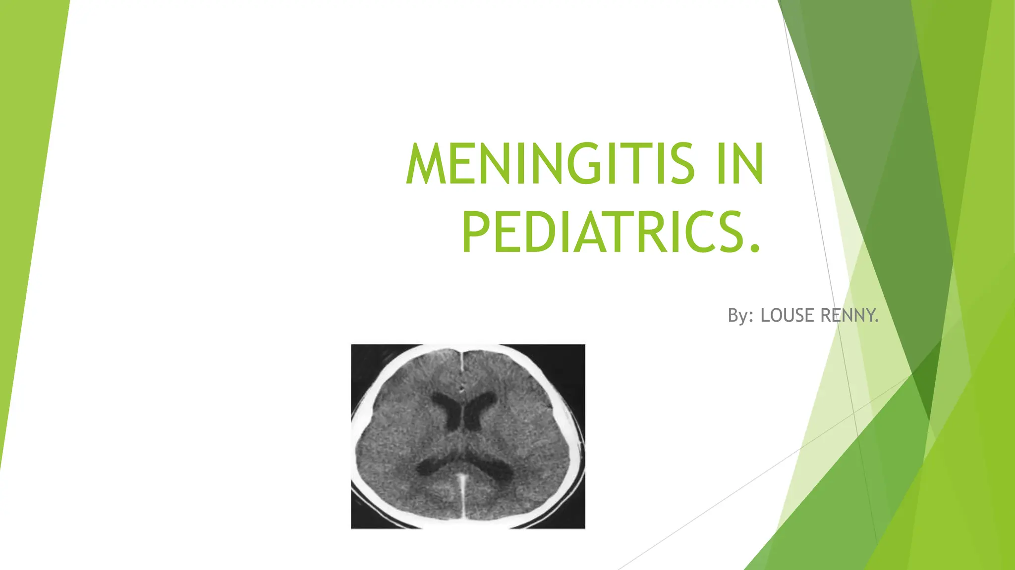
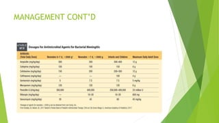
MENINGITIS IN
PEDIATRICS.
By: LOUSE RENNY.
COURSE OUTLINE.
 Definition of meningitis.
 Introduction to meningitis.
 Etiology of meningitis and risk factors.
 Modes of transmission.
 Pathophysiology of meningitis.
 Clinical presentation of meningitis.
 Investigations.
 Management of meningitis.
 Complications of meningitis.
 Differential diagnosis.
DEFINITION OF MENINGITIS.
 According to WHO.
 Meningitis refers to the inflammation of the tissues surrounding the brain and
the spinal cord.
 Almost always, Meningitis refers to the inflammation of leptomeninges(
arachnoid mater and pia mater) including the subarachnoid space leading to
constellation of signs and symptoms and presence of inflammatory cells in
CFS.
 However, inflammation of the dura mater is referred to as Pachymeningitis
and is usually manifested by thickening of the intracranial dura mater on
radiology.
Other definitions.
 Acute meningitis refers to onset of symptoms of meningeal inflammation over
the course of hours to several days.
 Chronic meningitis is defined as atleast 4 weeks of symptoms of inflammation
of meninges.
 Aseptic meningitis refers to a syndrome consistent with signs and symptoms of
meningeal inflammation but with negative routine CSF cultures.
 Recurrent meningitis refers to atleast 2 episodes of signs and symptoms of
meningeal inflammation with associated CSF findings separated by a period of
full recovery.
INTRODUCTION TO MENINGITIS.
 Infections of the CNS can be divided into 2 broad categories i.e.
 Those primarily involving the meninges (meningitis).
 Those primarily confined to the parenchyma (encephalitis).
INTRODUCTION TO MENINGITIS CONT’D.
 Meningitis almost always refers to inflammation of leptomeninges.
 Between the leptomeninges is the subarachnoid space that houses the CSF.
 In 1 microliter of CSF, there are only atmost 5 WBCs.
 In 1 deciliter of CSF, 70% of WBCs are lymphocytes while the other 30% are
monocytes. There are only very few PMNs( neutrophils).
o There are also about 15-50mg of proteins, 45-100mg of glucose which
approximates to that of plasma and the pressure is less than 200mmH2O or
15mmHg.
INTRODUCTION TO MENINGITIS CONT’D
 There is constantly about 150ml of CSF in the body.
 The CSF is constantly replenished with around 500ml of CSF daily. The excess
350ml is constantly being reabsorbed into blood to maintain CSF at 150ml.
 Triggers for meningitis include the following;
 Autoimmune disease e.g. lupus.
 Adverse drug reactions especially in intrathecal therapy.
 Infections are the most common triggers.
ETIOLOGY OF MENINGITIS AND RISK
FACTORS
a. BACTERIA.
 According to WHO, there are four main causes of acute bacterial meningitis
i.e.
 Neisseria Meningitidis (meningococcus).
 Streptococcus pneumoniae (pneumococcus).
 Haemophilus influenza.
 Streptococcus agalactiae (Group B streptococcus).
BACTERIA CONT’D
Note the following:
 Newborns are at most risk from Group B streptococcus.
 Young children are at higher risk from meningococcus, pneumococcus and
haemophilus influenza.
 Adolescents and young adults are at particular risk of meningococcal disease.
 The elderly are at particular risk of pneumococcal disease.
ETIOLOGY OF MENINGITIS AND RISK
FACTORS CONT’D
b. VIRUSES.
 They include;
 Enteroviruses.
 Herpes simplex virus type 2.
c. FUNGI.
 They include;
 Cryptococcus neoformans.
 Coccidiodes immitis.
ETIOLOGY OF MENINGITIS AND RISK
FACTORS CONT’D.
 The risk factors include;
 Skipping vaccination.
 Age.
 Living in a community setting.
 Pregnancy.
 Weakened immunity.
MODES OF TRANSMISSION.
 There are two main modes of spread of pathogens that cause meningitis i.e.
 Direct spread.
 Hematogenous spread.
 Direct spread is where pathogens spread to the subarachnoid space through
the overlying skin e.g. in head trauma leading to a skull fracture and direct
inoculation during intracranial manipulation or up through the nose e.g. in
sinusitis and otitis media or through an anatomical defect e.g. spina bifida.
 Hematogenous spread is where pathogens move in blood and to the
subarachnoid space by binding to surface receptors on endothelial cells or use
damaged parts or vulnerable spots of blood vessels like the choroid plexus.
MODES OF TRANSMISSION CONT’D.
 The route of transmission varies by organisms.
 Most bacteria that cause meningitis such as meningococcus, pneumococcus
and haemophilus influenza are carried in the human nose and throat. They
spread from person to person by respiratory droplets or throat secretions.
Note:
 Group B streptococcus is often carried in human gut or vagina and can spread
from mother to child around the time of birth.
PATHOPHYSIOLOGY OF MENINGITIS.
 The brain is naturally protected from the body’s immune system by the
barrier that the meninges create between the blood stream and the brain.
 Normally, this protection is an advantage because the barrier prevents the
immune system from attacking the brain.
PATHOPHYSIOLOGY CONT’D
 However, in meningitis, the blood brain barrier can become disrupted. Once
bacteria or other organisms have found their way to the brain, they are
somewhat isolated from the immune system and can spread.
 When the body tries to fight the infection, the problem can worsen. Blood
vessels become leaky and allow fluid, WBCs and other infection fighting
particles to enter the meninges and brain. This process in turn causes brain
swelling and can eventually result in decreasing blood flow to parts of the
brain worsening the symptoms of the infection.
PATHOPHYSIOLOGY CONT’D.
 Depending on the severity of bacterial meningitis, the inflammatory process
may remain confined to the subarachnoid space.
 In severe forms, the pial barrier is not penetrated and the underlying
parenchyma remains intact.
 However, in more severe forms of bacterial meningitis, the pial barrier is
breached and the underlying parenchyma is invaded by the inflammatory
process.
 Note that bacterial meningitis may therefore lead to widespread cortical
destruction, particularly when left untreated.
CLINICAL PRESENTATION OF MENINGITIS
 History.
 In younger children, bacterial meningitis symptoms are usually nonspecific
including fever, hypothermia, irritability and poor feeding as well as signs of
increased intracranial pressure, including seizures and apnea.
 Attention should be noted to the patient’s immunization status, birth history,
travel history, trauma, health status, geographic location and exposure to
high risk contacts.
History cont’d.
 Common chief complaints by the infants care givers include;
 Irritable or “sleeping all the time”.
 “Won’t take the bottle”.
 “Not acting right”.
 “Cries when moved or picked”.
History cont’d.
 Note that older children may complain of classic meningeal inflammation
signs including neck pain, headache, or back pain as well as photophobia,
anorexia and myalgias.
 Nausea and vomiting are common.
CLINICAL PRESENTATION OF MENINGITIS
CONT’D.
 Physical examination.
 Stiff neck in older children. Infants have a poor neck muscle tone and this
finding is commonly absent.
 Brudzinski and Kernig signs maybe present.
Brudzinski sign is where with the patient supine, flexion of the neck elicits
involuntary flexion of the hips and knees.
Kernig sign is where with the patient in supine, the legs are flexed 90 degrees at
the hip, extensions of the lower legs are unable to be accomplished beyond 135
degrees.
 Negative Brudzinski or Kernig sign does not rule out meningitis.
Physical examination cont’d.
 Younger children may not have nuchal rigidity and Kernig and/or Brudzinski
signs.
 Ant infant presenting with a sepsis-like picture needs to have meningitis as a
consideration.
 Classically, there maybe “paradoxical” crying i.e. crying that increases when
the child is picked up.
 Signs of increased intracranial pressure, including papilledema, asymmetric
pupils, bulging fontanelle, diplopia.
 Skin exam for erythema migrans from borreliosis(Lyme disease), petechiae or
purpura with invasive meningococcal disease or vesicles in an infant <6 weeks
old with HSV.
INVESTIGATIONS.
 Labaratory investigations.
 CSF analysis (cell count with differential, protein measurement, glucose
concentration and measurement of pressure).
 CSF gram staining.
 Blood culture.
 CBC, platelet count, electrolytes, BUN, creatinine, serum glucose.
 Consider prothrombin time(PT), partial thromboplastin time(PTT), liver
function tests, arterial blood gases.
INVESTIGATIONS CONT’D.
 Diagnostic procedure and others.
 Lumber puncture.
It is contraindicated in cardiopulmonary compromise, uncorrected coagulopathy,
signs of increased intracranial pressure or focal neurologic findings until head
imaging is obtained.
• If no etiology is discovered after the first lumber puncture and the child is not
responding to therapy, repeat lumber puncture at 36-48hrs.
• Opening pressure: normal is <200mmH2O in lateral recumbent position.
INVESTIGATIONS CONT’D.
 Depending on the presentation, age, history and physical exam findings, some
or all of the following tests should be requested for CSF analysis:
 Cell count with differential and gram staining.
• Bacterial meningitis is characterized by CSF pleocytosis(>1.0x10^3/microliter)
with predorminance of neutrophils, >100WBCs/microliter with >90% being
PMNs.
• Viral meningitis typically has a lower CSF count(0.005-0.5x10^3/microliter)
with a predominance of lymphocytes, >50% lymphocytes with <20% being
PMNs.
INVESTIGATIONS CONT’D.
 Glucose: compares to serum glucose, normal is >40mg/dL or 1/2-2/3 of serum
glucose.
 Protein: normal is 5-40mg/dL except in newborns, who may have protein
levels of 150-200mg/Dl.
• >1.0g/dL in bacterial meningitis and normal to slightly elevated in viral
meningitis.
 Cultures for bacteria, fungi, virus and mycobacteria.
• 80% of blood cultures are positive in children with bacterial meningitis.
MANAGEMENT OF MENINGITIS
MANAGEMENT CONT’D
MANAGEMENT CONT’D
DIFFERENTIAL DIAGNOSIS.
 Encephalitis.
 Toxic encephalopathy.
 Epidural abscess.
 Cerebral abscess.
COMPLICATIONS OF MENINGITIS.
 According to the WHO, 1 in 5 people surviving an episode of bacterial
meningitis may have long lasting after-effects.
 These after-effects include hearing loss, seizures, limb weakness, difficulties
with vision, speech, language, memory and communication as well as scaring
and limb amputations after sepsis. The above effects result from exudates
due to the inflammatory process which extend through the CSF particularly to
the basal cisterns resulting in damage to cranial nerves.
• Exudates also obliterate the CSF pathways causing obstructive hydrocephalus.
• Exudates also induce vasculitis and thrombophlebitis causing local brain
ischaemia.
COMPLICATIONS OF MENINGITIS CONT’D
 Increased intracranial pressure and cerebral edema are the other
complications of meningitis.
• Cerebral edema significantly contributes to intracranial hypertension and a
consequent decrease in cerebral flow.
THANK YOU
LOUSE RENNY

MENINGITIS IN PEDIATRICS [Autosaved].pptx

  • 1.
  • 2.
    COURSE OUTLINE.  Definitionof meningitis.  Introduction to meningitis.  Etiology of meningitis and risk factors.  Modes of transmission.  Pathophysiology of meningitis.  Clinical presentation of meningitis.  Investigations.  Management of meningitis.  Complications of meningitis.  Differential diagnosis.
  • 3.
    DEFINITION OF MENINGITIS. According to WHO.  Meningitis refers to the inflammation of the tissues surrounding the brain and the spinal cord.  Almost always, Meningitis refers to the inflammation of leptomeninges( arachnoid mater and pia mater) including the subarachnoid space leading to constellation of signs and symptoms and presence of inflammatory cells in CFS.  However, inflammation of the dura mater is referred to as Pachymeningitis and is usually manifested by thickening of the intracranial dura mater on radiology.
  • 4.
    Other definitions.  Acutemeningitis refers to onset of symptoms of meningeal inflammation over the course of hours to several days.  Chronic meningitis is defined as atleast 4 weeks of symptoms of inflammation of meninges.  Aseptic meningitis refers to a syndrome consistent with signs and symptoms of meningeal inflammation but with negative routine CSF cultures.  Recurrent meningitis refers to atleast 2 episodes of signs and symptoms of meningeal inflammation with associated CSF findings separated by a period of full recovery.
  • 5.
    INTRODUCTION TO MENINGITIS. Infections of the CNS can be divided into 2 broad categories i.e.  Those primarily involving the meninges (meningitis).  Those primarily confined to the parenchyma (encephalitis).
  • 6.
    INTRODUCTION TO MENINGITISCONT’D.  Meningitis almost always refers to inflammation of leptomeninges.  Between the leptomeninges is the subarachnoid space that houses the CSF.  In 1 microliter of CSF, there are only atmost 5 WBCs.  In 1 deciliter of CSF, 70% of WBCs are lymphocytes while the other 30% are monocytes. There are only very few PMNs( neutrophils). o There are also about 15-50mg of proteins, 45-100mg of glucose which approximates to that of plasma and the pressure is less than 200mmH2O or 15mmHg.
  • 7.
    INTRODUCTION TO MENINGITISCONT’D  There is constantly about 150ml of CSF in the body.  The CSF is constantly replenished with around 500ml of CSF daily. The excess 350ml is constantly being reabsorbed into blood to maintain CSF at 150ml.  Triggers for meningitis include the following;  Autoimmune disease e.g. lupus.  Adverse drug reactions especially in intrathecal therapy.  Infections are the most common triggers.
  • 8.
    ETIOLOGY OF MENINGITISAND RISK FACTORS a. BACTERIA.  According to WHO, there are four main causes of acute bacterial meningitis i.e.  Neisseria Meningitidis (meningococcus).  Streptococcus pneumoniae (pneumococcus).  Haemophilus influenza.  Streptococcus agalactiae (Group B streptococcus).
  • 9.
    BACTERIA CONT’D Note thefollowing:  Newborns are at most risk from Group B streptococcus.  Young children are at higher risk from meningococcus, pneumococcus and haemophilus influenza.  Adolescents and young adults are at particular risk of meningococcal disease.  The elderly are at particular risk of pneumococcal disease.
  • 10.
    ETIOLOGY OF MENINGITISAND RISK FACTORS CONT’D b. VIRUSES.  They include;  Enteroviruses.  Herpes simplex virus type 2. c. FUNGI.  They include;  Cryptococcus neoformans.  Coccidiodes immitis.
  • 11.
    ETIOLOGY OF MENINGITISAND RISK FACTORS CONT’D.  The risk factors include;  Skipping vaccination.  Age.  Living in a community setting.  Pregnancy.  Weakened immunity.
  • 12.
    MODES OF TRANSMISSION. There are two main modes of spread of pathogens that cause meningitis i.e.  Direct spread.  Hematogenous spread.  Direct spread is where pathogens spread to the subarachnoid space through the overlying skin e.g. in head trauma leading to a skull fracture and direct inoculation during intracranial manipulation or up through the nose e.g. in sinusitis and otitis media or through an anatomical defect e.g. spina bifida.  Hematogenous spread is where pathogens move in blood and to the subarachnoid space by binding to surface receptors on endothelial cells or use damaged parts or vulnerable spots of blood vessels like the choroid plexus.
  • 13.
    MODES OF TRANSMISSIONCONT’D.  The route of transmission varies by organisms.  Most bacteria that cause meningitis such as meningococcus, pneumococcus and haemophilus influenza are carried in the human nose and throat. They spread from person to person by respiratory droplets or throat secretions. Note:  Group B streptococcus is often carried in human gut or vagina and can spread from mother to child around the time of birth.
  • 14.
    PATHOPHYSIOLOGY OF MENINGITIS. The brain is naturally protected from the body’s immune system by the barrier that the meninges create between the blood stream and the brain.  Normally, this protection is an advantage because the barrier prevents the immune system from attacking the brain.
  • 15.
    PATHOPHYSIOLOGY CONT’D  However,in meningitis, the blood brain barrier can become disrupted. Once bacteria or other organisms have found their way to the brain, they are somewhat isolated from the immune system and can spread.  When the body tries to fight the infection, the problem can worsen. Blood vessels become leaky and allow fluid, WBCs and other infection fighting particles to enter the meninges and brain. This process in turn causes brain swelling and can eventually result in decreasing blood flow to parts of the brain worsening the symptoms of the infection.
  • 16.
    PATHOPHYSIOLOGY CONT’D.  Dependingon the severity of bacterial meningitis, the inflammatory process may remain confined to the subarachnoid space.  In severe forms, the pial barrier is not penetrated and the underlying parenchyma remains intact.  However, in more severe forms of bacterial meningitis, the pial barrier is breached and the underlying parenchyma is invaded by the inflammatory process.  Note that bacterial meningitis may therefore lead to widespread cortical destruction, particularly when left untreated.
  • 17.
    CLINICAL PRESENTATION OFMENINGITIS  History.  In younger children, bacterial meningitis symptoms are usually nonspecific including fever, hypothermia, irritability and poor feeding as well as signs of increased intracranial pressure, including seizures and apnea.  Attention should be noted to the patient’s immunization status, birth history, travel history, trauma, health status, geographic location and exposure to high risk contacts.
  • 18.
    History cont’d.  Commonchief complaints by the infants care givers include;  Irritable or “sleeping all the time”.  “Won’t take the bottle”.  “Not acting right”.  “Cries when moved or picked”.
  • 19.
    History cont’d.  Notethat older children may complain of classic meningeal inflammation signs including neck pain, headache, or back pain as well as photophobia, anorexia and myalgias.  Nausea and vomiting are common.
  • 20.
    CLINICAL PRESENTATION OFMENINGITIS CONT’D.  Physical examination.  Stiff neck in older children. Infants have a poor neck muscle tone and this finding is commonly absent.  Brudzinski and Kernig signs maybe present. Brudzinski sign is where with the patient supine, flexion of the neck elicits involuntary flexion of the hips and knees. Kernig sign is where with the patient in supine, the legs are flexed 90 degrees at the hip, extensions of the lower legs are unable to be accomplished beyond 135 degrees.  Negative Brudzinski or Kernig sign does not rule out meningitis.
  • 21.
    Physical examination cont’d. Younger children may not have nuchal rigidity and Kernig and/or Brudzinski signs.  Ant infant presenting with a sepsis-like picture needs to have meningitis as a consideration.  Classically, there maybe “paradoxical” crying i.e. crying that increases when the child is picked up.  Signs of increased intracranial pressure, including papilledema, asymmetric pupils, bulging fontanelle, diplopia.  Skin exam for erythema migrans from borreliosis(Lyme disease), petechiae or purpura with invasive meningococcal disease or vesicles in an infant <6 weeks old with HSV.
  • 22.
    INVESTIGATIONS.  Labaratory investigations. CSF analysis (cell count with differential, protein measurement, glucose concentration and measurement of pressure).  CSF gram staining.  Blood culture.  CBC, platelet count, electrolytes, BUN, creatinine, serum glucose.  Consider prothrombin time(PT), partial thromboplastin time(PTT), liver function tests, arterial blood gases.
  • 23.
    INVESTIGATIONS CONT’D.  Diagnosticprocedure and others.  Lumber puncture. It is contraindicated in cardiopulmonary compromise, uncorrected coagulopathy, signs of increased intracranial pressure or focal neurologic findings until head imaging is obtained. • If no etiology is discovered after the first lumber puncture and the child is not responding to therapy, repeat lumber puncture at 36-48hrs. • Opening pressure: normal is <200mmH2O in lateral recumbent position.
  • 24.
    INVESTIGATIONS CONT’D.  Dependingon the presentation, age, history and physical exam findings, some or all of the following tests should be requested for CSF analysis:  Cell count with differential and gram staining. • Bacterial meningitis is characterized by CSF pleocytosis(>1.0x10^3/microliter) with predorminance of neutrophils, >100WBCs/microliter with >90% being PMNs. • Viral meningitis typically has a lower CSF count(0.005-0.5x10^3/microliter) with a predominance of lymphocytes, >50% lymphocytes with <20% being PMNs.
  • 25.
    INVESTIGATIONS CONT’D.  Glucose:compares to serum glucose, normal is >40mg/dL or 1/2-2/3 of serum glucose.  Protein: normal is 5-40mg/dL except in newborns, who may have protein levels of 150-200mg/Dl. • >1.0g/dL in bacterial meningitis and normal to slightly elevated in viral meningitis.  Cultures for bacteria, fungi, virus and mycobacteria. • 80% of blood cultures are positive in children with bacterial meningitis.
  • 27.
  • 28.
  • 29.
  • 30.
    DIFFERENTIAL DIAGNOSIS.  Encephalitis. Toxic encephalopathy.  Epidural abscess.  Cerebral abscess.
  • 31.
    COMPLICATIONS OF MENINGITIS. According to the WHO, 1 in 5 people surviving an episode of bacterial meningitis may have long lasting after-effects.  These after-effects include hearing loss, seizures, limb weakness, difficulties with vision, speech, language, memory and communication as well as scaring and limb amputations after sepsis. The above effects result from exudates due to the inflammatory process which extend through the CSF particularly to the basal cisterns resulting in damage to cranial nerves. • Exudates also obliterate the CSF pathways causing obstructive hydrocephalus. • Exudates also induce vasculitis and thrombophlebitis causing local brain ischaemia.
  • 32.
    COMPLICATIONS OF MENINGITISCONT’D  Increased intracranial pressure and cerebral edema are the other complications of meningitis. • Cerebral edema significantly contributes to intracranial hypertension and a consequent decrease in cerebral flow.
  • 33.