Download to read offline
![IOSR Journal of Biotechnology and Biochemistry (IOSR-JBB)
e-ISSN: XXXX-XXXX, p-ISSN: XXXX-XXXX, Volume 1, Issue 2 (Jan. – Feb. 2015), PP 12-16
www.iosrjournals.org
www.iosrjournals.org 12 | Page
The effectS of Brassica oleracea plant extracts on tow type of
leukemia cells (Invitro study)
Bashar Oda Jawad *, Koloud W. AL Samaraie**
*Department of biology, college of sciences, Babylon University
**Department of biotechnology, college of sciences, AL Nahrain University
Abstract: Cold water hot water and ethanol extracts from Brassica oleracea plant they were prepared and
tested for their antioxidant activity and efficacy against leukemia cells. All of the extracts showed
significant antioxidant activity. All the extracts could kill the majority (50-75%) of abnormal
cell among primary cells harvested from 3 patients with acute lympho-blastic leukemia (ALL)
and 3 with acute myeloid leukemia (AML). DNA fragmentation patterns were detected within treated cells
and inferred targeted cell death by apoptosis.The metabolites within the ext-racts may act as tumor
inhibitors that promote apoptosis. In addition the plant extracts may be used to supplement or replace
established drugs treatments .
Key words: Plant Extract, leukemia cells, Invitro study
I. Introduction
In recent years, chemoprevention has attached two considerable attentions as a mean of blocking
malignant transformation in its early stages and disease progression in laterstages [1, 2]. Herbal medicin is
still the most common source for primary health care of about 65-80% of the world’s population ,mainly
in developing countries, because of better cultural acceptability, better compatibility with the human
body and fewer side effects. Leaves, flowers, stems, roots, seeds, fruit and bark can all be
constituents of herbal medicines [3]. The medicinal values of these plants lie in their components
which produce definite physiological actions on the human body [4, 5]. The most important of these
components are alkaloids, tannins, flavonoids and phenolic compounds [6].
Brassica genus is native in the wild in western Europe, the Mediterranean and temperate
regions of Asia. In addition to the cultivated species, which are grown worldwide, many of the wild species
grow as weeds, especially in North America South America, and Australia [7,8]. It is important
genus in the Brassicaceae ZWSZ2family, several species and types of Brassicas are significant oilseed
crops, vegetables ,forage crops, and are used in the production of condiments, such as mustard, Brassica
species are widely used in the cuisine of many cultures and recognized as a valuable source of dietary.
Fiber and contain little fat, and source of vitamins and minerals [9]. They contain a large number of
novel photochemical, some of which protect against carcinogenesis. Hence, Brassicas are believed to be useful
in the prevention of cancer [10,11, 12].
Aqueous extracts from willow (Salixsp.) (Saliceae) leaves prevented proliferation of three
cancer cell types acute myeloid leukemia (AML), acute lymphoblastic leukemia (ALL) and Ehrlich
a scites carcinoma cells [12]. Alcohol extra-cts of Ganoderma lucidum induced apoptosis in MCF-7
human breast cancer cells. In many cases the complex mixtures in crude extracts were more effective
than single purified compounds [13].
Leukemia is one of the most common cancers . The greater prevalence of leukemia in the modern
world may be due to the reduction of incidence of most infectious diseases and the increased life
span of humans. Treatments for cancer diseases are expensive with no assurance that even simple
leukemia can be cured .For developing countries the use of endogenous medicinal plants as cures against
leukemia and other cancers is attractive[14].
The objectives of the study were to study the cytotoxic effects of Brassica extract against two
leukemia cell types.
II. Materials And Methods
The plant collection
The plant Brassica oleracea identifid by Dr. Ali Hussein Al-Musawi (Department of Biology,
College of Science, University of Baghdad).The plant was collected from a botanical garden located in
Palestine Street (Baghdad) in September 2012.](https://image.slidesharecdn.com/b0121216-150625054822-lva1-app6892/85/The-effectS-of-Brassica-oleracea-plant-extracts-on-tow-type-of-leukemia-cells-Invitro-study-1-320.jpg)
![IOSR Journal of Biotechnology and Biochemistry (IOSR-JBB)
e-ISSN: XXXX-XXXX, p-ISSN: XXXX-XXXX, Volume 1, Issue 2 (Jan. – Feb. 2015), PP 12-16
www.iosrjournals.org
www.iosrjournals.org 12 | Page
The effectS of Brassica oleracea plant extracts on tow type of
leukemia cells (Invitro study)
Bashar Oda Jawad *, Koloud W. AL Samaraie**
*Department of biology, college of sciences, Babylon University
**Department of biotechnology, college of sciences, AL Nahrain University
Abstract: Cold water hot water and ethanol extracts from Brassica oleracea plant they were prepared and
tested for their antioxidant activity and efficacy against leukemia cells. All of the extracts showed
significant antioxidant activity. All the extracts could kill the majority (50-75%) of abnormal
cell among primary cells harvested from 3 patients with acute lympho-blastic leukemia (ALL)
and 3 with acute myeloid leukemia (AML). DNA fragmentation patterns were detected within treated cells
and inferred targeted cell death by apoptosis.The metabolites within the ext-racts may act as tumor
inhibitors that promote apoptosis. In addition the plant extracts may be used to supplement or replace
established drugs treatments .
Key words: Plant Extract, leukemia cells, Invitro study
I. Introduction
In recent years, chemoprevention has attached two considerable attentions as a mean of blocking
malignant transformation in its early stages and disease progression in laterstages [1, 2]. Herbal medicin is
still the most common source for primary health care of about 65-80% of the world’s population ,mainly
in developing countries, because of better cultural acceptability, better compatibility with the human
body and fewer side effects. Leaves, flowers, stems, roots, seeds, fruit and bark can all be
constituents of herbal medicines [3]. The medicinal values of these plants lie in their components
which produce definite physiological actions on the human body [4, 5]. The most important of these
components are alkaloids, tannins, flavonoids and phenolic compounds [6].
Brassica genus is native in the wild in western Europe, the Mediterranean and temperate
regions of Asia. In addition to the cultivated species, which are grown worldwide, many of the wild species
grow as weeds, especially in North America South America, and Australia [7,8]. It is important
genus in the Brassicaceae ZWSZ2family, several species and types of Brassicas are significant oilseed
crops, vegetables ,forage crops, and are used in the production of condiments, such as mustard, Brassica
species are widely used in the cuisine of many cultures and recognized as a valuable source of dietary.
Fiber and contain little fat, and source of vitamins and minerals [9]. They contain a large number of
novel photochemical, some of which protect against carcinogenesis. Hence, Brassicas are believed to be useful
in the prevention of cancer [10,11, 12].
Aqueous extracts from willow (Salixsp.) (Saliceae) leaves prevented proliferation of three
cancer cell types acute myeloid leukemia (AML), acute lymphoblastic leukemia (ALL) and Ehrlich
a scites carcinoma cells [12]. Alcohol extra-cts of Ganoderma lucidum induced apoptosis in MCF-7
human breast cancer cells. In many cases the complex mixtures in crude extracts were more effective
than single purified compounds [13].
Leukemia is one of the most common cancers . The greater prevalence of leukemia in the modern
world may be due to the reduction of incidence of most infectious diseases and the increased life
span of humans. Treatments for cancer diseases are expensive with no assurance that even simple
leukemia can be cured .For developing countries the use of endogenous medicinal plants as cures against
leukemia and other cancers is attractive[14].
The objectives of the study were to study the cytotoxic effects of Brassica extract against two
leukemia cell types.
II. Materials And Methods
The plant collection
The plant Brassica oleracea identifid by Dr. Ali Hussein Al-Musawi (Department of Biology,
College of Science, University of Baghdad).The plant was collected from a botanical garden located in
Palestine Street (Baghdad) in September 2012.](https://image.slidesharecdn.com/b0121216-150625054822-lva1-app6892/75/The-effectS-of-Brassica-oleracea-plant-extracts-on-tow-type-of-leukemia-cells-Invitro-study-1-2048.jpg)
![The effectS of Brassica oleracea plant extracts on tow type of leukemia cells (Invitro study)
www.iosrjournals.org 13 | Page
Extract preparation
The extraction used 1 g of dried, powdered leaves suspended in 10 ml of hot water, cold water,
or 80% (v/v)ethanol. Extracts were stirred mechanically for 12 h at room temperature (25ºC) except the
hot water extract (80ºC) that was made in 30 min . Solids were removed by centrifugation (4,000
g,10min)and the supernatant collected. The resulting extracts were completely dried in a rotary evaporator at
40 ºC and the lyophilized extracts stored at 4ºC for further process.
Antioxidant activity
The antioxidant activity of the plant extracts was evaluated by using the 2.2'diphenylpicrylhydrazyl
(DPPH) assay [15]. The extracts (5-20 g in 50 l) were added to 5 ml of a 0.004% (w/v) of DPHH in
methanol (100% v/v). After, a 30 min incubation period at room temperature the absorbance at 517 nm
was compared to DPPH in methanol without an extract sample (blank) and quercetin was used as
positive control. The percent inhibition of free radical formation (I %) was calculated as
I% = (A blank _ A sample / A blank) × 100 Where; A blank is the absorbance of the control
reaction (containing all reagents except the extract), and A sample is the absorbance of the mixture
containing the extract. The experiment was carried out out in triplicate.
Viability of tumor cells
The study was performed on cells harvested from adult leukemia patients or healthy relatives
admitted to the National Center for Treatment and Research of Blood Diseases.
International protocols governing the ethical treatment of patient were followed. The
experimental samples were taken from healthy volunteer relatives (3 samples) and leukemia patients
that included 3 ALL (acute lymphoblastic leukemia) and 3AML ( acute myeloid leukemia, immature
monocytes) patients. ALL and AML had been diagnosed by peripheral blood andbone marrow examination
and cytochemistry (with immunological markers used in two cases). Mononuclear cells were separated from
other blood cells by Ficoll hypaque density gradient(Pharmacia, Uppsala Sweden).
The cells were then washed with three changes of PBS. The cell counts were adjusted so there were
105 cells in 0.1 ml (counting both mature and immature cells). The culture medium was prepared using
modified Earle’s salt with 1.2g/l sodium carbonate and L-glutamine (Gibco, Grand, USA), 10%(v/v)
inactivated fetal bovine serum (Gib- co) , 100 g /ml penicillin and 100 g/ml streptomycin was added.
The medium was filtered through 0.22 m Millipore filter, one ml of which was transferred into a 1.8 ml
screw-capped sterile plastic tube. Next, 0.1 ml of the cell suspension containing 105 cells was added to
each of 5 tubes per extract. To three of the tubes, 0.1 ml of the extract was added, while the other two
tubes served as negative and positive controls. Culture medium was used instead of the extract for the
negative control and the extract was added to the cells from healthy volunteers as a positive control
.The tubes were incubated at 37°Cin the presence of 5% (v/v) CO2 for 24 h (dark condition, humid- ified air).
The cells were tested for their viability using the trypan blue exclusion test [16]. Two hundred cells
were counted, and the percentage of viable cells was estimated.
DNA Extraction
DNA was extracted from mature (normal cells) and immature white blood cells (leukemic cells) before and
after treatment with each extract. Cells were washed with PBS and then lysed in cold lysis solution (5
mM of Tris, pH 7.4, 20 mM of EDTA, 0.5% (v/v) Triton X- 100) for 20 min (Gao et al., 2002). Cell lysates
were centrifuged at 27,000 g for 15 min, and DNA was extracted from the aqueous phase with phenol:
chloroform: isoamyl alcohol (25:24:1, v:v:v) containing 0.1% (w/v) hydroxyquinoline. DNA was precipitated
with 0.3 mM of sodium acetate and 2 cm3 of cold 100% (v/v) ethanol. Agarose gel electrophoresis (1% w/v) at
30 mA for 2 h followed by UV fluorescence was used to determine the degree of DNA fragmentation
[17,18].
III. Results And Discation
DPPH radical scavenging activity
The plant extracts each showed a concentration dependent scavenging activity by quenching DPPH
radicals (Table 1).
As judged by this assay, the ethanol extract showed high antioxidant activity at 68% inhibitionof
radical formation compared to 37% for the cold water extract at 1g/ml (Table 1). In the other
hand, the positive control (quercetin) was tested and had the antioxidant activity at 58%
inhibition of radical formation. This high antioxidant capacity may be due to the high concentration of
phenolics and flavonoids in ethanol extracts.](https://image.slidesharecdn.com/b0121216-150625054822-lva1-app6892/85/The-effectS-of-Brassica-oleracea-plant-extracts-on-tow-type-of-leukemia-cells-Invitro-study-2-320.jpg)
![The effectS of Brassica oleracea plant extracts on tow type of leukemia cells (Invitro study)
www.iosrjournals.org 15 | Page
Table 2.c.The effect of the plant samples Ethanol on the percentage of viable AmL cells after 24 h of
incubation.
Figure 3. Agarose gel electrophoresis of DNA extracted from AML cells treated with plant extracts. Lane 1
shows a DNA ladder Lane 2 shows AML cells treated with culture fluid but no extract. Lane 3 shows the
cold water extract treatment effect. Lane 4 shows the hot water extract treatment effect. Lane 5 shows the
ethanol extract treatment effect.
In addition , the extracts were incubated with normal mononuclear cells from healthy
volunteer(was no significant difference in killing healthy cells (mean15.3%) when compared to the7%
caused by the media addition control (negative control; data not shown).
Therefore, leukemia cells were more vulnerable to the extract than healthy cells 3 samples). There
was no significant difference in killing healthy cells (mean15.3%) when compared to the 7% caused by the
media addition control (negative control; data not shown).Therefore, leukemia cells were more
vulnerable to the extract than healthy cells .Phenolic and flavonoid compounds are common in medicinal
plants, spices, vegetables, fruits ,grains , pulses and other seeds. These compounds are an important
group of natural antioxidants with beneficial effects on human health [19] . They can participate in
protection against the harmful action of Reactive oxygen species, mainly oxygen free radicals.
Phytochemicals, especially the phenolics found in medicinal plants, fruits and vegetables,
have been proposed as the major bioactive compounds providing the health benefits associated with diets
rich in plant foods [20].
In this context, redox and antioxidant systems are among the most promising targets for
functional food science . For this reason, many functional foods aim to increase human intake of
antioxidants to reduce the risk of chronic diseases linked to oxidative stress. Among the most common
dietary sources of natural antioxidants are grapes and berries that are rich in phenolic compounds
and particularly flavonoids [20,21].
The results of this study suggest extracts of the herbs like Brassica oleracea can substitute for
grapes and berries .In the earlier reports anti leukemic plant extracts, the all amandine derivatives that
are extracted with water and/or ethanol from All Amanda catharica, L (Apocynaceae) showed
significant activity in vivo against the p-388 leukemia in the mouse [22]. In addition, willow leaves showed
highly active against ALL and AML cells [23,24]. probably related to salicylic acid derivatives . The
resveratrol induced DNA fragmentation in 32Dp210 leukemic cells. Resveratrol (a phenolic) induced
apoptosis in 32Dp210 cell as shown by the induction of internuleosomal DNA fragmentation and the
cleavage of procaspase3 in resveratrol treated leukemic cells. Here, a major destructive effect on AML
and ALL cells was obtained by the ethanol extract (Tables 2 and 3). That extract could be used as a
natural antitumor medicine.The active ingredient(s) may be phenolic compounds because most glycosides
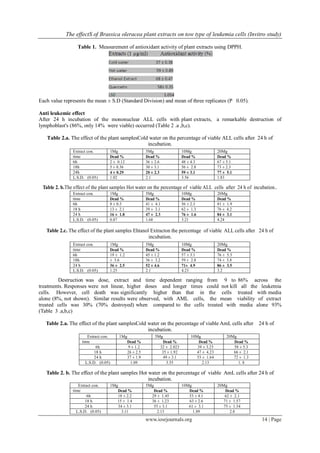
Extract con. 1Mg 5Mg 10Mg 20Mg
time Dead % Dead % Dead % Dead %
6h 22 ± 2.2 36 ± 1.25 59 ± 2.12 64 ± 1.23
18 h 31± 2.1 39 ± 0.87 68 ± 3.2 77 ± 4.21
24 h 28± 3.1 51 ± 3.2 73 ± 3.12 82 ± 5.2
L.S.D. (0.05) 3.23 2.21 4.2 3.26](https://image.slidesharecdn.com/b0121216-150625054822-lva1-app6892/85/The-effectS-of-Brassica-oleracea-plant-extracts-on-tow-type-of-leukemia-cells-Invitro-study-4-320.jpg)
![The effectS of Brassica oleracea plant extracts on tow type of leukemia cells (Invitro study)
www.iosrjournals.org 16 | Page
and many types of tannin will dissolve in ethanol solutions [25,26,27]. Whatever the active factor of the
extract appears to promote cell apoptosis since DNA damaged in leukemic cells incubated with ethanol
extract (Figure 3).
In comparison to established treatments for leukemia,th extract were equally effective in killing
80% of diseased cells within a few hours .Therefore, this plant extract used alone or in combination with
other extracts or other drugs has the potential to kill all leukemia cells whilst leaving healthy cells viable. The
plant extracts may provide low cost treatments for cancers and new treatments for drug resistant cancers.
Refrences
[1]. Tracy, K.S.; Richard, M. and Ian, T.J:Effect of Brassica vegetable juice on the induction of apoptosis and aberrant crypt foci in
rat colonicmucosal crypts in vivo.Carcinogenesis, 24, 3, 491-95. 2003.
[2]. Blok, G.; Patterson, B. and Subar, A.Fruits, vegetables, and cancer prevention: a review of the epidemiological evidence. Nutr.
Cancer, 18: 1-29. 1992.
[3]. Verhoeveven, D. T. H.; Verhagen,H. H.; Goldbohm, R. A.; Van der Brandt, P. A. and Van Poppel, G. A review of
mechanisms underlying Anticarcinogenicity by Brassica vegetables. Chem. Biol. Interact. 103: 79-129. 1997.
[4]. Zhung, Y. Anticarcinogenic activities of organic isothiocyanytes: chemistry and mechanisms. Cancer Res., 54, 1976s-
1981s. 1994.
[5]. Johnson, I.T. Mechanisms and Anticarcinogenic effects of diet related apoptosis in the intestinal mucosa. Nutra. Res.
Rev. 14, 229-256. 2001.
[6]. Musk, S.R. and Johnson, I. T. Allyl isothiocyanate is selectively toxic transformed cells of human colorectal
tumor line HT29. Carcinogenesis, 14, 2079-2083. 1993.
[7]. Huang, M. T.; Ferrero, T. and Ho, C. T. Cancer chemoprevention by phytochemicals in fruit and vegetables. In:
M.T. Huang, T. Osawa, C. T. Ho and R. T. Rosen, Editors, Food phytochemicals for cancer prevention I. Fruits and
vegetables, American Chemical Society, Washington, DC,pp. 2–16. 1994.
[8]. Cohen, J.H.; Kristal A.R. and Stanford,J.L. Fruit and vegetable intakes and prostate cancer risk. J. Natl. Cancer Inst. 92:
61-68. 2000.
[9]. Yu, R.; Mandlekar, S.; Harvey, K J.; Ucker, D. S. and Kong, A. N. T Chemopreventive isothiocyanate induce
apoptosis and caspase-3-like protease activity. Cancer Res. 58: 402-408. 1998.
[10]. Cohen, J.H.; Kristal, A.R. and Stanford,J.L. Fruit and vegetable intakes and prostate cancer risk, J. Natl. Cancer
Inst., 92 (1), 61–68. 2000.
[11]. Huang, M. T.; Ferrero, T. and Ho, C.T. Cancer chemoprevention by phytochemicals in fruit and vegetables. American
Chemical Society, pp. 2-16. 1994.
[12]. Zhang, Y.; Talalay, P.; Cho C. G. and Posner, G. H. A major inducer of anticarcinogenic protective enzymes from
broccoli: isolation and elucidation of structure, Proc Natl Acad Sci USA 89 (6), pp. 2399–2403.1992.
[13]. McCullough, B.D. and Wilson, B On the accuracy of statistical procedures in Microsoft Excel 2003.”Computational
Statistics and Data Analysis. Vol. 49,1244-125, 2005.
[14]. Freshney, R. I. Culture of animal cells. Third Edition. Wiley-Liss. New York.1994.
[15]. Mahony, D. E.; Gilliat, E.; Dawson, S.; Stockdale, E. and Lee, S. H. Vero Cell Assay for Rapid Detection of Clostridium
perfringens Enterotoxin. Applied and Enviromental Microbiology.pp:2141-2143. 1989.
[16]. Gao, S.; Yu, B.; Li, Y.; Dong, W. and Luo, H.Antiproliferative effect of Octreotide on gastric cells mediated by inhibition of
Akt/PKB and telomerase. World J. Gastroenterol., 9: 2362-28. 2003.
[17]. Kobori, M.; Yoshida, M.; Ohnishi, M.;Takei, T. and Shinmoto, H. 5α8 α Epidioxy -22E- ergoEdible Mushroom
suppresses growth of HL60 Leukemia and HT29 colon Adenocar- cinoma cells. j.Biol. Pharm. Bull., 29 (4):755-759, 2006.
[18]. Yaniv,Z.; Shabelsky, E. and Schafferman, D. Colocynth Potential arid land oilseed from an ancient cucurbit. Journal
Janick (ED). 257-261. 1999.
[19]. Talalay, P.; Fahey, J. W.; Healy, Z. R.; Wehage, S. L.; Benedict, A. L. andMin C. Sulforaphane mobilizes cellular defenses
that protect skin against damage by UV radiation, Proc Natl Acad Sci USA 104 (44) pp. 17500–17505, 2007.
[20]. Prestera, T. and Talalay, P. Electrophile and antioxidant regulation of enzymes that detoxify carcinogens, Proc Natl Acad Sci
USA 92 (19) pp. 8965–8969. 1995. [21]. Yokata, J. Tumor progression and metastasis. Carcinogeneis.
[21]. (3): 497-503. 2000.
[22]. Fahey, J. W.; Haristoy, X.; Dolan, P.M.;Kensler, T. W.; Scholtus, I. and Stephenson K. K. Sulforaphane inhibits
extracellular, intracellular and antibiotic resistant strains of Helicobacter pylori and prevents benzo [a] pyrene-induced
stomach tumors, Proc Natl Acad Sci USA 99 (11) 7610–7615. 2002.
[23]. Yoshizawa, Y.; Sakurai, K.; Kawaii, S.; Asari, M.; Soejima, J. and Murofushi, N. Comparison of antiproliferative and
antioxidant properties among nineteen apple cultivars Hort Science 40 (5) pp. 1204–1207. 2005.
[24]. Hwang, E. S. and Bowen, P. DNA damage, a biomarker of carcinogenesis:Its measurement and modulation by diet and
environment , Crit Rev Food Sci Nutr 47 (1) pp. 27–50. 2007.
[25]. Williamson, G.; Plumb, G. W.; Uda, Y.;Price K. R. and Rhodes, M. J. Dietary quercetin glycosides: antioxidant activity and
induction of the anticarcinogenic phase II marker enzyme quinine reductase in Hepalclc7 cells, Carcinogenesis 17
(11) pp. 2385–2387. 1996.
[26]. Letavayova, L.; Vlckova, V. and Brozmanova, J. Selenium: from cancer prevention to DNA damage, Toxicology 227 (1–2)
pp. 1–14. 2006.
[27]. Ge, X.; Yannai, S.; Rennet, G.; Gruener, N. and Fares, F.A. 3, 3-Diindolylmethane induces apoptosis in human cancer cells.
Biochem. Biophys. Res. Comm. 228: 153-158. 1996.](https://image.slidesharecdn.com/b0121216-150625054822-lva1-app6892/85/The-effectS-of-Brassica-oleracea-plant-extracts-on-tow-type-of-leukemia-cells-Invitro-study-5-320.jpg)

1. Brassica oleracea plant extracts were prepared using cold water, hot water, and ethanol and tested for antioxidant activity and effects on leukemia cells. 2. All extracts showed significant antioxidant effects in a DPPH radical scavenging assay. 3. The extracts were able to kill 50-75% of abnormal cells from patients with acute lympho-blastic leukemia (ALL) and acute myeloid leukemia (AML) after 24 hours of treatment. DNA fragmentation indicated the extracts induced apoptosis or programmed cell death.


