2007 antiproliferative, cytotoxic and antitumour activity
•
1 like•314 views

The document describes a study that evaluated the biological effects of coumarins isolated from Calophyllum brasiliense on cell survival, cell cycle, and apoptosis. Specifically: - Coumarins from C. brasiliense reduced survival of BMK cells by inducing apoptosis and necrosis, and arrested the cell cycle in S-phase, inhibiting cell division. - In mice studies, coumarins caused reduction of experimental tumors in 83% of animals by the end of treatment. - The study isolated and identified two coumarins - mammea A/BA and mammea A/BB - from C. brasiliense leaves, which have previously shown cytotoxic activity against tumor cell lines.
![720 César Ruiz-Marcial et al
A/BA, A/BB, B/BB, C/OA, C/OB, B/BA cyclo F, B/BB cyclo F
and isomammeigin (Reyes-Chilpa etal 2004). Recent studies
have shown that mammea A/BA and A/BB have high cytotoxic
activity against some tumour cell lines (Reyes-Chilpa et al 2004).
We have studied the biological effects of the antiprolifera-tive
activity of mammea A/BA and A/BB, isolated from
C. brasiliense, on the survival, cell cycle and apoptosis of BMK
cells. We attempted to define their antitumour effects in mice.
Chemicals
Vincristine was obtained from Lemery Laboratories (Mexico).
DMEM, fetal calf serum, MTT ((3-(4,5-dimethyl-2-thia-zolyl)-
2,5-diphenyl-2H-tetrazolium bromide), trypsin, trypsin
inhibitor, ribonuclease A (RNase), and propidium iodide
were obtained from Sigma Chemical Co. (USA).
Extraction and isolation of mammea A/BA and
A/BB from C. brasiliense
C. brasiliense (Clusiaceae) was collected at Ejido Benigno
Mendoza, Sierra de Santa Marta, Veracruz, Mexico. A voucher
specimen was deposited in the Herbarium of the Instituto de
Investigaciones Biologicas, Universidad Veracruzana at Xalapa,
Mexico, labelled number 435. Dried leaves (2075g) were
extracted at room temperature over one week with hexane, ace-tone,
and methanol, successively. Compounds were isolated after
spontaneous crystallization or by column chromatography on
Silica Gel-60, and individualized by their spectroscopy (1H
NMR, 13C NMR, IR, UV), EIMS and CIMS. The results of these
experiments were compared with Reyes-Chilpa etal (2004).
Hexane extracts (70.3 g)
While in solution, the extract showed spontaneous precipita-tion,
giving a white powder (19.8 g). Part of this material
(5.1 g) was treated with CH2Cl2. The insoluble part was a
mixture (583 mg) of friedelin and canophyllol, and the solu-ble
part afforded a mixture (4 g) of coumarins of two mam-mea
types: mammea A/BA and mammea A/BB. The extract
was then concentrated in-vacuo (35.3 g), and 9.4 g subjected
to column chromatography (200 g). Elution with hexane first
yielded friedelin (45.9mg), then a mixture of coumarins
(5 mg; mammea A/BA and mammea A/BB).
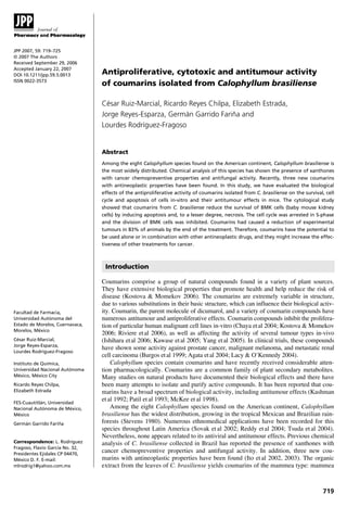
Identification and chemical structure of
mammea A/BA and mammea A/BB
The compounds isolated after spontaneous crystallization or
column chromatography were individually identified. The
purification of compounds (mp 122–124°C) was obtained by
HPLC using an ODS-column and elution with 90% MeOH.
Chemical structures are shown in Figure 1. The identification
of the mammeas was as follows:
1H NMR (200 MHz, CDCl3/TMS): 7.56 m, 3H (Ar);
7.41 m, 2H (Ar); 6.0 s, 1H (H-3); 5.95 s, 1H (OH-5). Isopre-nyl
on C-6: 5.09 tm, J = 6.9Hz, 1H (CH); 3.29 d, J = 6.9Hz,
2H (CH2); 1.65 and 1.70, both s and 3H, (2CH3).
HO
OH
6
7
5 4
3
2 6
O O
8 1
O
Mammea A/BA
HO
OH
5 4
7 8 1
O
3
2
O O
Mammea A/BB
1: 14.61 s, 1H (OH-7). R= 3-methylbutyryl: 3.19 d, J =
6.7Hz, 2H (CH2); 2.32 m, J = 6.7Hz, 1H (CH); 1.06 d,
J = 6.6Hz, 6H, (2CH3).
2: 14.57 s, 1H (OH-7). R= 2-methylbutyryl (A/BB):
3.95 m, J = 6.6Hz, 1H (CH-CH3); 1.29 d, J = 6.7Hz, 3H (CH-CH3),
1.94 m, 2H (CH2); 1.01 t, J = 7.2Hz, 3H (CH3).
EMIE: 70 eV m/z (%): 406M+ (96.4%) [C25H26O5]+;
363 (55.6%); 351 (76.0%); 293 (100%). IR n max (KBr)
3985 (OH); 2964, 2932, 2872 (C-H); 1729 (C= O); 1724
(C=O); 1614 (C = C); 1556 (C = C); 1390 (C-O).
For the pharmacological studies, a mixture of coumarins,
mammea A/BA and mammea A/BB, at a ratio of 2:1 was
used. The mix of mammeas (5mgmL−1) was prepared in
0.1% dimethyl sulfoxide (DMSO) (Aldrich, USA) at room
temperature.
Cell culture and pharmacological treatments
For in-vitro studies BMK cells were used, derived from baby
mouse kidney. These cells were kindly donated by Professor
Sophie Hallez from Brussels University, Belgium. Cells were
cultured in minimal essential medium (Gibco-BRL Inc.,
Grand Island, NY), supplemented with nonessential amino acids
(Gibco-BRL Inc., Grand Island, NY), 10% fetal calf serum
(Gibson-BRL Inc., Grand Island, NY), 2mol L−1 L-glutamine
and antibiotics. Cells were plated in 100-mm culture dishes
(106 cells/dish), and maintained at 37°C in an atmosphere of
5% CO2 in humidified air. Subcultures were obtained by
trypsinization (0.025% trypsin solution containing 0.01%
N,N,-diethyldithiocarbamic acid, sodium salt, and EDTA).
For cytological investigation, 105 cells were plated and
allowed to adhere overnight. The medium was then aspirated
and replaced with medium containing vehicle alone or
medium containing the mixture of mammeas (3, 5, 8, 10, 16,
20 and 24 mgmL−1), and the cells were then incubated for
24 h. After incubation, the cells were collected for further
cytological investigation.
Assessment of cell viability
BMK cells were incubated in 96-well plates. After 24 h, the
medium was removed; the cells were washed twice with
phosphate-buffered saline (PBS) and then incubated with the
mixture of coumarins (3, 5, 8, 10, 16, 20 and 24 mgmL−1).
Materials and Methods
Figure 1 Coumarins isolated from Calophyllum brasiliense leaves.](data:image/gif;base64,R0lGODlhAQABAIAAAAAAAP///yH5BAEAAAAALAAAAAABAAEAAAIBRAA7)
Recommended
Recommended
More Related Content
What's hot
What's hot (19)
Viewers also liked
Viewers also liked (20)
Similar to 2007 antiproliferative, cytotoxic and antitumour activity
Similar to 2007 antiproliferative, cytotoxic and antitumour activity (20)
More from Ricardo Reyes Chilpa. Instituto de Química. UNAM
More from Ricardo Reyes Chilpa. Instituto de Química. UNAM (20)
Recently uploaded
Recently uploaded (20)
2007 antiproliferative, cytotoxic and antitumour activity
- 1. JPP 2007, 59: 719–725 © 2007 The Authors Received September 29, 2006 Accepted January 22, 2007 DOI 10.1211/jpp.59.5.0013 ISSN 0022-3573 719 Antiproliferative, cytotoxic and antitumour activity of coumarins isolated from Calophyllum brasiliense César Ruiz-Marcial, Ricardo Reyes Chilpa, Elizabeth Estrada, Jorge Reyes-Esparza, Germán Garrido Fariña and Lourdes Rodríguez-Fragoso Abstract Among the eight Calophyllum species found on the American continent, Calophyllum brasiliense is the most widely distributed. Chemical analysis of this species has shown the presence of xanthones with cancer chemopreventive properties and antifungal activity. Recently, three new coumarins with antineoplastic properties have been found. In this study, we have evaluated the biological effects of the antiproliferative activity of coumarins isolated from C. brasiliense on the survival, cell cycle and apoptosis of cells in-vitro and their antitumour effects in mice. The cytological study showed that coumarins from C. brasiliense reduce the survival of BMK cells (baby mouse kidney cells) by inducing apoptosis and, to a lesser degree, necrosis. The cell cycle was arrested in S-phase and the division of BMK cells was inhibited. Coumarins had caused a reduction of experimental tumours in 83% of animals by the end of the treatment. Therefore, coumarins have the potential to be used alone or in combination with other antineoplastic drugs, and they might increase the effec-tiveness of other treatments for cancer. Introduction Coumarins comprise a group of natural compounds found in a variety of plant sources. They have extensive biological properties that promote health and help reduce the risk of disease (Kostova & Momekov 2006). The coumarins are extremely variable in structure, due to various substitutions in their basic structure, which can influence their biological activ-ity. Coumarin, the parent molecule of dicumarol, and a variety of coumarin compounds have numerous antitumour and antiproliferative effects. Coumarin compounds inhibit the prolifera-tion of particular human malignant cell lines in-vitro (Chaya et al 2004; Kostova & Momekov 2006; Riviere et al 2006), as well as affecting the activity of several tumour types in-vivo (Ishihara et al 2006; Kawase et al 2005; Yang et al 2005). In clinical trials, these compounds have shown some activity against prostate cancer, malignant melanoma, and metastatic renal cell carcinoma (Burgos et al 1999; Agata et al 2004; Lacy & O’Kennedy 2004). Calophyllum species contain coumarins and have recently received considerable atten-tion pharmacologically. Coumarins are a common family of plant secondary metabolites. Many studies on natural products have documented their biological effects and there have been many attempts to isolate and purify active compounds. It has been reported that cou-marins have a broad spectrum of biological activity, including antitumour effects (Kashman et al 1992; Patil et al 1993; McKee et al 1998). Among the eight Calophyllum species found on the American continent, Calophyllum brasiliense has the widest distribution, growing in the tropical Mexican and Brazilian rain-forests (Stevens 1980). Numerous ethnomedical applications have been recorded for this species throughout Latin America (Sovak et al 2002; Reddy et al 2004; Tsuda et al 2004). Nevertheless, none appears related to its antiviral and antitumour effects. Previous chemical analysis of C. brasiliense collected in Brazil has reported the presence of xanthones with cancer chemopreventive properties and antifungal activity. In addition, three new cou-marins with antineoplastic properties have been found (Ito et al 2002, 2003). The organic extract from the leaves of C. brasiliense yields coumarins of the mammea type: mammea Facultad de Farmacia, Universidad Autónoma del Estado de Morelos, Cuernavaca, Morelos, México César Ruiz-Marcial, Jorge Reyes-Esparza, Lourdes Rodríguez-Fragoso Instituto de Química, Universidad Nacional Autónoma México, México City Ricardo Reyes Chilpa, Elizabeth Estrada FES-Cuautitlán, Universidad Nacional Autónoma de México, México Germán Garrido Fariña Correspondence: L. Rodríguez Fragoso, Flavio García No. 32, Presidentes Ejidales CP 04470, México D. F. E-mail: mlrodrig1@yahoo.com.mx
- 2. 720 César Ruiz-Marcial et al A/BA, A/BB, B/BB, C/OA, C/OB, B/BA cyclo F, B/BB cyclo F and isomammeigin (Reyes-Chilpa etal 2004). Recent studies have shown that mammea A/BA and A/BB have high cytotoxic activity against some tumour cell lines (Reyes-Chilpa et al 2004). We have studied the biological effects of the antiprolifera-tive activity of mammea A/BA and A/BB, isolated from C. brasiliense, on the survival, cell cycle and apoptosis of BMK cells. We attempted to define their antitumour effects in mice. Chemicals Vincristine was obtained from Lemery Laboratories (Mexico). DMEM, fetal calf serum, MTT ((3-(4,5-dimethyl-2-thia-zolyl)- 2,5-diphenyl-2H-tetrazolium bromide), trypsin, trypsin inhibitor, ribonuclease A (RNase), and propidium iodide were obtained from Sigma Chemical Co. (USA). Extraction and isolation of mammea A/BA and A/BB from C. brasiliense C. brasiliense (Clusiaceae) was collected at Ejido Benigno Mendoza, Sierra de Santa Marta, Veracruz, Mexico. A voucher specimen was deposited in the Herbarium of the Instituto de Investigaciones Biologicas, Universidad Veracruzana at Xalapa, Mexico, labelled number 435. Dried leaves (2075g) were extracted at room temperature over one week with hexane, ace-tone, and methanol, successively. Compounds were isolated after spontaneous crystallization or by column chromatography on Silica Gel-60, and individualized by their spectroscopy (1H NMR, 13C NMR, IR, UV), EIMS and CIMS. The results of these experiments were compared with Reyes-Chilpa etal (2004). Hexane extracts (70.3 g) While in solution, the extract showed spontaneous precipita-tion, giving a white powder (19.8 g). Part of this material (5.1 g) was treated with CH2Cl2. The insoluble part was a mixture (583 mg) of friedelin and canophyllol, and the solu-ble part afforded a mixture (4 g) of coumarins of two mam-mea types: mammea A/BA and mammea A/BB. The extract was then concentrated in-vacuo (35.3 g), and 9.4 g subjected to column chromatography (200 g). Elution with hexane first yielded friedelin (45.9mg), then a mixture of coumarins (5 mg; mammea A/BA and mammea A/BB). Identification and chemical structure of mammea A/BA and mammea A/BB The compounds isolated after spontaneous crystallization or column chromatography were individually identified. The purification of compounds (mp 122–124°C) was obtained by HPLC using an ODS-column and elution with 90% MeOH. Chemical structures are shown in Figure 1. The identification of the mammeas was as follows: 1H NMR (200 MHz, CDCl3/TMS): 7.56 m, 3H (Ar); 7.41 m, 2H (Ar); 6.0 s, 1H (H-3); 5.95 s, 1H (OH-5). Isopre-nyl on C-6: 5.09 tm, J = 6.9Hz, 1H (CH); 3.29 d, J = 6.9Hz, 2H (CH2); 1.65 and 1.70, both s and 3H, (2CH3). HO OH 6 7 5 4 3 2 6 O O 8 1 O Mammea A/BA HO OH 5 4 7 8 1 O 3 2 O O Mammea A/BB 1: 14.61 s, 1H (OH-7). R= 3-methylbutyryl: 3.19 d, J = 6.7Hz, 2H (CH2); 2.32 m, J = 6.7Hz, 1H (CH); 1.06 d, J = 6.6Hz, 6H, (2CH3). 2: 14.57 s, 1H (OH-7). R= 2-methylbutyryl (A/BB): 3.95 m, J = 6.6Hz, 1H (CH-CH3); 1.29 d, J = 6.7Hz, 3H (CH-CH3), 1.94 m, 2H (CH2); 1.01 t, J = 7.2Hz, 3H (CH3). EMIE: 70 eV m/z (%): 406M+ (96.4%) [C25H26O5]+; 363 (55.6%); 351 (76.0%); 293 (100%). IR n max (KBr) 3985 (OH); 2964, 2932, 2872 (C-H); 1729 (C= O); 1724 (C=O); 1614 (C = C); 1556 (C = C); 1390 (C-O). For the pharmacological studies, a mixture of coumarins, mammea A/BA and mammea A/BB, at a ratio of 2:1 was used. The mix of mammeas (5mgmL−1) was prepared in 0.1% dimethyl sulfoxide (DMSO) (Aldrich, USA) at room temperature. Cell culture and pharmacological treatments For in-vitro studies BMK cells were used, derived from baby mouse kidney. These cells were kindly donated by Professor Sophie Hallez from Brussels University, Belgium. Cells were cultured in minimal essential medium (Gibco-BRL Inc., Grand Island, NY), supplemented with nonessential amino acids (Gibco-BRL Inc., Grand Island, NY), 10% fetal calf serum (Gibson-BRL Inc., Grand Island, NY), 2mol L−1 L-glutamine and antibiotics. Cells were plated in 100-mm culture dishes (106 cells/dish), and maintained at 37°C in an atmosphere of 5% CO2 in humidified air. Subcultures were obtained by trypsinization (0.025% trypsin solution containing 0.01% N,N,-diethyldithiocarbamic acid, sodium salt, and EDTA). For cytological investigation, 105 cells were plated and allowed to adhere overnight. The medium was then aspirated and replaced with medium containing vehicle alone or medium containing the mixture of mammeas (3, 5, 8, 10, 16, 20 and 24 mgmL−1), and the cells were then incubated for 24 h. After incubation, the cells were collected for further cytological investigation. Assessment of cell viability BMK cells were incubated in 96-well plates. After 24 h, the medium was removed; the cells were washed twice with phosphate-buffered saline (PBS) and then incubated with the mixture of coumarins (3, 5, 8, 10, 16, 20 and 24 mgmL−1). Materials and Methods Figure 1 Coumarins isolated from Calophyllum brasiliense leaves.
- 3. Antitumour effect of Calophyllum brasiliense 721 After 24 h, both cells and conditioned media were collected and processed. Cell viability was measured by the MTT assay (Wang et al 1996). Briefly, 20 mL MTT (5 g L−1) was added to each well and incubated for 4 h at 37°C, 5% CO2. The culture media was then discarded and 200 mL DMSO with 25 mL Sorensen’s buffer (glycine 0.1M, NaCl 0.1 M, pH 10.5) was added to each well. Once the blue crystals had dissolved, the optical density was determined on a microreader (Bio-Rad Co.) at 450 nm. Mitotic activity BMK cells were fixed in 3% paraformaldehyde with 0.25 M mannitol (45.54 g L−1) for 2 h, rinsed in PBS and stained with DAPI (4′,6-diamidino-2-phenylindol, 480 nm). After rinsing in PBS, the cells were embedded in Citifluor mounting medium. The mitotic index was counted with a fluorescent microscope (Optiphot 2 Nikon). For each experiment, the indices were determined per 1000 cells and with four replicates. Survival of cells and detection of percentage of apoptosis and necrosis Survival of cells was evaluated based on propidium iodide (PI; Sigma P4170) staining (Koopman et al 1994). PI (final concentration 5 mgmL−1) was added to each sample for 10 min (room temperature, in the dark). The samples (105) were analysed using a FACScalibur flow cytometer at 488nm (Becton Dickinson, USA, equipped with an argon laser). Ten thousand cells were analysed using four replicates. The results were analysed using the CELLQuest program. Based on the penetration of PI and the size of the cells, the percent-age of apoptotic and necrotic cells was determined. Analysis of cell cycle Cells (105) were fixed in 75% ethanol for 24 h and then washed in PBS and resuspended in 0.1% Nonidet P40 (Bio-chemica Fluka) and DNase-free RNase (10 mgmL−1) for 20 min at room temperature (Darzynkiewicz et al 2001). PI was then added (final concentration 5 mgmL−1) and incubated for 12 h at 4°C in the dark. Samples were analysed using a FACScalibur flow cytometer (Becton Dickinson). For each sample, 10 000 cells were analysed using four replicates. The results were analysed using the CELLQuest program. Tumour formation in mice Female BALB/c mice (6–8-weeks-old; purchased from Harlan Mexico, Mexico) were fed using a standard basal diet (24% protein) and exposed to a daily cycle of alternating 12-h light periods. The animals were housed in a temperature-and-humidity- controlled environment and food and water were freely available. The experiments were conducted according to the principles set forth in “Guide for the Care and Use of Laboratory Animals” (National Institutes of Health Publica-tion Number 86-23; Committee on Care and Use of Labora-tory Animals of the Institute for Laboratory Animal Resources 1986) and the Animal Welfare Act of 1966, as amended. Tumour formation in mice was performed as described by Dehenhardt et al (2002). Briefly, 2.5 × 10⁷ BMK cells were injected into each mouse and tumours were measured regularly. Pharmacological evaluation of coumarins Mice were randomly divided into four groups, each group containing 10 animals. Group I, control animals that received no treatment; Group II, mice with BMK-cell tumours; Group III, mice with BMK-cell tumours and treated with coumarins (20mgkg−1, i.p., three times per week, for three weeks); Group IV, mice with BMK-cell tumours and treated with vin-cristine (1 mgm−2, i.p., twice per week, for two weeks). Cou-marins and vincristine treatments started two weeks after the BMK cells were injected, when the tumour size was 5 mm. To assess the pharmacological effect of coumarins on tumour growth, the size of the tumour was measured every third day during the treatment. After completion of the treat-ment period, animals were anaesthetized with diethyl ether and killed by exsanguination. A complete post-mortem exam-ination was performed on each animal. Samples of liver, kid-ney, thymus, spleen and lungs were weighed and sliced, and several sections were fixed by immersion in alcohol and embedded in paraffin for histological analysis to identify the presence of metastasis. Statistical analysis Data were reported as means ± s.d. of three independent experiments conducted in quadruplicate. Statistical analysis was performed using a nonparametric analysis of variance and by Chi-squared testing. Individual differences between treatments were analysed using Tukey’s test. Significant differences were established at P < 0.001. Results Cell survival and induction of apoptosis and necrosis The coumarin extract caused a significant decrease in the survival of BMK cells (Figure 2). The coumarins (mammea A/BA and mammea A/BB) showed a concentration-dependent inhibitory effect on the growth of BMK cells (Figure 2) (P < 0.001). The lowest concentration produced a 20% reduc-tion in cell survival (P < 0.001). Concentrations of 5, 8 and 10mgmL−1 reduced the survival by approximately 30% (P<0.001). A reduction in cell survival greater than 50% was observed with concentrations higher than 20mgmL−1 (P<0.001). During incubation with all concentrations of coumarins, the decrease in the percentage of live cells was accompanied by a proportional increase in the percentage of apoptotic and necrotic cells (Table 1). Treatment with the lowest concentra-tion did not produce any important change in the relationship between live and dead cells. Treatment of cells with concen-trations higher than 5 mgmL−1 gave a higher increase in the percentage of apoptotic cells than the increase in necrotic cells (Table 1). All concentrations gave a higher percentage of apoptotic cells than necrotic cells (P < 0.001).
- 4. 722 César Ruiz-Marcial et al 120 100 80 60 40 20 0 Survival (% of control) # # Control 3 5 8 10 16 20 24 Concn (μg mL–1) ∗ ∗ ∗ ∗ ∗ Cell cycle analysis Cell cycle analysis following treatment with coumarins (mammea A/BA and mammea A/BB) showed that, during incubation with the extract, the proportion of the BMK cell population in each of G1 and S phases varied with the con-centration of the extract (Figure 3). The proportion of G1 phase cells reduced, while the percentage of cells at S phase of the cell cycle increased (Figure 3). This change was dependent on the concentration of the coumarins, and was accompanied by the appearance of apoptotic cells at the sub-phase G1 (data not shown). Treatment with the lower concentrations of coumarins (3 and 5 mgmL−1) caused a reduction in G1 and G2/M phases after 24 h incubation (Figure 3; P < 0.05). Concentrations of coumarins higher than 5 mgmL−1 caused an almost 50% reduction of G1 phase cells (P < 0.05). In contrast, a signific-ant increase of the percentage in S phase cells was observed with all concentrations, P < 0.001. Mitotic index after treatment with coumarins The coumarins (mammea A/BA and mammea A/BB), at all chosen concentrations, reduced the percentage of cell divisions, Cell population (%) 80 60 40 20 0 ∗ ∗ ∗ ∗ ∗ ∗ ∗ ∗ ∗ ∗ ∗ ∗ ∗ ∗ ∗ Control 3 5 8 10 16 20 24 Concn (μg mL–1) G1 S G2/M 6 5 4 3 2 1 0 Mitotic index (%) ∗ ∗ ∗ ∗ ∗& & & ∗ ∗ # # # Control 3 5 8 10 16 20 24 Concn (μg mL–1) or led to their almost total inhibition, in a concentration-dependent manner (Figure 4). The lowest concentrations of coumarins (3 and 5 mgmL−1) lowered the mitotic activity of the BMK cells by 18% and 30%, respectively, after incuba-tion for 24 h. However, concentrations higher than 8 mgmL−1 produced a reduction of cell division in a dose-dependent manner. The two highest concentrations used (20 and 24 mgmL−1) caused almost total inhibition of cell division (P < 0.001). Tumour formation in mice The subcutaneous injection of viable BMK cells led to the development of tumours in mice. Tumour growth did not pro-duce any significant change in body weight compared with the control group. Similarly, it did not lead to the death of any animals. Treatment with coumarins caused a reduction in the devel-opment of tumours; this was evident seven days after the treatment started (Figure 5). Tumours reached an average size Figure 2 Changes in cell survival after incubation for 24 h in differ-ent concentrations of coumarins from Calophyllum brasiliense (control = 100% survival). The results are presented as means ± s.d. of three independent experiments, *P < 0.001 as compared with all groups; #P < 0.001 as compared with control, 3, 16, 20 and 24 mgmL−1 Table 1 Changes in the percentage of normal, apoptotic and necrotic BMK cells after incubation for 24 h in different concentrations of coumarins from Calophyllum brasiliense extract Concn extracted coumarins (mgmL-1) Normal cells (%) Apoptotic cells (%) Necrotic cells (%) Control 87± 6.5 8.5 ± 1.2 4.5 ± 1.2 3 86± 3.2 10 ± 2.1 4.3 ± 1.4 5 79± 3.3 16 ± 2.1 5.5 ± 1.2 8 74± 3.7 18 ± 2.8 8.7 ± 3.0 10 70± 3.4 21 ± 2.9 9.0 ± 2.1 16 64± 6.5 24 ± 2.5 10.5 ± 2.6 20 62± 4.1 28 ± 3.3 9.7 ± 3.8 24 52± 5.3 30 ± 4.0 20.1 ± 2.0 Figure 3 Percentage of cells in G1, S, G2/M phases of the cell cycle after incubation for 24 h in different concentrations of coumarins from Calophyllum brasiliense. The results are presented as means ± s.d. of three independent experiments. *P < 0.001 vs control values. Figure 4 Changes in the mitotic index of cells after incubation for 24 h in different concentrations of coumarins from Calophyllum brasiliense. The results are presented as means ± s.d. of three independent experi-ments. *P < 0.001 as compared with all groups; #P < 0.001 as compared with 3, 16, 20 and 24 mg mL−1; &P < 0.001 as compared with 3, 5, 8 and 10 mg mL−1.
- 5. Antitumour effect of Calophyllum brasiliense 723 20 16 12 8 4 0 Tumour Tumour + vincristine Tumour + coumarins 3 6 9 12 15 18 21 24 27 30 33 36 39 Time (day) BMK cells injection Mean tumour diameter (mm) Figure 5 Effect of coumarins on tumour size. Viable BMK cells were injected subcutaneously into mice. The size of the tumours was measured every third day during the treatment. Mean tumour diameter (mm) was expressed relative to time after cell injection. The results are presented as means ± s.d., n = 10 animals. of 8 mm in animals treated with coumarins. Compared with animals with no treatment, a reduction of 83% was observed in tumour size at the end of treatment with coumarins and the tumour size was approximately 2mm when the animals were killed. In contrast, vincristine stopped the development of tumours, which was evident after the first dose (Figure 5). The tumours reached a size of 6mm (average) during the treatment with vincristine and they continued to reduce in size after the treatment. A reduction of 100% was observed in tumour size at the end of four weeks. Histopathological analysis revealed the presence of meta-static cells in hepatic tissue and, to a lesser degree, in kidney tissue in tumour-bearing mice receiving no treatment (data not shown). Animals with tumours and treated with cou-marins did not show any evidence of metastasis to any of the tissues analysed, including the liver. Animals with tumour and treated with vincristine had livers showing large areas of metastatic cells (data not shown). Cancer is one of the main causes of death in the world. Two related fields are being explored through basic and clinical research: the identification of the molecular mechanisms involved in the illness, and the discovery of drugs for preven-tion or treatment (Kirsner 2003). In the area of drug discov-ery, there is increasing interest in cancer-prevention agents of plant origin (Newman et al 2003). Coumarins comprise a vast array of biologically active compounds ubiquitous in plants, many of which have been used in traditional medicine for thousands of years. The cou-marins constitute an important class of compounds with sev-eral types of pharmacological actions, possessing anticancer, anti-HIV, anticoagulant, spasmolytic and antibacterial activ-ity among others. However, their most prominent actions are their antioxidant and antiproliferative effects (Mattern et al 1999; Kostova & Momekov 2006). Antineoplastic drugs are designed either to inhibit abnor-mal cell proliferation or to cause the death of abnormal cells. Due to the complex biochemical pathways and the specific phases of cellular life cycles, there are numerous opportuni-ties for these drugs to exert a beneficial effect. Some types of coumarin show antitumour activity in-vivo and in-vitro (Guilet et al 2000; Kawaii et al 2001; Finn et al 2002); however, there have been no investigations regarding the effect of coumarins from C. brasiliense on the mitotic index, survival, apoptosis induction or cell cycle of cells in-vitro. The results of our investigation have shown that the coumarins isolated from C. brasiliense—a mixture of mammea A/BA and A/BB— were capable of producing antiproliferative and cytotoxic effects in-vitro, and reduced tumour development in mice. Our results showed that coumarins at concentrations of 5–10 mgmL−1 had a cytotoxic effect on BMK cells (30%). However, with higher concentrations (15–24 mgmL−1) the cytotoxic effect was the greatest. The cytotoxicity produced by other types of coumarin have been reported using a non-small- cell bronchial carcinoma line (Kofinas et al 1998; López-González et al 2000), carcinoma epidermoid cells (Guilet et al 2001), human renal cell lines (Kawase et al 2005), and cells from the uterine cervix (HeLa), larynx (Hep2), prostate gland (PC3), lymphoma (K562) and neural tissue (U251) (Reyes-Chilpa et al 2004). One aspect of neoplastic cells is their proclivity to replicate. Depending on their mechanism of action, antineoplastic agents may exert their effects specifically during one of the phases or they may act during any phase of the cell cycle to cause cell death (Dictor et al 2004). We analysed the cell cycle in cells treated with coumarins. Interestingly, in BMK cells we observed arrest in the S-phase of the cell cycle. Damage to DNA may pre-vent replication by altering DNA, RNA or their function. Agents that work by this mechanism inhibit the proliferation of neoplas-tic cells (they cannot divide). Other drugs may cause damage to DNA, which can result in cell death. After treatment, inhibition of the mitotic division of BMK cells occurred. The inhibition depended on the concentration of the coumarins. This inhibition was accompanied by the appearance of large numbers of apop-totic cells. Therefore, it can be speculated that the cells enter apoptosis at the border of the G1 phase and cells that have alter-ations in their DNA are arrested so to repair the damage. The effects of coumarins on the cell cycle have been stud-ied previously. Coumarins and 7-hydroxycoumarin have anti-tumour actions in-vitro and in-vivo (Marshall et al 1999; Lacy & O’Kennedy 2004; Elinos-Baez et al 2005). Coumarins and 7-hydroxycoumarin inhibit cell growth by inducing cell cycle arrest in the G1 phase in lung carcinoma cell lines (López- González et al 2004). Jimenez-Orozco et al (2001) observed that 7-hydroxycoumarin 1mM inhibited the G1/S transition of the cell cycle in the human lung adenocarcinoma cell line A-427. They also found that 7-hydroxycoumarin significantly reduced cyclin D1 expression, which appeared to indicate an action of 7-hydroxycoumarin in early events of phase G1. Finally, studies using nitro-derivatives of coumarins showed that they caused dose-dependent inhibition of the S-phase regulatory protein, cyclin A, in an irreversible manner. There-fore, these and other nitro-derivatives of 7-hydroxycoumarin possess selective and irreversible cytotoxicity (Finn et al 2004). Recent studies using C. brasiliense collected in Brazil have shown that it inhibited the growth of leukaemic cells and induced caspase-mediated and p53-independent apoptosis Discussion Vincristine Start Coumarins Killed
- 6. 724 César Ruiz-Marcial et al (Kimura et al 2005). Our results agreed with those previous studies; we found that coumarins isolated from C. brasiliense collected in Mexico displayed potent cytotoxic and antiprolif-erative activity in-vitro. Tumorigenesis is an extremely complex multistep process that leads to the pathological expansion of a tissue. Tumour cells gain an unlimited replicative potential because of an imbal-ance between finely tuned proliferate, growth-inhibitory and apoptotic signals. Animal models that accurately represent the cellular and molecular changes associated with the initiation and progression of cancer have significant potential to facilitate the development of better methods for the early detection and treatment of this illness. Chemotherapeutic drugs can be divided into several categories, based on the way they act. Antineoplas-tic agents act primarily on rapidly dividing and growing cells. To date, there are no reports regarding the assay of the effects of coumarins in animal models. In this study, we have evaluated the antitumoral effects of coumarins in tumours induced by subcutaneous injection of BMK cells into mice. Our in-vitro results demonstrated that coumarins isolated from C. brasiliense had cytotoxic and antiproliferative effects. Additionally, we demonstrated that coumarins were better than vincristine in inhibiting tumour growth. Conclusion Coumarins isolated from C. brasiliense had cytotoxic effects and were able to alter the G1/S transition of the cell cycle in-vitro. In addition, they had antitumour effects. Therefore, coumarins have the potential to be used alone or in combina-tion with other antineoplastic drugs, and they might increase the effectiveness of other treatments against cancer. Agata, N., Nogi, H., Milhollen, M., Kharbanda, S., Kufe, D. (2004) 2-(8-Hydroxy-6-methoxy-1-oxo-1H-2-benzopyran-3-yl)propionic acid, a small molecule isocoumarin, potentiates dexamethasone-induced apoptosis of human multiple myeloma cells. Cancer Res. 64: 8512–8516 Burgos, A., Alcaide, A., Alcoba, C., Azcona, J. M., Garrido, J., Lorente, C., Moreno, E., Murillo, E., Olsina-Pavia, J., Olsina- Kissler, J., Samaniego, E., Serra, M. (1999) Comparative study of the clinical efficacy of two different coumarin dosages in the man-agement of arm lymphedema after treatment for breast cancer. Lymphology 32: 1–2 Chaya, N., Terauchi, K., Yamagata, Y., Kinjo, J., Okabe, H. (2004) Antiproliferative constituents in plants 14.1) Coumarins and acri-done alkaloids from Boenninghausenia japonica NAKAI. Biol. Pharm. Bull. 27: 1312–1316 Committee on Care and Use of Laboratory Animals of the Institute for Laboratory Animal Resources (1986) Commission on Life Sci-ences, National Research Council: Guide for the care and use of laboratory animals. Publication 86-23, 18 and the Animal Welfare Act of 1966, as amended. Darzynkiewicz, J., Robinson, P., Crissman, H. A. (2001) Analysis of cell cycle. In: Flow cytometry. Part A. Vol. 41, Plenum Press, London, pp 223–254 Dehenhardt, K., Cheng, G., Lindsten, T., White E. (2002) Bax and Bak mediate p53-independent suppression of tumorigenesis. Can-cer Cell 2: 193–203 Dictor, M., Ehribger, M., Mertens, F., Akervall, J., Wenneberg, J., Dorai, T., Aggarwal, B. B. (2004) Role of chemopreventive agents in cancer therapy. Cancer Lett. 215: 129–140 Elinos-Baez, C. M., Leon, F., Santos, E. (2005) Effects of coumarin and 7 OH-coumarin on bcl-2 and Bax expression in two human lung cancer cell lines in vitro. Cell. Biol. 29: 703–708 Finn, G. J., Kenealy, E., Creaven, B. S., Egan, D. A. (2002) In vitro cytotoxic potential and mechanism of action of selected cou-marins, using human renal cell lines. Cancer Lett. 183: 61–68 Finn, G. J., Creaven, B. S., Egan, D. A. (2004) A study of the role of cell cycle events mediating the action of coumarin derivatives in human malignant melanoma cells. Cancer Lett. 214: 43–54 Guilet, D., Hélesbeux, J. J., Séraphin, D., Sévenet, T., Richomme, P., Bruneton, J. (2000) Novel cytotoxic 4-phenylfuranocoumarins from Calophyllum dispar. J. Nat. Prod. 64: 10–15 Guilet, D., Séraphin, D., Rondeau, D., Richomme, P., Bruneton, J. (2001) Cytotoxic coumarins from Calophyllum dispar. Phyto-chemistry 58: 571–575 Ishihara, M., Yokote, Y., Sakagami, M. (2006) Quantitative struc-ture- cytotoxicity relationship analysis of coumarin and its deriva-tives by semiempirical molecular orbital method. Anticancer Res. 26: 2883–2886 Ito, C., Itoigawa, M., Mishina, Y., Filho, V. C., Mukainaka, T., Tokuda, H., Nishino, H., Furukawa, H. (2002) Chemical constitu-ents of Calophyllum brasiliense: structure elucidation of seven new xanthones and their cancer chemopreventive activity. J. Nat. Prod. 65: 267–272 Ito, C., Itoigawa, M., Mishina, Y., Filho, V. C., Enjo, F., Tokuda, H., Nishino, H., Furukawa, H. (2003) Chemical constituents of Calo-phyllum brasiliense. 2. Structure of three new coumarins and can-cer chemopreventive activity of 4-substituted coumarins. J. Nat. Prod. 66: 368–371 Jimenez-Orozco, F. A., López-González, J. S., Nieto-Rodriguez, A., Velasco-Velazquez, M. A., Molina-Guarneros, J. A., Mendoza- Patino, N., Garcia-Mondragon, M. J., Elizalde-Galvan, P., Leon- Cedeno, F., Mandoki, J. J. (2001) Decrease of cyclin D1 in the human lung adenocarcinoma cell line A-427 by 7-hydroxycou-marin. Lung Cancer 34: 185–194 Kashman, Y., Gustafson, K. R., Fuller, R. W., Cardellina, J. H., McMahon, J. B., Currens, M. J., Buckheit, R. W., Hughes, S. H., Cragg, G. M., Boyd, M. R. (1992) The calanolides, a novel HIV-inhibitory class of coumarin derivatives from the tropical rainforest tree, Calophyllum lanigerum. J. Med. Chem. 35: 2735–2743 Kawaii, S., Tomono, Y., Ogawa, K., Sugiura, M., Yano, M., Yoshizawa, Y. (2001) The antiproliferative effect of coumarins on several cancer cell lines. Anticancer Res. 21: 917–924 Kawase, M., Sakagami, H., Motohashi, N., Hauer, H., Chatterjee, S. S., Spengler, G., Vigyikanne, A. V., Molnar, A., Molnar, J. (2005) Coumarin derivatives with tumor-specific cytotoxicity and multi-drug resistance reversal activity. In Vivo 19: 705–711 Kimura, S., Ito, C., Jyoko, N., Segawa, H., Kuroda, J., Okada, M., Adachi, S., Nakahata, T., Yuasa, T., Filho, V. C., Furukawa, H., Maekawa, T. (2005) Inhibition of leukemic cell growth by a novel anti-cancer drug (GUT-70) from Calophyllum brasiliense that acts by induction of apoptosis. Int. J. Cancer 113: 158–165 Kirsner, M. K. (2003) Cancer: new therapies and new approaches to recurring problems. AANA J. 71: 55–62 Kofinas, C., Chinou, I., Loukis, A., Harvala, C., Roussakis, C., Maillard, M., Hostettman, K. (1998) Cytotoxic coumarins from the aerial parts of Tordylium apulum and their effects on a non-small- cell bronchial carcinoma line. Planta Med. 64: 174–176 Koopman, G., Reutelingsperger, C. P. M., Kuijten, G. A. M., Keehnen, R. M. J., Pals, S. T., van Oers, M. H. J. (1994) Annexin V for flow cytometric detection of phosphatidylserine expression on B cells undergoing apoptosis. Blood 84: 1415–1420 References
- 7. Antitumour effect of Calophyllum brasiliense 725 Kostova, I., Momekov, G. (2006) New zirconium (IV) complexes of coumarins with cytotoxic activity. Eur. J. Med. Chem. 41: 717–726 Lacy, A., O’Kennedy, R. (2004) Studies on coumarins and cou-marin- related compounds to determine their therapeutic role in the treatment of cancer. Curr. Pharm. Des. 10: 3797–3811 Liu, R. H. (2004) Potential synergy of phytochemicals in cancer pre-vention: mechanism of action. J. Nutr. 134: 3479S–3485S López-González, J. S., Garcia, H. P., Cázares, D. A., Guarneros, J. M., Mandoki, J. J., Morales, F. M. (2000) Efecto en el ciclo celular de líneas de adenocarcinoma pulmonar por cumarina y 7-hidroxicumarina. Rev. Inst. Nal. Enf. Resp. Mex. 13: 92–197 López-González, J. S., Prado-García, H., Aguilar-Cázares, D., Molina- Guarneros, J. A., Morales-Fuentes, J., Mandoki, J. J. (2004) Apoptosis and cell cycle disturbances induced by coumarin and 7-hydroxycou-marin on human lung carcinoma cell lines. Lung Cancer 43: 275–283 Marshall, M. E., Kervin, K., Benefield, C., Umerani, A., Albainy-Jenei, S., Zhao, Q. (1994) Growth-inhibitor effects of coumarin (1,2-benzo-pyrone) and 7-hydroxycoumarin in human malignant cell lines in vitro. J. Cancer Res. Clin. Oncol. 120: 3–10 Mattern, U., Luer, P., Kreusch, D. (1999) Biosynthesis of coumarin. Taken from: Comprehensive Natural Products Chemistry, 1st edn. Elsevier, pp 623–637 McKee, T. C., Covington, C. D., Fuller, R. W., Bokesch, H. R., Young, S., Cardellina, J. H. I., Kadushin, M. R., Soejarto, D. D., Stevens, P. F., Cragg, G. M., Boyd, M. R. (1998) Pyranocoumarins from tropical spe-cies of the genus Calophyllum: a chemotaxonomic study of extracts in the National Cancer Institute Collection. J. Nat. Prod. 6: 1252–1256 Newman, D. J., Cragg, G. M., Snader, K. M. (2003) Natural products as sources of new drugs over the period 1981–2002. J. Nat. Prod. 66: 1022–1037 Patil, A. D., Freyer, A. J., Eggleston, D. S., Haltiwanger, R. C., Bean,M. F., Taylor, P. B., Caranfa, M. J., Breen, A. L., Bartus, H. R., Johnson, R. K. (1993) The inophyllums, novel inhibitors of HIV-1 reverse transcriptase isolated from Malaysian tree, Calophyllum inophyllum Linn. J. Med. Chem. 36: 4131–4138 Reddy, N. S., Reddy, M. M., Cosenza, S., Gumireddy, K., Bell, S. C., Reddy, E. P., Ramana, M. V. (2004) Synthesis of new coumarin 3-(N-aryl) sulfonamides and their anticancer activity. Bioorgan. Med. Chem. Lett. 14: 4093–4097 Reyes-Chilpa, R., Estrada-Muñiz, E., Ramírez-Apan, T., Amekraz, B., Aumelas, A., Jankowski, Ch., Vazquez-Torres, M. (2004) Cyto-toxic effects of mammea type coumarins from Calophyllum bra-siliense. Life Sci. 75: 1635–1647 Riviere, C., Goossens, L., Pommery, N., Fourneau, C., Delelis, A., Henichart, J. P. (2006) Antiproliferative effects of isopentenylated coumarins isolated from Phellolophium madagascariense Baker. Nat. Prod. Res. 20: 909–916 Sovak, M., Seligson, A. L., Konas, M., Hajduch, M., Dolezal, M., Machala, M., Nagourney, R. (2002) Herbal composition PC-SPES for management of prostate cancer: identification of active princi-ples. J. Natl. Cancer Inst. 94: 1275–1281 Stevens, P. F. (1980) A revision of the old world species of Calo-phyllum (Guttiferae). J. Arnold Arb. 61: 117–171 Tsuda, H., Ohshima, Y., Nomoto, H., Fujita, K., Matsuda, E., Iigo, M., Takasuka, N., Moore, M. A. (2004) Cancer prevention by natural compounds. Drug Metab. Pharmacokinet. 19: 245–263 Wang, H. Z., Chang, C. H., Lin, C. P., Tsai, M. C. (1996) Using MTT viability assay to test the cytotoxicity of antibiotics and ster-oid to cultured porcine corneal endothelial cells. J. Ocul. Pharma-col. Ther. 12: 35–43 Yang, H., Protiva, P., Gil, R. R., Jiang, B., Bagget, T. S., Basile, M. J., Reynertson, K. A., Westein, I. B., Kennelly, E. J. (2005) Antioxi-dant and cytotoxic isoprenylated coumarins from Mammea ameri-cana. Planta Med. 71: 852–860