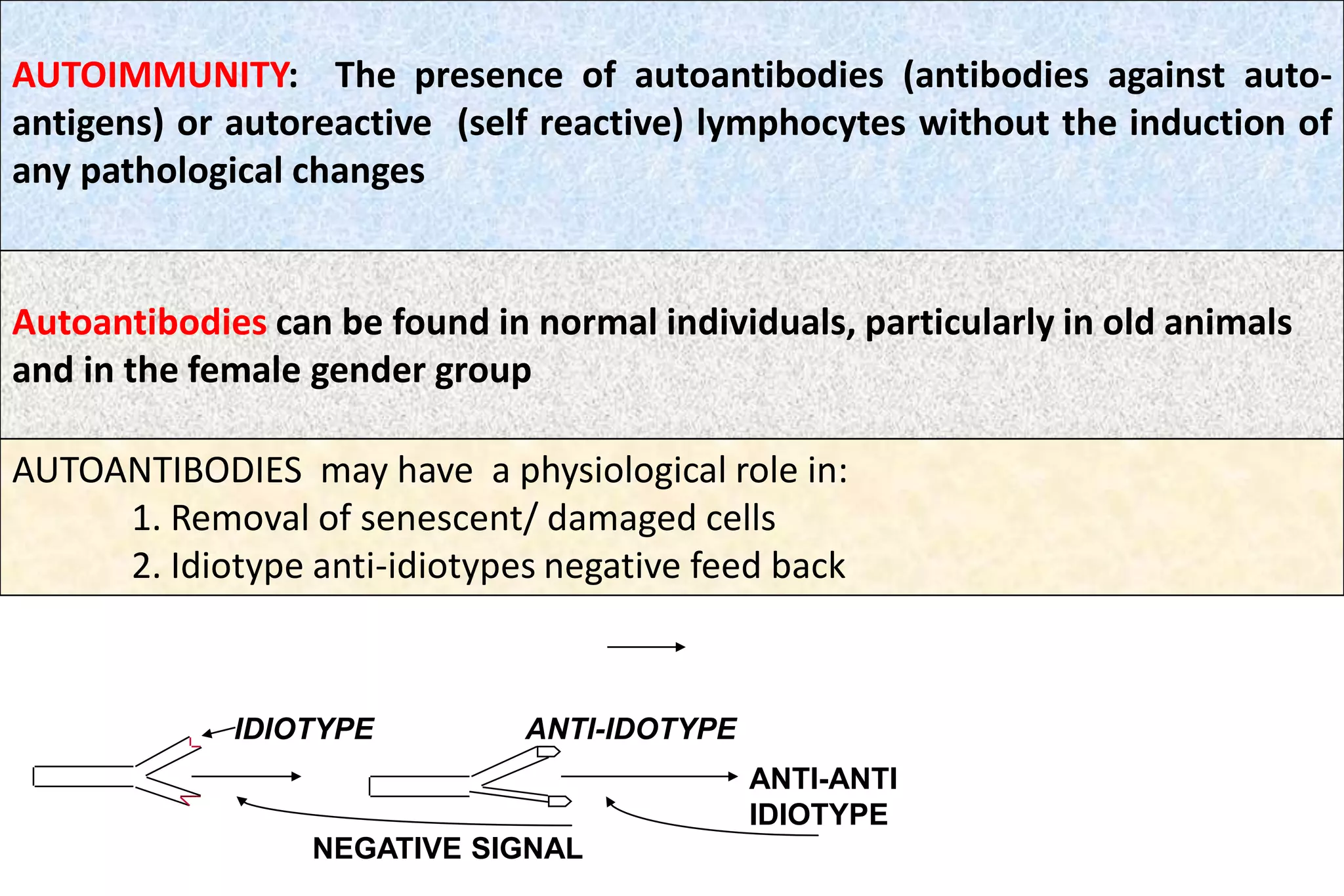
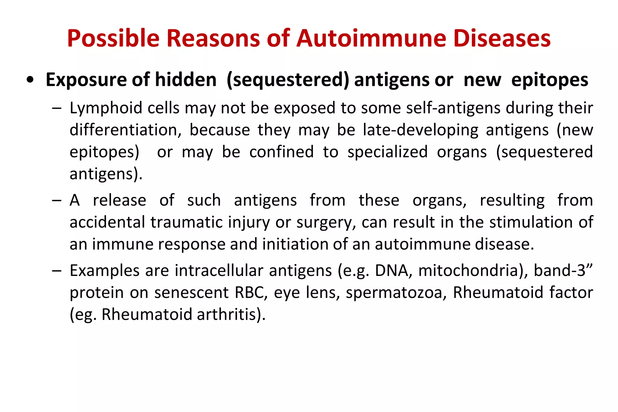
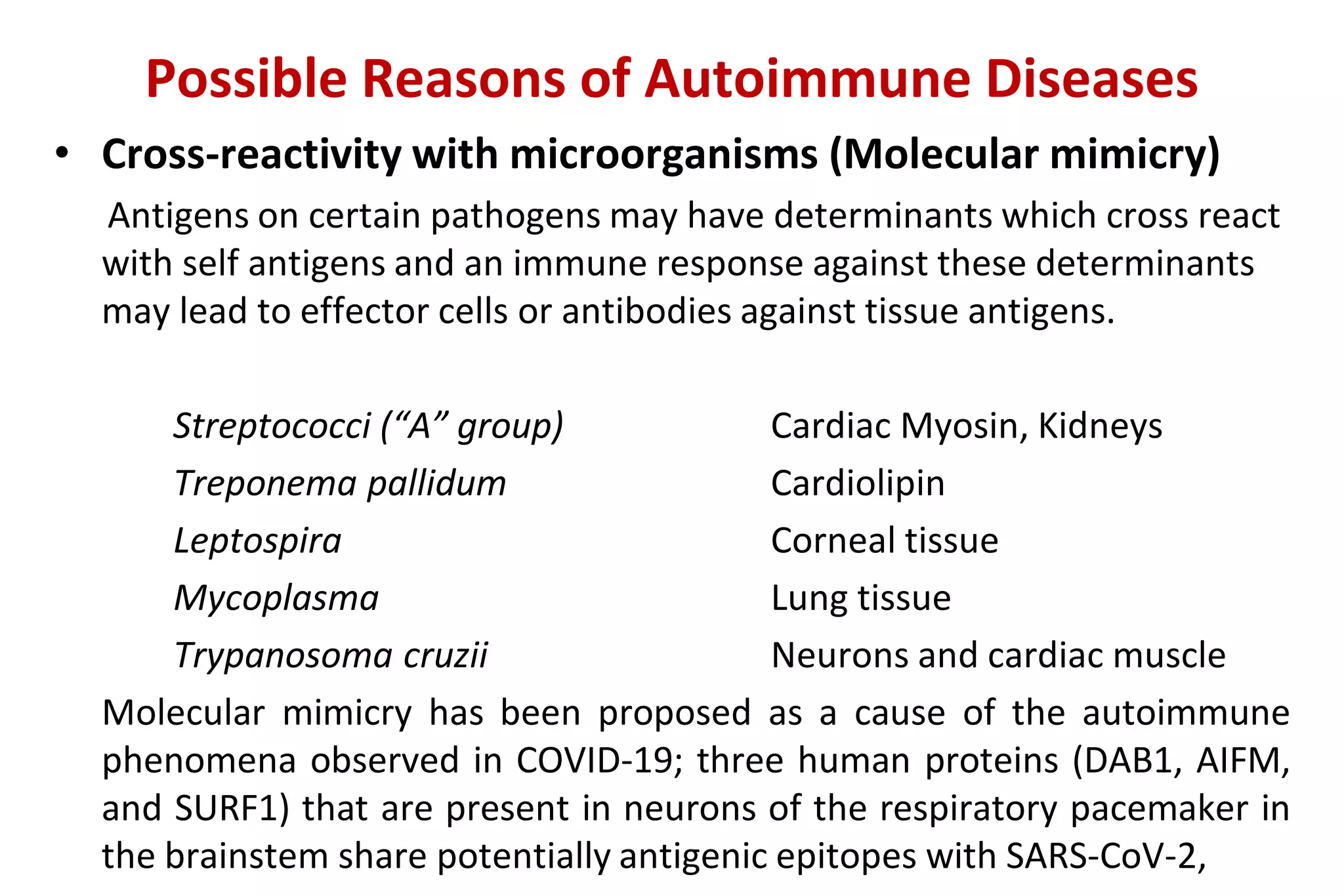
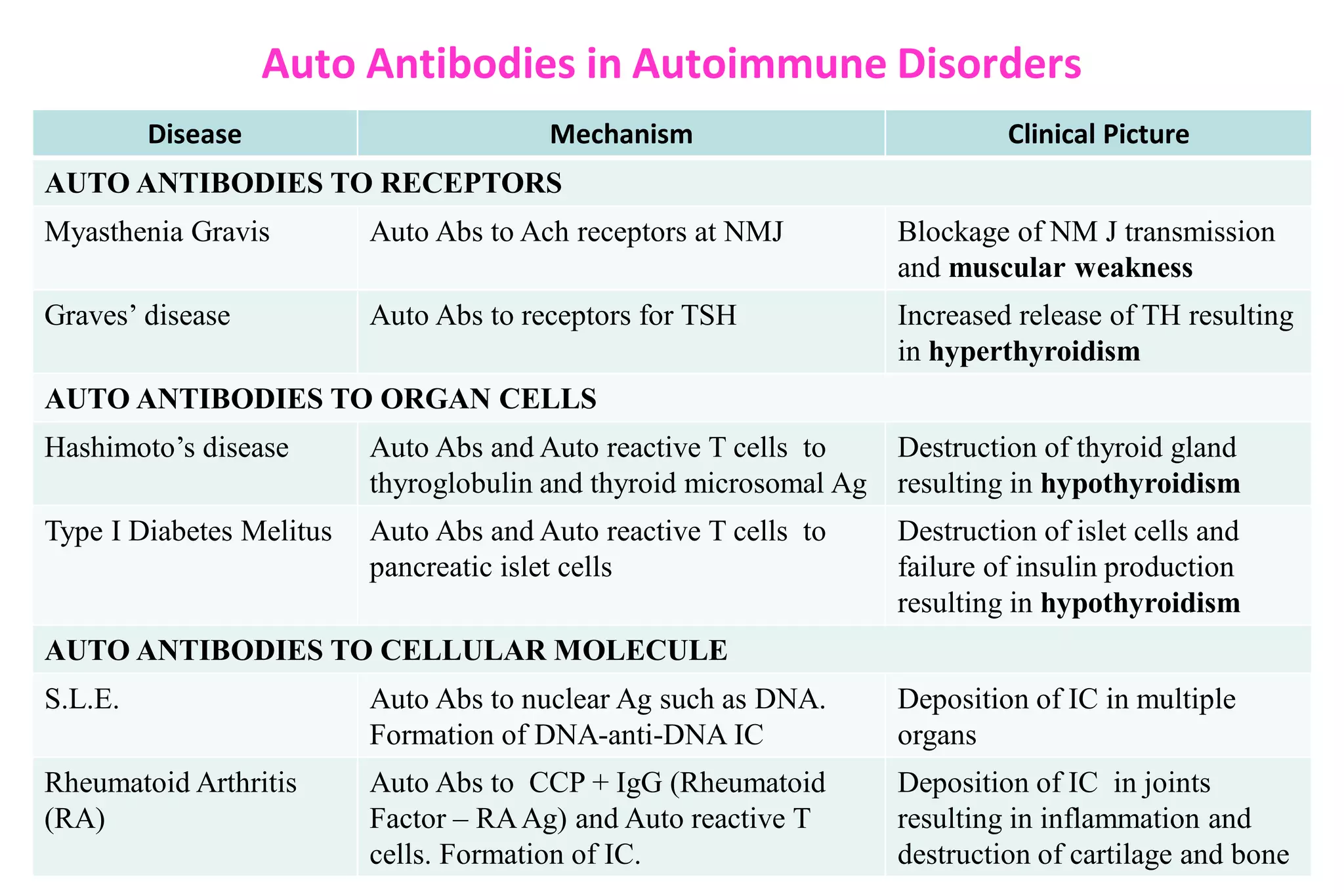
SlideShare a Scribd company logo
1 of 39
Download to read offline
AUTOIMMUNITY
AND
AUTOIMMUNE DISEASES
RAKESH SHARDA
Department of Veterinary Microbiology
NDVSU College of Veterinary Science & A.H.,
MHOW
Spleen
Lymph
Nodes
Thymus
Bone Marrow
T cell Development
In normal individuals, majority of lymphocytes
reacting with auto-antigens are deleted in primary
lymphoid organs by CLONAL DELETION (Central
Tolerance)
Few self-reactive lymphocytyes which escape and
reach secondary lymphoid tissues are down regulated
there by CLONAL ANERGY, Tregs or other
mechanisms (Peripheral Tolerance)
HORROR AUTOTOXICUS: Ehlrich proposed that
immune system should not be toxic to auto (self)
tissues.
Thus, normal individuals are TOLERANT to AUTO
ANTIGENS
IDIOTYPE ANTI-IDOTYPE
ANTI-ANTI
IDIOTYPE
Autoantibodies can be found in normal individuals, particularly in old animals
and in the female gender group
NEGATIVE SIGNAL
AUTOIMMUNITY: The presence of autoantibodies (antibodies against auto-
antigens) or autoreactive (self reactive) lymphocytes without the induction of
any pathological changes
AUTOANTIBODIES may have a physiological role in:
1. Removal of senescent/ damaged cells
2. Idiotype anti-idiotypes negative feed back
AUTOIMMUNE DISEASES: Pathological changes (structural and/or functional)
in tissues and/or organs as a consequence of autoreactive cells
CRITERIA FOR AUTOIMMUNE DISEASE OR WITEBSKY’S POSTULATES
1. The presence of autoantibodies or autoreactive lymphocytes ( in association
with pathological changes )
2. Experimental reproduction of the disease in laboratory animals
3. The features of the experimental disease must closely match with the
clinical disease.
4. Ability to passively transfer the disease by autoantibodies or lymphocytes
• Autoimmunity results from a failure or breakdown of the mechanisms
normally responsible for maintaining self-tolerance in B cells, T cells, or
both.
• Although underlying molecular etiologies remain elusive for most
autoimmune diseases, it is thought that autoimmunity is multifactorial,
resulting from a complex interplay between genetic susceptibility,
environmental triggers, and aberrant immune regulation
• Autoimmune diseases may be either systemic or organ specific.
• Various effector mechanisms are responsible for tissue injury in
different autoimmune diseases.
• It is not uncommon for a patient to have symptoms of more than one
autoimmune disease (known as ‘overlap’, or undifferentiated collagen
vascular syndrome).
• On contrary, a patient with one autoimmune disease may have
serologic markers – but no clinical manifestations – of another
General Characteristics
What Causes Autoimmune Diseases?
The Mosaic of Autoimmunity
• The exact etiology of autoimmune diseases still remains unknown.
• Several factors are presumed to contribute to the emergence of
an autoimmune disease in a host, which are:
– the genetic predisposition,
– the environmental triggers such as bacterial (including the gut microbiota),
viral, fungal and parasitic infections; physical and environmental agents;
hormonal factors, and
– the hosts immune system dysregulation.
Shoenfeld et al. (1989) coined the term “The Mosaic of
Autoimmunity” to include all these factors interplay.
(Bellanti JA (Ed). Immunology IV: Clinical Applications in Health and Disease. I Care Press, Bethesda, MD, 2012].)
The Three Etiopathogenic Factors Involved In Autoimmune
Diseases.
Possible Reasons of Autoimmune Diseases
• Genetics - Certain autoimmune disorders are familial. Many autoimmune diseases
are associated with MHC-I or II, including ankylosing spondylitis (HLA-B27), SLE
(HLA-DR2, DR3), RA (HLA-DR4), type 1 DM (HLA-DR3, DR4), and MS (HLA-DR2).
Gene mutations may also result in risk of autoimmune disorders
• Escape of auto-reactive clones: The negative selection in the thymus may not be
fully functional to eliminate self reactive cells. Not all self antigens may be
represented in the thymus or certain antigens may not be properly processed and
presented.
• Abnormal expression of class II antigens - Class II antigens begin to be expressed
on cells that normally do not bear these antigens. Eg. Epithelial cells of the thyroid
and salivary glands.
• Sex - Most autoimmune diseases such as Lupus, primarily affect women; estrogen
promotes autoimmunity
• Defective apoptosis and impaired Treg activity- For example, Autoimmune
polyendocrine syndrome type I (APSI) or (APECED) due to defects in AIRE induced
apoptosis in central tolerance and Immune dysregulation, polyendocrinopathy,
enteropathy (X-linked) (IPEX) syndrome due to Foxp3 mutation leading to and
absence of Treg
Association of autoimmune diseases and MHC
Possible Reasons of Autoimmune Diseases
• Exposure of hidden (sequestered) antigens or new epitopes
– Lymphoid cells may not be exposed to some self-antigens during their
differentiation, because they may be late-developing antigens (new
epitopes) or may be confined to specialized organs (sequestered
antigens).
– A release of such antigens from these organs, resulting from
accidental traumatic injury or surgery, can result in the stimulation of
an immune response and initiation of an autoimmune disease.
– Examples are intracellular antigens (e.g. DNA, mitochondria), band-3”
protein on senescent RBC, eye lens, spermatozoa, Rheumatoid factor
(eg. Rheumatoid arthritis).
Possible Reasons of Autoimmune Diseases
• Cross-reactivity with microorganisms (Molecular mimicry)
Antigens on certain pathogens may have determinants which cross react
with self antigens and an immune response against these determinants
may lead to effector cells or antibodies against tissue antigens.
Streptococci (“A” group) Cardiac Myosin, Kidneys
Treponema pallidum Cardiolipin
Leptospira Corneal tissue
Mycoplasma Lung tissue
Trypanosoma cruzii Neurons and cardiac muscle
Molecular mimicry has been proposed as a cause of the autoimmune
phenomena observed in COVID-19; three human proteins (DAB1, AIFM,
and SURF1) that are present in neurons of the respiratory pacemaker in
the brainstem share potentially antigenic epitopes with SARS-CoV-2,
Possible Reasons of Autoimmune Diseases
• Viral involvement
– Retroviral infection in Sjogren’s syndrome (Dry eye / Dry mouth) and in
Systemic lupus erythematosus (a multi-organ disease).
– Epstein-Barr virus infection and Systemic lupus erythematosus
– Coxsackie viral infection in type I diabetes
Possible mechanisms:
 Molecular mimicry - HCV and platelet glycoprotein IIIa (ITP)
 Interference with the regulatory control mechanisms - “bystander
activation” whereby the infection may lead to activation of APCSs that may
in turn activate pre-primed auto-reactive T-cells, leading to the production
of pro-inflammatory mediators, which in turn may lead to tissue damage
 Epitope spreading: Autoimmune reactions initiated against one self antigen
that injure tissues may result in the release and alterations of other tissue
antigens, activation of lymphocytes specific for these other antigens, and
exacerbation of the disease.
 Induction of new antigens
 Polyclonal B cell activation
Mechanisms of Tissue Damage in
Autoimmune Diseases
Mechanisms of issue damage
• Mechanisms involved in tissue damage are very complex.
• In a number of autoimmune diseases more than one mechanism are involved.
• IgE autoantibodies ( Type I ): Rare. A possible example: “Milk allergy” in Jersey
cattle; IgE autoantibody response to α-casein.
• Cytotoxic (Type II ):
Autoantibodies to cell surface, e.g.
RBC........................autoimmune hemolytic anemia
Thrombocytes....... autoimmune thrombocytopenia
• Immunecomplex (Type III):
Autoantibodies (Ab) + Antigen (Ag) [ Ab-Ag ] Immunecomplex, e.g. SLE, RA
• T-Cell Mediated Damage (Type IV):
T cells are primarily involved in tissue damage, e.g. autoimmune thyroiditis,
ulcerative colitis, Insulin-dependent Juvenile-onset Diabetes, autoimmune
encephalitis
 T cells are involved in almost all autoimmune diseases, particularly in organ
specific autoimmune disorders (probably due a lack of T regulatory cells or the
participation of Th17 cells).
 Macrophages, which are activated by T cells are also involved in tissue damage
(release of toxic enzymes and oxidative free-radicals; Type IV).
 Most autoimmune diseases are characterized by the production of antibodies
directed to self antigens, known as auto-antibodies
 The physical properties of an autoantibody that determines its pathogenicity
include:
 its affinity to the antigen that plays a role in immune complexes
formation, and
 its charge that helps antibody attach to tissues, e.g. in SLE, positively
charged auto-antibodies (anti-double stranded DNA) associate with
the negatively charged basement membrane in the kidney, where
they can form complexes in situ with DNA and lead to local
inflammation.
 Most of the pathogenic auto-antibodies are of IgG isotype.
Mechanisms of Tissue damage
Auto Antibodies in Autoimmune Disorders
Disease Mechanism Clinical Picture
AUTO ANTIBODIES TO RECEPTORS
Myasthenia Gravis Auto Abs to Ach receptors at NMJ Blockage of NM J transmission
and muscular weakness
Graves’ disease Auto Abs to receptors for TSH Increased release of TH resulting
in hyperthyroidism
AUTO ANTIBODIES TO ORGAN CELLS
Hashimoto’s disease Auto Abs and Auto reactive T cells to
thyroglobulin and thyroid microsomal Ag
Destruction of thyroid gland
resulting in hypothyroidism
Type I Diabetes Melitus Auto Abs and Auto reactive T cells to
pancreatic islet cells
Destruction of islet cells and
failure of insulin production
resulting in hypothyroidism
AUTO ANTIBODIES TO CELLULAR MOLECULE
S.L.E. Auto Abs to nuclear Ag such as DNA.
Formation of DNA-anti-DNA IC
Deposition of IC in multiple
organs
Rheumatoid Arthritis
(RA)
Auto Abs to CCP + IgG (Rheumatoid
Factor – RAAg) and Auto reactive T
cells. Formation of IC.
Deposition of IC in joints
resulting in inflammation and
destruction of cartilage and bone
AUTOANTIBODIES
RBC
NUCLEAR COMPONENTS
( DNA )
IMMUNE COMPLEXES
Deposit in joints, kidneys, skin
Arthritis
Glomerulonephritis
Skin lesions “Lupus Band”
PLATELETS
Thrombocytopenia
Hemolytic anemia
MUSCLE
Myositis
CARDIAC
Myocarditis
Hypertension
SUSPECT SLE IN AN ANIMAL WITH MULTIPLE ABNORMALITIES
(TOGETHER WITH POSITIVE SEROLOGY
eg. anti-nuclear antibodies and/or anti-DNA antibodies)
CNS
Examples of diseases caused by cell and tissue specific antibodies
Examples of T cell-mediated Immunological diseases
Types of Autoimmune Diseases
Autoimmune Diseases
ORGAN SPECIFIC
NON-ORGAN SPECIFIC
AUTOIMMUNE THYROIDITIS
AUTOIMMUNE SKIN DISEASES
AUTOIMMUNE REPRODUCTIVE
DISEASES
AUTOIMMUNE HEMOLYTIC
ANEMIA
MYESTHENIA GRAVIS
AUTOIMMUNE OPTHALMIC DISEASES
INSULIN-DEPENDENT
DIABETES
AUTOIMMUNE ADRENALITIS
AUTOIMMUNE NEUROLOGICAL DISEASES
SLE
RHEUMATOIDARTHRITIS
POLYARTHRITIS
( NON-RA )
DERMATOMYOSITIS
POSSIBLE
AUTOIMMUNE
• Alopecia atrea
• STEROID
RESPONSIVE
MENINGITIS
MULTIPLE SCLEROSIS
Organ Specific Autoimmune Diseases
AUTOIMMUNE REPRODUCTIVE DISEASES
• ORCHITIS: WITH ANTI-SPERM AUTOANTIBODIES
• AUTOANTIBODIES TO HORMONES - Progesterone, Estrogen, LH, GnRH
“IMMUNOLOGICAL CONTRACEPTION”
BOVINE
OVINE LH GnRH
Antibodies
cross react
with Feline LH
Antibodies
cross react
with Feline
GnRH
Female
DOG
Protein from zona
pellucida of Ovary
Infertility
Net result: Sterility
Autoantibodies to sperm antigens will agglutinate the sperms - Decreased Fertility
(Non-Surgical Contraception)
MYAESTHENIA GRAVIS - It is a disorder of signal transmission between the nerves and
muscles, which is characterized by muscular weakness and excessive fatigue. MG occurs due
to formation of autoantibodies to acetylcholine receptor. The antibodies, which are of IgG
type, initiate a complement mediated lysis of muscle cells. In dogs, cats and humans this
disorder is either congenital or acquired . In any breed of Dog, the disease can develop, but
German shepherds, Golden retrievers, and Labradors appear to be more susceptible to the
disease. Disease can be classified as focal myasthenia gravis and generalized myasthenia
gravis. In focal MG, an animal presents with mega esophagus and various degrees of facial
paralysis with limb muscle weakness. In generalized MG, limb muscle weakness is
associated with facial paralysis and mega esophagus.
AUTOIMMUNE OPTHALMIC DISEASES
Equine recurrent uveitis - periodic ophthalmia/moon blindness in horses. The inciting
factors in horses include microbial agents such as Leptospira spp.. Inter-photoreceptor
retinoid binding protein is the major autoantigen involved, with subsequent epitope
spreading to the S. protein. Affected animals (horses) have circulating antibodies to L.
interrogans, recurrent attacks of uveitis, retinitis, and vasculitis. They have
blepharospasms, lacrimation and photophobia in severe cases. The eye lesions are
infiltrated with Th-1 cells and neutrophils with extensive fibrin and C3 deposition
AUTOIMMUNE HEMOLYTIC ANEMIA (AHIA)
• AHIA is characterized by production of auto-antibodies against red blood cell
antigens resulting in their destruction.
• The disease is reported from humans, dogs, cattle, horses, cats, mice, rabbits.
• It can occur independently or in association with SLE or thrombocytopenia with
Feline Leukemia.
• The autoantibodies are produced against red cell glycophorins, cytoskeletal
protein spectrin and the membrane anion exchange protein (CD 233 or band 3)
in case of dogs.
• The destruction of red blood cells may result from either intra vascular
hemolysis that is mediated by compliment or by the removal of antibody
coated red cells by the macrophages of spleen and liver.
• The cause of red cell destruction is the alteration in red cell surface antigens
(neo-antigens) that is induced by drugs and viruses.
• Its onset may be associated with other immunological abnormalities, with
stress (vaccination), viral diseases, or with hormonal imbalances (pregnancy or
pyometra).
Type I Diabetes Melitus (TIDM)
• Autoimmune type 1 diabetes mellitus (T1DM) is a multi-stage disease of
undetermined aetiology characterised by an aggressive and selective
autoimmune response against β-cells of islets of Langerhans of pancreas.
• Genetic factors include both MHC and non-MHC genes.
• The most common environmental factors are viral infections (such as
CMV and Coxsackie) and vitamin D deficiency.
• Three major auto-antigens have been identified in T1DM: insulin, GAD65
and IA2.
• T1DM is primarily a cell-mediated autoimmune disease.
• The CD8+ auto-reactive Tlymphocytes are the most abundant and the
most active in β-cell destruction.
• The islet autoantibodies are predictive markers of an ongoing
autoimmune response, yet their exact role in beta cell destruction
remains to be clarified.
Organ-specific autoimmune diseases
Non-organ Specific or Systemic
Autoimmune Diseases
SYSTEMIC LUPUS ERYTHEMATOSUS ( SLE )
• SLE is an autoimmune disease characterized by multi-organ inflammation,
with a diverse array of clinical manifestations and production of
pathogenic auto antibodies directed against nucleic acids.
• The exact patho-aetiology of SLE remains elusive. Defective clearance of
apoptotic cells and immune complexes, are important contributors to the
development of SLE.
• Aberrant innate immune responses also play a significant role in the
pathogenesis of SLE.
• The central immunological disturbance in patients with SLE is
autoantibody production
• The basic pathological features of SLE are inflammation and blood vessel
abnormalities, which include band or occlusive vasculopathy, vasculitis,
and immune complex deposition.
• The most characterized organ pathology is in the kidney.
SYSTEMIC LUPUS ERYTHEMATOSUS ( SLE )
DOGS, CATS AND HORSES
MULTI-ORGAN INVOLVEMENT:
Kidneys, Joints, Skin, Salivary Glands, Hemopoetic cells etc.
Possible involvement of infectious agents:
Epstein- Barr virus (EBV)
Parainfluenza
Measles
Myxovirus
Retrovirus
B cell Hyperactivity
Abnormalities in T cells
Decreased regulatory T cells
Rheumatoid arthritis (RA)
• Rheumatoid arthritis (RA) is a common autoimmune disease affecting
approximately 1% of the world’s population.
• It is also observed in dogs (especially small), cats and horses.
• RA disease is characterized by a symmetric, polyarthritis of the small
joints of the hands and feet, but almost any joint can become involved.
• Affects not only joints but also other parts of the body .
• Pathologically, the synovial tissue becomes hypertrophied, highly
vascularized, and infiltrated with leukocytes, principally macrophages
• The proliferating fibroblasts and infiltrating macrophages, develops into
an inflammatory mass, termed the pannus.
• The synovia proliferates and release destructive proteolytic enzymes,
which cause erosion and dectruction of cartilage and bone.
• The subsynovial area is comprised of infiltrating macrophages and
dendritic cells and lymphocytes.
Rheumatoid arthritis (RA)
• The precise cause of RA is unknown but genetic, epigenetic and environmental
factors are involved.
• Possible infections: Mycobacteria, Lyme’s Disease, Parvovorus, Mycoplasma,
EBV
• Possible autoantigens:
– (1) Rhuematoid factor: IgM antibody against an abnormally glycosylated
IgG .
– (2) Type II- Collagen
• Macrophages have been most clearly implicated in pathogenesis and are the
major synovial producers of TNF-α. Other cells include:
– neutrophils, present in the synovial fluid, synthesize inflammatory PGs, proteases, and
ROS.
– Mast cells release cytokines, chemokines, proteases, and vasoactive amines.
• It is hypothesised that a T-cell dependent inflammatory reaction to an
unknown antigen underlies the pathology (Th1 and ?Th17)
Systemic autoimmune diseases
Treatment of autoimmune diseases
Available treatment thus far is non-specific
IMURAN ( AZATHIOPRINE )
CORTICOSTEROIDS
GOLD SALTS (e.g. Auranofin)
In the early course of the disease (e.g. RA) Asprin or steroidal and Non-steroidal anti-
inflammatory drugs (NSAID)
EXPERIMENTAL APPROACHES:
• Bone Marrow Transplantation
• Stem cell therapy
• Intravenous immunoglobulin therapy
• Fab ANTIBODIES FRAGMENTS TO T and B cells
• Anti- CD4, B cells OR Anti- IL-2 RECEPTOR antibodies
• Pentraxin 3 – a humoral soluble PRR
• Antigen-based immunotherapy - overloading the immune system by gradual but
ever-increasing doses of the same molecule that it is attacking to make reactive
T cells tolerant (T cell switching)
Monoclonal Antibodies – e.g. Rituximab for MS, Belimumab for anti-phospholipd Ab

More Related Content

What's hot

Mechanisms Of Defense Immune System.ppt
Mechanisms Of Defense Immune System.pptMechanisms Of Defense Immune System.ppt
Mechanisms Of Defense Immune System.ppt
Shama
 

What's hot (20)

Autoimmunity - pathophysiology (pharm D)
Autoimmunity - pathophysiology (pharm D)Autoimmunity - pathophysiology (pharm D)
Autoimmunity - pathophysiology (pharm D)
 
AUTOIMMUNITY
AUTOIMMUNITY AUTOIMMUNITY
AUTOIMMUNITY
 
Autoimmune Disease By Bishajit debnath
Autoimmune Disease By Bishajit debnath Autoimmune Disease By Bishajit debnath
Autoimmune Disease By Bishajit debnath
 
Auto immune disease
Auto immune diseaseAuto immune disease
Auto immune disease
 
Secondary immunodeficiency
Secondary immunodeficiencySecondary immunodeficiency
Secondary immunodeficiency
 
Autoimmunity - Microbiology - Immunology - Dr.Someshwaran Rajamani December 2015
Autoimmunity - Microbiology - Immunology - Dr.Someshwaran Rajamani December 2015Autoimmunity - Microbiology - Immunology - Dr.Someshwaran Rajamani December 2015
Autoimmunity - Microbiology - Immunology - Dr.Someshwaran Rajamani December 2015
 
Autoimmune disorders
Autoimmune disordersAutoimmune disorders
Autoimmune disorders
 
Hypersensitivity
Hypersensitivity Hypersensitivity
Hypersensitivity
 
Allergy 2016
Allergy 2016Allergy 2016
Allergy 2016
 
Immunity
Immunity Immunity
Immunity
 
Autoimmunity
AutoimmunityAutoimmunity
Autoimmunity
 
Mechanisms Of Defense Immune System.ppt
Mechanisms Of Defense Immune System.pptMechanisms Of Defense Immune System.ppt
Mechanisms Of Defense Immune System.ppt
 
Antigen
AntigenAntigen
Antigen
 
Allergy
AllergyAllergy
Allergy
 
Hypersensitivity type -3 reactions
Hypersensitivity type -3 reactions Hypersensitivity type -3 reactions
Hypersensitivity type -3 reactions
 
Allergy & hypersensitivity
Allergy & hypersensitivityAllergy & hypersensitivity
Allergy & hypersensitivity
 
Immunity
ImmunityImmunity
Immunity
 
Hypersensitivity
HypersensitivityHypersensitivity
Hypersensitivity
 
Antifungal chemotherapy
Antifungal chemotherapyAntifungal chemotherapy
Antifungal chemotherapy
 
An Overview of the Immune System
An Overview of the Immune SystemAn Overview of the Immune System
An Overview of the Immune System
 

Similar to Autoimmunity-and-Autoimmune-disorders.pdf

seminar presentation of general pathology by Ahmed ppt 2.pptx
seminar presentation of general pathology by Ahmed ppt 2.pptxseminar presentation of general pathology by Ahmed ppt 2.pptx
seminar presentation of general pathology by Ahmed ppt 2.pptx
AbasAhmed7
 
Tolerence and autoimmun diseases.pptx
Tolerence and autoimmun diseases.pptxTolerence and autoimmun diseases.pptx
Tolerence and autoimmun diseases.pptx
ssuser9976be
 

Similar to Autoimmunity-and-Autoimmune-disorders.pdf (20)

Autoimmunity engl
Autoimmunity englAutoimmunity engl
Autoimmunity engl
 
Tolerance Auto-immune Diseases and Tumor Immunity
Tolerance Auto-immune Diseases and Tumor ImmunityTolerance Auto-immune Diseases and Tumor Immunity
Tolerance Auto-immune Diseases and Tumor Immunity
 
Systemic Lupus Erythematosus, Sle
Systemic Lupus Erythematosus, SleSystemic Lupus Erythematosus, Sle
Systemic Lupus Erythematosus, Sle
 
seminar presentation of general pathology by Ahmed ppt 2.pptx
seminar presentation of general pathology by Ahmed ppt 2.pptxseminar presentation of general pathology by Ahmed ppt 2.pptx
seminar presentation of general pathology by Ahmed ppt 2.pptx
 
Autoimmune Diseases,including involved cells
Autoimmune Diseases,including involved cellsAutoimmune Diseases,including involved cells
Autoimmune Diseases,including involved cells
 
Autoimmunity and Autoimmune diseases.pptx
Autoimmunity and Autoimmune diseases.pptxAutoimmunity and Autoimmune diseases.pptx
Autoimmunity and Autoimmune diseases.pptx
 
Auto immunity and immunodeficiencies
Auto immunity and immunodeficienciesAuto immunity and immunodeficiencies
Auto immunity and immunodeficiencies
 
Tolerence and autoimmun diseases.pptx
Tolerence and autoimmun diseases.pptxTolerence and autoimmun diseases.pptx
Tolerence and autoimmun diseases.pptx
 
Autoimmunity
AutoimmunityAutoimmunity
Autoimmunity
 
Autoimmunity and autoimmune diseases dr. ihsan alsaimary
Autoimmunity and autoimmune diseases dr. ihsan alsaimaryAutoimmunity and autoimmune diseases dr. ihsan alsaimary
Autoimmunity and autoimmune diseases dr. ihsan alsaimary
 
Basic Principles of Immunology
Basic Principles of ImmunologyBasic Principles of Immunology
Basic Principles of Immunology
 
Autoimmunity
AutoimmunityAutoimmunity
Autoimmunity
 
Autoimmunity ''when good turns bad ''
Autoimmunity  ''when good turns bad ''Autoimmunity  ''when good turns bad ''
Autoimmunity ''when good turns bad ''
 
Autoimmune forum
Autoimmune forumAutoimmune forum
Autoimmune forum
 
Autoimmunity
AutoimmunityAutoimmunity
Autoimmunity
 
Immunology essay
Immunology essayImmunology essay
Immunology essay
 
3. autoimmunity
3. autoimmunity3. autoimmunity
3. autoimmunity
 
3. Autoimmunity.pptx
3. Autoimmunity.pptx3. Autoimmunity.pptx
3. Autoimmunity.pptx
 
Advances-health tech biocltechologyy recomminant dna
Advances-health tech biocltechologyy  recomminant dnaAdvances-health tech biocltechologyy  recomminant dna
Advances-health tech biocltechologyy recomminant dna
 
Autoimmunity.pptx
Autoimmunity.pptxAutoimmunity.pptx
Autoimmunity.pptx
 

Recently uploaded

Spellings Wk 4 and Wk 5 for Grade 4 at CAPS
Spellings Wk 4 and Wk 5 for Grade 4 at CAPSSpellings Wk 4 and Wk 5 for Grade 4 at CAPS
Spellings Wk 4 and Wk 5 for Grade 4 at CAPS
AnaAcapella
 

Recently uploaded (20)

NO1 Top Black Magic Specialist In Lahore Black magic In Pakistan Kala Ilam Ex...
NO1 Top Black Magic Specialist In Lahore Black magic In Pakistan Kala Ilam Ex...NO1 Top Black Magic Specialist In Lahore Black magic In Pakistan Kala Ilam Ex...
NO1 Top Black Magic Specialist In Lahore Black magic In Pakistan Kala Ilam Ex...
 
FICTIONAL SALESMAN/SALESMAN SNSW 2024.pdf
FICTIONAL SALESMAN/SALESMAN SNSW 2024.pdfFICTIONAL SALESMAN/SALESMAN SNSW 2024.pdf
FICTIONAL SALESMAN/SALESMAN SNSW 2024.pdf
 
AIM of Education-Teachers Training-2024.ppt
AIM of Education-Teachers Training-2024.pptAIM of Education-Teachers Training-2024.ppt
AIM of Education-Teachers Training-2024.ppt
 
HMCS Vancouver Pre-Deployment Brief - May 2024 (Web Version).pptx
HMCS Vancouver Pre-Deployment Brief - May 2024 (Web Version).pptxHMCS Vancouver Pre-Deployment Brief - May 2024 (Web Version).pptx
HMCS Vancouver Pre-Deployment Brief - May 2024 (Web Version).pptx
 
Beyond_Borders_Understanding_Anime_and_Manga_Fandom_A_Comprehensive_Audience_...
Beyond_Borders_Understanding_Anime_and_Manga_Fandom_A_Comprehensive_Audience_...Beyond_Borders_Understanding_Anime_and_Manga_Fandom_A_Comprehensive_Audience_...
Beyond_Borders_Understanding_Anime_and_Manga_Fandom_A_Comprehensive_Audience_...
 
FSB Advising Checklist - Orientation 2024
FSB Advising Checklist - Orientation 2024FSB Advising Checklist - Orientation 2024
FSB Advising Checklist - Orientation 2024
 
Simple, Complex, and Compound Sentences Exercises.pdf
Simple, Complex, and Compound Sentences Exercises.pdfSimple, Complex, and Compound Sentences Exercises.pdf
Simple, Complex, and Compound Sentences Exercises.pdf
 
latest AZ-104 Exam Questions and Answers
latest AZ-104 Exam Questions and Answerslatest AZ-104 Exam Questions and Answers
latest AZ-104 Exam Questions and Answers
 
Understanding Accommodations and Modifications
Understanding  Accommodations and ModificationsUnderstanding  Accommodations and Modifications
Understanding Accommodations and Modifications
 
Basic Civil Engineering first year Notes- Chapter 4 Building.pptx
Basic Civil Engineering first year Notes- Chapter 4 Building.pptxBasic Civil Engineering first year Notes- Chapter 4 Building.pptx
Basic Civil Engineering first year Notes- Chapter 4 Building.pptx
 
Spellings Wk 4 and Wk 5 for Grade 4 at CAPS
Spellings Wk 4 and Wk 5 for Grade 4 at CAPSSpellings Wk 4 and Wk 5 for Grade 4 at CAPS
Spellings Wk 4 and Wk 5 for Grade 4 at CAPS
 
Basic Intentional Injuries Health Education
Basic Intentional Injuries Health EducationBasic Intentional Injuries Health Education
Basic Intentional Injuries Health Education
 
How to Manage Call for Tendor in Odoo 17
How to Manage Call for Tendor in Odoo 17How to Manage Call for Tendor in Odoo 17
How to Manage Call for Tendor in Odoo 17
 
SOC 101 Demonstration of Learning Presentation
SOC 101 Demonstration of Learning PresentationSOC 101 Demonstration of Learning Presentation
SOC 101 Demonstration of Learning Presentation
 
On_Translating_a_Tamil_Poem_by_A_K_Ramanujan.pptx
On_Translating_a_Tamil_Poem_by_A_K_Ramanujan.pptxOn_Translating_a_Tamil_Poem_by_A_K_Ramanujan.pptx
On_Translating_a_Tamil_Poem_by_A_K_Ramanujan.pptx
 
Jamworks pilot and AI at Jisc (20/03/2024)
Jamworks pilot and AI at Jisc (20/03/2024)Jamworks pilot and AI at Jisc (20/03/2024)
Jamworks pilot and AI at Jisc (20/03/2024)
 
Sensory_Experience_and_Emotional_Resonance_in_Gabriel_Okaras_The_Piano_and_Th...
Sensory_Experience_and_Emotional_Resonance_in_Gabriel_Okaras_The_Piano_and_Th...Sensory_Experience_and_Emotional_Resonance_in_Gabriel_Okaras_The_Piano_and_Th...
Sensory_Experience_and_Emotional_Resonance_in_Gabriel_Okaras_The_Piano_and_Th...
 
Tatlong Kwento ni Lola basyang-1.pdf arts
Tatlong Kwento ni Lola basyang-1.pdf artsTatlong Kwento ni Lola basyang-1.pdf arts
Tatlong Kwento ni Lola basyang-1.pdf arts
 
Food safety_Challenges food safety laboratories_.pdf
Food safety_Challenges food safety laboratories_.pdfFood safety_Challenges food safety laboratories_.pdf
Food safety_Challenges food safety laboratories_.pdf
 
Unit 3 Emotional Intelligence and Spiritual Intelligence.pdf
Unit 3 Emotional Intelligence and Spiritual Intelligence.pdfUnit 3 Emotional Intelligence and Spiritual Intelligence.pdf
Unit 3 Emotional Intelligence and Spiritual Intelligence.pdf
 

Autoimmunity-and-Autoimmune-disorders.pdf

  • 1. AUTOIMMUNITY AND AUTOIMMUNE DISEASES RAKESH SHARDA Department of Veterinary Microbiology NDVSU College of Veterinary Science & A.H., MHOW
  • 2. Spleen Lymph Nodes Thymus Bone Marrow T cell Development In normal individuals, majority of lymphocytes reacting with auto-antigens are deleted in primary lymphoid organs by CLONAL DELETION (Central Tolerance) Few self-reactive lymphocytyes which escape and reach secondary lymphoid tissues are down regulated there by CLONAL ANERGY, Tregs or other mechanisms (Peripheral Tolerance) HORROR AUTOTOXICUS: Ehlrich proposed that immune system should not be toxic to auto (self) tissues. Thus, normal individuals are TOLERANT to AUTO ANTIGENS
  • 3. IDIOTYPE ANTI-IDOTYPE ANTI-ANTI IDIOTYPE Autoantibodies can be found in normal individuals, particularly in old animals and in the female gender group NEGATIVE SIGNAL AUTOIMMUNITY: The presence of autoantibodies (antibodies against auto- antigens) or autoreactive (self reactive) lymphocytes without the induction of any pathological changes AUTOANTIBODIES may have a physiological role in: 1. Removal of senescent/ damaged cells 2. Idiotype anti-idiotypes negative feed back
  • 4. AUTOIMMUNE DISEASES: Pathological changes (structural and/or functional) in tissues and/or organs as a consequence of autoreactive cells CRITERIA FOR AUTOIMMUNE DISEASE OR WITEBSKY’S POSTULATES 1. The presence of autoantibodies or autoreactive lymphocytes ( in association with pathological changes ) 2. Experimental reproduction of the disease in laboratory animals 3. The features of the experimental disease must closely match with the clinical disease. 4. Ability to passively transfer the disease by autoantibodies or lymphocytes
  • 5. • Autoimmunity results from a failure or breakdown of the mechanisms normally responsible for maintaining self-tolerance in B cells, T cells, or both. • Although underlying molecular etiologies remain elusive for most autoimmune diseases, it is thought that autoimmunity is multifactorial, resulting from a complex interplay between genetic susceptibility, environmental triggers, and aberrant immune regulation • Autoimmune diseases may be either systemic or organ specific. • Various effector mechanisms are responsible for tissue injury in different autoimmune diseases. • It is not uncommon for a patient to have symptoms of more than one autoimmune disease (known as ‘overlap’, or undifferentiated collagen vascular syndrome). • On contrary, a patient with one autoimmune disease may have serologic markers – but no clinical manifestations – of another General Characteristics
  • 7. The Mosaic of Autoimmunity • The exact etiology of autoimmune diseases still remains unknown. • Several factors are presumed to contribute to the emergence of an autoimmune disease in a host, which are: – the genetic predisposition, – the environmental triggers such as bacterial (including the gut microbiota), viral, fungal and parasitic infections; physical and environmental agents; hormonal factors, and – the hosts immune system dysregulation. Shoenfeld et al. (1989) coined the term “The Mosaic of Autoimmunity” to include all these factors interplay.
  • 8. (Bellanti JA (Ed). Immunology IV: Clinical Applications in Health and Disease. I Care Press, Bethesda, MD, 2012].) The Three Etiopathogenic Factors Involved In Autoimmune Diseases.
  • 9. Possible Reasons of Autoimmune Diseases • Genetics - Certain autoimmune disorders are familial. Many autoimmune diseases are associated with MHC-I or II, including ankylosing spondylitis (HLA-B27), SLE (HLA-DR2, DR3), RA (HLA-DR4), type 1 DM (HLA-DR3, DR4), and MS (HLA-DR2). Gene mutations may also result in risk of autoimmune disorders • Escape of auto-reactive clones: The negative selection in the thymus may not be fully functional to eliminate self reactive cells. Not all self antigens may be represented in the thymus or certain antigens may not be properly processed and presented. • Abnormal expression of class II antigens - Class II antigens begin to be expressed on cells that normally do not bear these antigens. Eg. Epithelial cells of the thyroid and salivary glands. • Sex - Most autoimmune diseases such as Lupus, primarily affect women; estrogen promotes autoimmunity • Defective apoptosis and impaired Treg activity- For example, Autoimmune polyendocrine syndrome type I (APSI) or (APECED) due to defects in AIRE induced apoptosis in central tolerance and Immune dysregulation, polyendocrinopathy, enteropathy (X-linked) (IPEX) syndrome due to Foxp3 mutation leading to and absence of Treg
  • 10. Association of autoimmune diseases and MHC
  • 11. Possible Reasons of Autoimmune Diseases • Exposure of hidden (sequestered) antigens or new epitopes – Lymphoid cells may not be exposed to some self-antigens during their differentiation, because they may be late-developing antigens (new epitopes) or may be confined to specialized organs (sequestered antigens). – A release of such antigens from these organs, resulting from accidental traumatic injury or surgery, can result in the stimulation of an immune response and initiation of an autoimmune disease. – Examples are intracellular antigens (e.g. DNA, mitochondria), band-3” protein on senescent RBC, eye lens, spermatozoa, Rheumatoid factor (eg. Rheumatoid arthritis).
  • 12. Possible Reasons of Autoimmune Diseases • Cross-reactivity with microorganisms (Molecular mimicry) Antigens on certain pathogens may have determinants which cross react with self antigens and an immune response against these determinants may lead to effector cells or antibodies against tissue antigens. Streptococci (“A” group) Cardiac Myosin, Kidneys Treponema pallidum Cardiolipin Leptospira Corneal tissue Mycoplasma Lung tissue Trypanosoma cruzii Neurons and cardiac muscle Molecular mimicry has been proposed as a cause of the autoimmune phenomena observed in COVID-19; three human proteins (DAB1, AIFM, and SURF1) that are present in neurons of the respiratory pacemaker in the brainstem share potentially antigenic epitopes with SARS-CoV-2,
  • 13. Possible Reasons of Autoimmune Diseases • Viral involvement – Retroviral infection in Sjogren’s syndrome (Dry eye / Dry mouth) and in Systemic lupus erythematosus (a multi-organ disease). – Epstein-Barr virus infection and Systemic lupus erythematosus – Coxsackie viral infection in type I diabetes Possible mechanisms:  Molecular mimicry - HCV and platelet glycoprotein IIIa (ITP)  Interference with the regulatory control mechanisms - “bystander activation” whereby the infection may lead to activation of APCSs that may in turn activate pre-primed auto-reactive T-cells, leading to the production of pro-inflammatory mediators, which in turn may lead to tissue damage  Epitope spreading: Autoimmune reactions initiated against one self antigen that injure tissues may result in the release and alterations of other tissue antigens, activation of lymphocytes specific for these other antigens, and exacerbation of the disease.  Induction of new antigens  Polyclonal B cell activation
  • 14. Mechanisms of Tissue Damage in Autoimmune Diseases
  • 15.
  • 16. Mechanisms of issue damage • Mechanisms involved in tissue damage are very complex. • In a number of autoimmune diseases more than one mechanism are involved. • IgE autoantibodies ( Type I ): Rare. A possible example: “Milk allergy” in Jersey cattle; IgE autoantibody response to α-casein. • Cytotoxic (Type II ): Autoantibodies to cell surface, e.g. RBC........................autoimmune hemolytic anemia Thrombocytes....... autoimmune thrombocytopenia • Immunecomplex (Type III): Autoantibodies (Ab) + Antigen (Ag) [ Ab-Ag ] Immunecomplex, e.g. SLE, RA • T-Cell Mediated Damage (Type IV): T cells are primarily involved in tissue damage, e.g. autoimmune thyroiditis, ulcerative colitis, Insulin-dependent Juvenile-onset Diabetes, autoimmune encephalitis
  • 17.  T cells are involved in almost all autoimmune diseases, particularly in organ specific autoimmune disorders (probably due a lack of T regulatory cells or the participation of Th17 cells).  Macrophages, which are activated by T cells are also involved in tissue damage (release of toxic enzymes and oxidative free-radicals; Type IV).  Most autoimmune diseases are characterized by the production of antibodies directed to self antigens, known as auto-antibodies  The physical properties of an autoantibody that determines its pathogenicity include:  its affinity to the antigen that plays a role in immune complexes formation, and  its charge that helps antibody attach to tissues, e.g. in SLE, positively charged auto-antibodies (anti-double stranded DNA) associate with the negatively charged basement membrane in the kidney, where they can form complexes in situ with DNA and lead to local inflammation.  Most of the pathogenic auto-antibodies are of IgG isotype. Mechanisms of Tissue damage
  • 18. Auto Antibodies in Autoimmune Disorders Disease Mechanism Clinical Picture AUTO ANTIBODIES TO RECEPTORS Myasthenia Gravis Auto Abs to Ach receptors at NMJ Blockage of NM J transmission and muscular weakness Graves’ disease Auto Abs to receptors for TSH Increased release of TH resulting in hyperthyroidism AUTO ANTIBODIES TO ORGAN CELLS Hashimoto’s disease Auto Abs and Auto reactive T cells to thyroglobulin and thyroid microsomal Ag Destruction of thyroid gland resulting in hypothyroidism Type I Diabetes Melitus Auto Abs and Auto reactive T cells to pancreatic islet cells Destruction of islet cells and failure of insulin production resulting in hypothyroidism AUTO ANTIBODIES TO CELLULAR MOLECULE S.L.E. Auto Abs to nuclear Ag such as DNA. Formation of DNA-anti-DNA IC Deposition of IC in multiple organs Rheumatoid Arthritis (RA) Auto Abs to CCP + IgG (Rheumatoid Factor – RAAg) and Auto reactive T cells. Formation of IC. Deposition of IC in joints resulting in inflammation and destruction of cartilage and bone
  • 19. AUTOANTIBODIES RBC NUCLEAR COMPONENTS ( DNA ) IMMUNE COMPLEXES Deposit in joints, kidneys, skin Arthritis Glomerulonephritis Skin lesions “Lupus Band” PLATELETS Thrombocytopenia Hemolytic anemia MUSCLE Myositis CARDIAC Myocarditis Hypertension SUSPECT SLE IN AN ANIMAL WITH MULTIPLE ABNORMALITIES (TOGETHER WITH POSITIVE SEROLOGY eg. anti-nuclear antibodies and/or anti-DNA antibodies) CNS
  • 20. Examples of diseases caused by cell and tissue specific antibodies
  • 21. Examples of T cell-mediated Immunological diseases
  • 23. Autoimmune Diseases ORGAN SPECIFIC NON-ORGAN SPECIFIC AUTOIMMUNE THYROIDITIS AUTOIMMUNE SKIN DISEASES AUTOIMMUNE REPRODUCTIVE DISEASES AUTOIMMUNE HEMOLYTIC ANEMIA MYESTHENIA GRAVIS AUTOIMMUNE OPTHALMIC DISEASES INSULIN-DEPENDENT DIABETES AUTOIMMUNE ADRENALITIS AUTOIMMUNE NEUROLOGICAL DISEASES SLE RHEUMATOIDARTHRITIS POLYARTHRITIS ( NON-RA ) DERMATOMYOSITIS POSSIBLE AUTOIMMUNE • Alopecia atrea • STEROID RESPONSIVE MENINGITIS MULTIPLE SCLEROSIS
  • 24.
  • 26.
  • 27. AUTOIMMUNE REPRODUCTIVE DISEASES • ORCHITIS: WITH ANTI-SPERM AUTOANTIBODIES • AUTOANTIBODIES TO HORMONES - Progesterone, Estrogen, LH, GnRH “IMMUNOLOGICAL CONTRACEPTION” BOVINE OVINE LH GnRH Antibodies cross react with Feline LH Antibodies cross react with Feline GnRH Female DOG Protein from zona pellucida of Ovary Infertility Net result: Sterility Autoantibodies to sperm antigens will agglutinate the sperms - Decreased Fertility (Non-Surgical Contraception)
  • 28. MYAESTHENIA GRAVIS - It is a disorder of signal transmission between the nerves and muscles, which is characterized by muscular weakness and excessive fatigue. MG occurs due to formation of autoantibodies to acetylcholine receptor. The antibodies, which are of IgG type, initiate a complement mediated lysis of muscle cells. In dogs, cats and humans this disorder is either congenital or acquired . In any breed of Dog, the disease can develop, but German shepherds, Golden retrievers, and Labradors appear to be more susceptible to the disease. Disease can be classified as focal myasthenia gravis and generalized myasthenia gravis. In focal MG, an animal presents with mega esophagus and various degrees of facial paralysis with limb muscle weakness. In generalized MG, limb muscle weakness is associated with facial paralysis and mega esophagus. AUTOIMMUNE OPTHALMIC DISEASES Equine recurrent uveitis - periodic ophthalmia/moon blindness in horses. The inciting factors in horses include microbial agents such as Leptospira spp.. Inter-photoreceptor retinoid binding protein is the major autoantigen involved, with subsequent epitope spreading to the S. protein. Affected animals (horses) have circulating antibodies to L. interrogans, recurrent attacks of uveitis, retinitis, and vasculitis. They have blepharospasms, lacrimation and photophobia in severe cases. The eye lesions are infiltrated with Th-1 cells and neutrophils with extensive fibrin and C3 deposition
  • 29. AUTOIMMUNE HEMOLYTIC ANEMIA (AHIA) • AHIA is characterized by production of auto-antibodies against red blood cell antigens resulting in their destruction. • The disease is reported from humans, dogs, cattle, horses, cats, mice, rabbits. • It can occur independently or in association with SLE or thrombocytopenia with Feline Leukemia. • The autoantibodies are produced against red cell glycophorins, cytoskeletal protein spectrin and the membrane anion exchange protein (CD 233 or band 3) in case of dogs. • The destruction of red blood cells may result from either intra vascular hemolysis that is mediated by compliment or by the removal of antibody coated red cells by the macrophages of spleen and liver. • The cause of red cell destruction is the alteration in red cell surface antigens (neo-antigens) that is induced by drugs and viruses. • Its onset may be associated with other immunological abnormalities, with stress (vaccination), viral diseases, or with hormonal imbalances (pregnancy or pyometra).
  • 30.
  • 31. Type I Diabetes Melitus (TIDM) • Autoimmune type 1 diabetes mellitus (T1DM) is a multi-stage disease of undetermined aetiology characterised by an aggressive and selective autoimmune response against β-cells of islets of Langerhans of pancreas. • Genetic factors include both MHC and non-MHC genes. • The most common environmental factors are viral infections (such as CMV and Coxsackie) and vitamin D deficiency. • Three major auto-antigens have been identified in T1DM: insulin, GAD65 and IA2. • T1DM is primarily a cell-mediated autoimmune disease. • The CD8+ auto-reactive Tlymphocytes are the most abundant and the most active in β-cell destruction. • The islet autoantibodies are predictive markers of an ongoing autoimmune response, yet their exact role in beta cell destruction remains to be clarified.
  • 33. Non-organ Specific or Systemic Autoimmune Diseases
  • 34. SYSTEMIC LUPUS ERYTHEMATOSUS ( SLE ) • SLE is an autoimmune disease characterized by multi-organ inflammation, with a diverse array of clinical manifestations and production of pathogenic auto antibodies directed against nucleic acids. • The exact patho-aetiology of SLE remains elusive. Defective clearance of apoptotic cells and immune complexes, are important contributors to the development of SLE. • Aberrant innate immune responses also play a significant role in the pathogenesis of SLE. • The central immunological disturbance in patients with SLE is autoantibody production • The basic pathological features of SLE are inflammation and blood vessel abnormalities, which include band or occlusive vasculopathy, vasculitis, and immune complex deposition. • The most characterized organ pathology is in the kidney.
  • 35. SYSTEMIC LUPUS ERYTHEMATOSUS ( SLE ) DOGS, CATS AND HORSES MULTI-ORGAN INVOLVEMENT: Kidneys, Joints, Skin, Salivary Glands, Hemopoetic cells etc. Possible involvement of infectious agents: Epstein- Barr virus (EBV) Parainfluenza Measles Myxovirus Retrovirus B cell Hyperactivity Abnormalities in T cells Decreased regulatory T cells
  • 36. Rheumatoid arthritis (RA) • Rheumatoid arthritis (RA) is a common autoimmune disease affecting approximately 1% of the world’s population. • It is also observed in dogs (especially small), cats and horses. • RA disease is characterized by a symmetric, polyarthritis of the small joints of the hands and feet, but almost any joint can become involved. • Affects not only joints but also other parts of the body . • Pathologically, the synovial tissue becomes hypertrophied, highly vascularized, and infiltrated with leukocytes, principally macrophages • The proliferating fibroblasts and infiltrating macrophages, develops into an inflammatory mass, termed the pannus. • The synovia proliferates and release destructive proteolytic enzymes, which cause erosion and dectruction of cartilage and bone. • The subsynovial area is comprised of infiltrating macrophages and dendritic cells and lymphocytes.
  • 37. Rheumatoid arthritis (RA) • The precise cause of RA is unknown but genetic, epigenetic and environmental factors are involved. • Possible infections: Mycobacteria, Lyme’s Disease, Parvovorus, Mycoplasma, EBV • Possible autoantigens: – (1) Rhuematoid factor: IgM antibody against an abnormally glycosylated IgG . – (2) Type II- Collagen • Macrophages have been most clearly implicated in pathogenesis and are the major synovial producers of TNF-α. Other cells include: – neutrophils, present in the synovial fluid, synthesize inflammatory PGs, proteases, and ROS. – Mast cells release cytokines, chemokines, proteases, and vasoactive amines. • It is hypothesised that a T-cell dependent inflammatory reaction to an unknown antigen underlies the pathology (Th1 and ?Th17)
  • 39. Treatment of autoimmune diseases Available treatment thus far is non-specific IMURAN ( AZATHIOPRINE ) CORTICOSTEROIDS GOLD SALTS (e.g. Auranofin) In the early course of the disease (e.g. RA) Asprin or steroidal and Non-steroidal anti- inflammatory drugs (NSAID) EXPERIMENTAL APPROACHES: • Bone Marrow Transplantation • Stem cell therapy • Intravenous immunoglobulin therapy • Fab ANTIBODIES FRAGMENTS TO T and B cells • Anti- CD4, B cells OR Anti- IL-2 RECEPTOR antibodies • Pentraxin 3 – a humoral soluble PRR • Antigen-based immunotherapy - overloading the immune system by gradual but ever-increasing doses of the same molecule that it is attacking to make reactive T cells tolerant (T cell switching) Monoclonal Antibodies – e.g. Rituximab for MS, Belimumab for anti-phospholipd Ab