Download to read offline
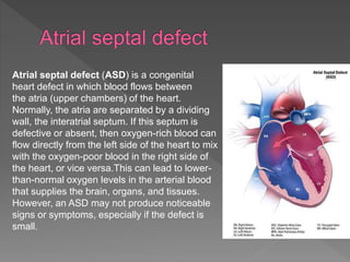
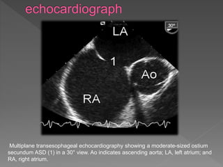
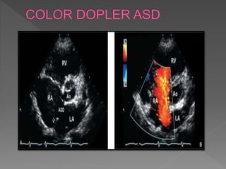


An atrial septal defect is a congenital heart defect where a hole exists in the wall separating the two upper chambers of the heart, the atria. This allows oxygen-rich blood from the left side of the heart to mix with oxygen-poor blood from the right side. This mixing of blood can lead to lower than normal oxygen levels in blood flowing to the brain, organs, and tissues. However, a small atrial septal defect may not produce noticeable symptoms. Multiplane transesophageal echocardiography can be used to image a moderate-sized atrial septal defect between the two atria.


