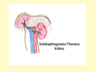
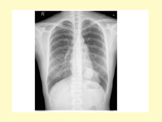
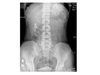
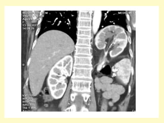
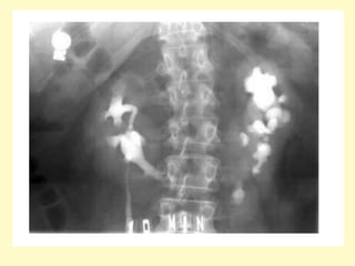
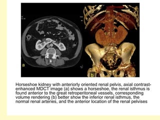
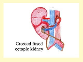
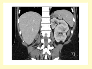
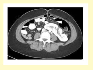
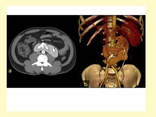
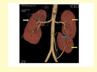
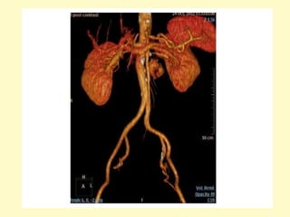
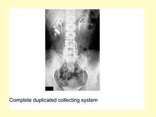
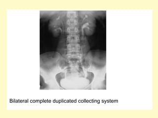
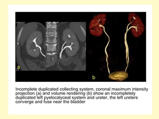
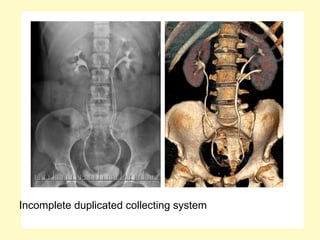
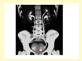
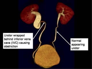
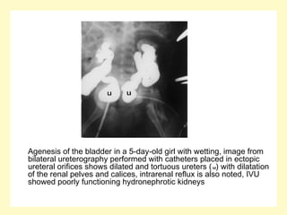
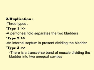
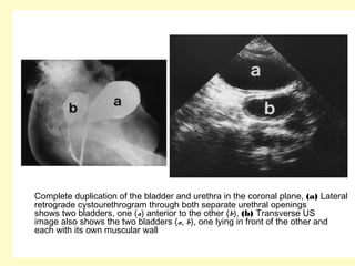
Genito-Urinary System
Congenital Renal Anomalies
Subject : visual diagonistics
Congenital Anomalies
a) Anomalies related to Position
b) Anomalies related to Form
c) Anomalies related to Number
d) Anomalies related to Vascular Development
e) Anomalies related to Bladder Development
a) Anomalies related to Position :
1-Plevic Kidney
2-Ectopic Kidney
1-Plevic Kidney :
-Failure of the kidney to ascend during
embryologic development results in a pelvic
kidney
-These kidneys are often small and abnormally
rotated
-Blood supply is from the iliac artery or the aorta
-These kidneys are prone to poor drainage and
may develop dilation of the collecting system
with susceptibility to infection & stone formation
Ectopic pelvic kidney, Volume rendering (a) demonstrates absence of the left
kidney at the left renal fossa, instead, an ectopic left pelvic kidney is seen,
the axial contrast-enhanced MDCT image (b) shows an engorged left iliac
vein (asterisk), due to left aberrant renal vein (not shown), the ectopic
kidney receives blood supply from two renal arteries arising form the aorta
(arrowhead) and left iliac artery (arrow)
2-Ectopic Kidney :
-If the kidney ascends too high , it may pass
through the foramen of Bochdalek and
become a true thoracic kidney
-Can also be presacral or at the lower
lumbar level
b) Anomalies related to Form :
1-Horseshoe Kidney
2-Pacake / Discoid Kidney
3-Crossed Renal Ectopia
1-Horseshoe Kidney :
-Most common fusion anomaly
-Two kidneys joined by parenchymal/fibrous
isthmus
-Fusion of right & left kidneys at lower pole in 90 %
-Bilateral malrotation
-Associated with other anomalies in 50 % (e.g.
vesicoureteric reflux , ureteral duplication ,
genital anomalies & Turner’s syndrome)
-Radiographic Features :
1-Abnormal axis of each kidney with lower
calyx more medial than upper calyx
2-Bilateral malrotation of renal pelvises in
anterior position
3-Isthmus lies anterior to aorta and inferior
vena cava (IVC) but behind inferior
mesenteric artery (IMA)
Horseshoe kidney with anteriorly oriented renal pelvis, axial contrast-
enhanced MDCT image (a) shows a horseshoe, the renal isthmus is
found anterior to the great retroperitoneal vessels, corresponding
volume rendering (b) better show the inferior renal isthmus, the
normal renal arteries, and the anterior location of the renal pelvises
I = isthmus, IMA (arrow)
-Other anomalies related to orientation :
Renal malrotation
Malrotated kidney about the horizontal axis, axial contrast-enhanced
MDCT image (a) and volume renderings (b, c) show a malrotated
right kidney about the horizontal axis, with a medially located pelvis
Anterior malrotated kidney, axial (a) and sagittal (b) contrast-enhanced
MDCT images show an anterior malrotated left kidney with
pelvicaliceal stones, volume rendering (c) shows stones in anteriorly
oriented renal pelvis
Lateral malrotated kidney, axial maximum intensity projection (a) and
volume rendering (b) show a lateral malrotated right kidney, the
renal pelvis shows abnormally lateral location
2-Pacake / Discoid Kidney :
-Bilateral fused pelvic kidneys , usually near
the aortic bifurcation
Pancake kidney with APKD
3-Crossed Renal Ectopia :
-Both kidneys are found in the same side
-In 85-90 %of cases , the ectopic kidney will
be fused to the other side
-The upper pole of the ectopic kidney is
usually fused to the lower pole of the other
kidney , although fusion may occur
anywhere
c) Anomalies related to Number :
1-Unilateral Agenesis
2-Bilateral Renal Agenesis
3-Supernumerary Kidney
4-Duplex Collecting System & Ureterocele
1-Unilateral Agenesis :
-Increased incidence of extrarenal
abnormalities (meningomyelocele , VSD ,
intestinal tract strictures , imperforate anus
, unicornuate uterus & skeletal
abnormalities)
-The other kidney shows compensatory
hypertrophy
Renal agenesis, Coronal maximum intensity projection (a) and volume
rendering (b) demonstrate absence of the left kidney and
compensatory hypertrophy of the right kidney, which shows a
cortical cyst, the left main renal artery was absent
2-Bilateral Renal Agenesis :
-Potter syndrome
-Fatal in first few days of life due to
pulmonary hypoplasia secondary to the
associated oligohydramnios
3-Supernumerary Kidney :
-Very rare
-Most common on the left side caudal to
normal kidney
4-Duplex Collecting System & Ureterocele :
-Duplex collecting system is the most common
congenital anomaly of the urinary tract
- The degree of duplication is variable , duplication
is complete when there are two separate
collecting systems and two separate ureters ,
each with their own ureteral orifice , duplication
is incomplete when the ureters join and enter the
bladder through a single ureteral orifice
Complete duplicated collecting system
Bilateral complete duplicated collecting system
Incomplete duplicated collecting system, coronal maximum intensity
projection (a) and volume rendering (b) show an incompletely
duplicated left pyelocalyceal system and ureter, the left ureters
converge and fuse near the bladder
Incomplete duplicated collecting system
-In complete obstruction , the ureter draining
the lower pole has a more perpendicular
course through the bladder wall making it
more prone to reflux , the ectopic ureter
from the upper pole is prone to obstruction
, reflux or both , if obstruction is present it
can result in cystic dilatation of the
intramural portion of the ureter giving rise
to a ureterocele
-Ureterocele :
a) Definition
b) Types
c) Radiographic Features
a) Definition :
Herniation of the distal ureter into the bladder
b) Types :
1-Simple : 25 %
-Normal location of ureter (at a VUJ)
-In adults
2-Ectopic : 75 %
-Abnormal location of ureter
-Unilateral with duplication
c) Radiographic Features :
1-IVP :
-Filling defect in the bladder on IVP (cobra head sign)
2-Ultrasound :
-cystic structure projecting into the bladder, often near the
normal location of the vesicoureteric junction (VUJ)
-This is ectopic in the majority of cases and therefore not at
the expected location of the ureteric orifice, the
associated ureter is usually noticeably dilated
3-MCUG :
-Round or oval lucency near the trigone
Cobra head sign
Ureterocele, early anteroposterior voiding cystourethrography (VCUG)
demonstrates small ureterocele
Ureterocele, coronal curved (a) and axial (b) contrast-enhanced MDCT
images show distal ureter balloons at its opening into the bladder,
forming a sac-like pouch
d) Anomalies related to Vascular
Development :
1-Aberrant vessels
2-Retrocaval ureter
1-Aberrant vessels
-As the kidney ascends during embryologic
development , it derives its blood supply from
the aorta at successively higher levels with
regression of the lower level vessels
-If the lower level vessels persist , aberrant renal
arteries will be present
-Aberrant vessels can compress the ureter
anywhere along its course , giving rise to
obstruction
-With color Doppler , aberrant vessels may be
seen crossing the ureter at the level of ureteric
obstruction
(a) Excretory urogram after 30 min shows severe dilatation of the right pelvocalyceal
system with cortical thinning and smooth obstruction of the right ureteropelvic
junction, (b) CTA with a maximum intensity projection technique demonstrates two
renal arteries originating from the aorta, an inferior aberrant artery (arrow) is crossing
over the ureteropelvic junction
2-Retrocaval ureter
-Also known as circumcaval ureter
-Rare but well recognized congenital anomaly ,
there is a 3:1 predominance in men , with most
patients presenting with pain in the 2 to 4nd th
decade of life
-Right ureter swings medially over pedicle of L3/4,
passes behind IVC, then exits anteriorly
between IVC and aorta returning to its normal
position, produces varying degrees of proximal
hydroureteronephrosis
Intravenous urogram showing right-sided hydronephrosis and the
dilation of the proximal ureter up to the level of the L3 transverse
process, the medial deviation of the ureter at this level (arrow) gives
rise to the typical fish hook or reversed S appearance
e) Anomalies related to
Bladder
Development :1-Agenesis
2-Duplication
3-Exstrophy
4-Prune Belly Syndrome
5-Bladder Diverticula
1-Agenesis :
-Is a rare anomaly , most infants with
bladder agenesis are stillborn with virtually
all surviving infants being female
-Often , many associated anomalies are
present
-U/S :
*The bladder is absent
Agenesis of the bladder in a 5-day-old girl with wetting, image from
bilateral ureterography performed with catheters placed in ectopic
ureteral orifices shows dilated and tortuous ureters (u) with dilatation
of the renal pelves and calices, intrarenal reflux is also noted, IVU
showed poorly functioning hydronephrotic kidneys
2-Duplication :
-Three types :
*Type 1 >>
-A peritoneal fold separates the two bladders
*Type 2 >>
-An internal septum is present dividing the bladder
*Type 3 >>
-There is a transverse band of muscle dividing the
bladder into two unequal cavities
Complete duplication of the bladder and urethra in the coronal plane, (a) Lateral
retrograde cystourethrogram through both separate urethral openings
shows two bladders, one (a) anterior to the other (b), (b) Transverse US
image also shows the two bladders (a, b), one lying in front of the other and
each with its own muscular wall
3-Exstrophy :
-There is 2:1 predominance in males
-Also known as ectopia vesicae, refers to a herniation of
the urinary bladder through an anterior abdominal wall
defect
-Failure in development of the mesoderm below the
umbilicus leads to absence of the lower abdominal and
anterior bladder wall
-Imaging findings include a soft-tissue mass extending from
a large infra-umbilical anterior wall defect which may be
close to the umbilical arterial exits, the absence of a
normal urinary bladder and a low-lying umbilical cord
insertion may also indicate the diagnosis
-Failure of the pubic bones to meet in the midline
(widened pubic symphysis), this appearance on AP plain
radiograph of the pelvis (manta ray sign)
4-Prune Belly Syndrome :
-Rare anomaly comprising a specific constellation of
features
-It consists of three major findings :
1-Gross ureteric dilatation
2-Anterior abdominal wall underdevelopment (resulting in
the prune belly appearance)
3-Bilateral undescended testes (cryptorchidism) in males
-There is often an association with other respiratory,
gastrointestinal, musculoskeletal, and cardiovascular
anomalies
-It is also known as Eagle Barrett syndrome or triad
syndrome
-Urinary tract abnormalities include :
1-Bilateral hydroureteronephrosis: often with
extremely dilated, tortuous ureters
2-Varying degrees of renal dysplasia
3-Enlarged urinary bladder, often with urachal
diverticulum
4-Vesicoureteral reflux is common
5-Poor bladder contractility
6-Dilated posterior urethra without urethral
obstruction
Supine frontal view of abdomen, the right flank is bulging with very little
abdominal wall tissue covering the air-filled right flank bowel loops.,
the ribs have a transverse orientation, sometimes referred to as a
'coat-hanger' deformity
The abdomen of an infant with prune belly syndrome shows marked
distention of the abdomen and bulging flanks secondary to a large
urinary system and the absence of abdominal wall musculature
(a) Oblique radiograph bladder, (b) voiding urethral film, a urachal
diverticulum (arrow) is seen at the dome of the urinary bladder, the
urethra is tortuous with a dilated prostatic urethra (arrowhead)
Prune-belly syndrome, IVU (a) and VCUG (b) images show the characteristic
large floppy bladder (★) and marked bilateral hydronephrosis and
hydroureter (u) with renal parenchymal wasting, note the anomalies in the
pelvic bones and both femurs due to renal osteodystrophy in b
5-Bladder Diverticula :
-Outpouchings from the bladder wall, whereby mucosa
herniates through the bladder wall
-They may be solitary or multiple in nature and can very
considerably in size
-May be congenital (primary) or acquired (secondary)
-A Hutch diverticulum is a congenital bladder diverticulum,
seen at vesicoureteric junction
-Acquired diverticula are more common, usually occurring
the context of a trabeculated bladder, resulting from
chronic bladder outlet obstruction
A view from a voiding cystourethrogram demonstrates two large
bladder diverticula (D) flanking the bladder lumen (B), these
diverticula are in the region of the insertion of the ureters, they are
frequently associated with reflux (Hutch diverticula) but no reflux
was demonstrated in this patient
Congenital anomalies radiology

Congenital anomalies radiology

  • 1.
    Genito-Urinary System Congenital RenalAnomalies Subject : visual diagonistics
  • 2.
    Congenital Anomalies a) Anomaliesrelated to Position b) Anomalies related to Form c) Anomalies related to Number d) Anomalies related to Vascular Development e) Anomalies related to Bladder Development
  • 3.
    a) Anomalies relatedto Position : 1-Plevic Kidney 2-Ectopic Kidney
  • 4.
    1-Plevic Kidney : -Failureof the kidney to ascend during embryologic development results in a pelvic kidney -These kidneys are often small and abnormally rotated -Blood supply is from the iliac artery or the aorta -These kidneys are prone to poor drainage and may develop dilation of the collecting system with susceptibility to infection & stone formation
  • 7.
    Ectopic pelvic kidney,Volume rendering (a) demonstrates absence of the left kidney at the left renal fossa, instead, an ectopic left pelvic kidney is seen, the axial contrast-enhanced MDCT image (b) shows an engorged left iliac vein (asterisk), due to left aberrant renal vein (not shown), the ectopic kidney receives blood supply from two renal arteries arising form the aorta (arrowhead) and left iliac artery (arrow)
  • 11.
    2-Ectopic Kidney : -Ifthe kidney ascends too high , it may pass through the foramen of Bochdalek and become a true thoracic kidney -Can also be presacral or at the lower lumbar level
  • 16.
    b) Anomalies relatedto Form : 1-Horseshoe Kidney 2-Pacake / Discoid Kidney 3-Crossed Renal Ectopia
  • 17.
    1-Horseshoe Kidney : -Mostcommon fusion anomaly -Two kidneys joined by parenchymal/fibrous isthmus -Fusion of right & left kidneys at lower pole in 90 % -Bilateral malrotation -Associated with other anomalies in 50 % (e.g. vesicoureteric reflux , ureteral duplication , genital anomalies & Turner’s syndrome)
  • 18.
    -Radiographic Features : 1-Abnormalaxis of each kidney with lower calyx more medial than upper calyx 2-Bilateral malrotation of renal pelvises in anterior position 3-Isthmus lies anterior to aorta and inferior vena cava (IVC) but behind inferior mesenteric artery (IMA)
  • 23.
    Horseshoe kidney withanteriorly oriented renal pelvis, axial contrast- enhanced MDCT image (a) shows a horseshoe, the renal isthmus is found anterior to the great retroperitoneal vessels, corresponding volume rendering (b) better show the inferior renal isthmus, the normal renal arteries, and the anterior location of the renal pelvises
  • 25.
    I = isthmus,IMA (arrow)
  • 26.
    -Other anomalies relatedto orientation : Renal malrotation
  • 27.
    Malrotated kidney aboutthe horizontal axis, axial contrast-enhanced MDCT image (a) and volume renderings (b, c) show a malrotated right kidney about the horizontal axis, with a medially located pelvis
  • 28.
    Anterior malrotated kidney,axial (a) and sagittal (b) contrast-enhanced MDCT images show an anterior malrotated left kidney with pelvicaliceal stones, volume rendering (c) shows stones in anteriorly oriented renal pelvis
  • 29.
    Lateral malrotated kidney,axial maximum intensity projection (a) and volume rendering (b) show a lateral malrotated right kidney, the renal pelvis shows abnormally lateral location
  • 30.
    2-Pacake / DiscoidKidney : -Bilateral fused pelvic kidneys , usually near the aortic bifurcation
  • 32.
  • 33.
    3-Crossed Renal Ectopia: -Both kidneys are found in the same side -In 85-90 %of cases , the ectopic kidney will be fused to the other side -The upper pole of the ectopic kidney is usually fused to the lower pole of the other kidney , although fusion may occur anywhere
  • 38.
    c) Anomalies relatedto Number : 1-Unilateral Agenesis 2-Bilateral Renal Agenesis 3-Supernumerary Kidney 4-Duplex Collecting System & Ureterocele
  • 39.
    1-Unilateral Agenesis : -Increasedincidence of extrarenal abnormalities (meningomyelocele , VSD , intestinal tract strictures , imperforate anus , unicornuate uterus & skeletal abnormalities) -The other kidney shows compensatory hypertrophy
  • 40.
    Renal agenesis, Coronalmaximum intensity projection (a) and volume rendering (b) demonstrate absence of the left kidney and compensatory hypertrophy of the right kidney, which shows a cortical cyst, the left main renal artery was absent
  • 42.
    2-Bilateral Renal Agenesis: -Potter syndrome -Fatal in first few days of life due to pulmonary hypoplasia secondary to the associated oligohydramnios
  • 43.
    3-Supernumerary Kidney : -Veryrare -Most common on the left side caudal to normal kidney
  • 46.
    4-Duplex Collecting System& Ureterocele : -Duplex collecting system is the most common congenital anomaly of the urinary tract - The degree of duplication is variable , duplication is complete when there are two separate collecting systems and two separate ureters , each with their own ureteral orifice , duplication is incomplete when the ureters join and enter the bladder through a single ureteral orifice
  • 47.
  • 48.
  • 49.
    Incomplete duplicated collectingsystem, coronal maximum intensity projection (a) and volume rendering (b) show an incompletely duplicated left pyelocalyceal system and ureter, the left ureters converge and fuse near the bladder
  • 50.
  • 51.
    -In complete obstruction, the ureter draining the lower pole has a more perpendicular course through the bladder wall making it more prone to reflux , the ectopic ureter from the upper pole is prone to obstruction , reflux or both , if obstruction is present it can result in cystic dilatation of the intramural portion of the ureter giving rise to a ureterocele
  • 53.
    -Ureterocele : a) Definition b)Types c) Radiographic Features
  • 54.
    a) Definition : Herniationof the distal ureter into the bladder b) Types : 1-Simple : 25 % -Normal location of ureter (at a VUJ) -In adults 2-Ectopic : 75 % -Abnormal location of ureter -Unilateral with duplication
  • 55.
    c) Radiographic Features: 1-IVP : -Filling defect in the bladder on IVP (cobra head sign) 2-Ultrasound : -cystic structure projecting into the bladder, often near the normal location of the vesicoureteric junction (VUJ) -This is ectopic in the majority of cases and therefore not at the expected location of the ureteric orifice, the associated ureter is usually noticeably dilated 3-MCUG : -Round or oval lucency near the trigone
  • 56.
  • 58.
    Ureterocele, early anteroposteriorvoiding cystourethrography (VCUG) demonstrates small ureterocele
  • 59.
    Ureterocele, coronal curved(a) and axial (b) contrast-enhanced MDCT images show distal ureter balloons at its opening into the bladder, forming a sac-like pouch
  • 60.
    d) Anomalies relatedto Vascular Development : 1-Aberrant vessels 2-Retrocaval ureter
  • 61.
    1-Aberrant vessels -As thekidney ascends during embryologic development , it derives its blood supply from the aorta at successively higher levels with regression of the lower level vessels -If the lower level vessels persist , aberrant renal arteries will be present -Aberrant vessels can compress the ureter anywhere along its course , giving rise to obstruction -With color Doppler , aberrant vessels may be seen crossing the ureter at the level of ureteric obstruction
  • 62.
    (a) Excretory urogramafter 30 min shows severe dilatation of the right pelvocalyceal system with cortical thinning and smooth obstruction of the right ureteropelvic junction, (b) CTA with a maximum intensity projection technique demonstrates two renal arteries originating from the aorta, an inferior aberrant artery (arrow) is crossing over the ureteropelvic junction
  • 63.
    2-Retrocaval ureter -Also knownas circumcaval ureter -Rare but well recognized congenital anomaly , there is a 3:1 predominance in men , with most patients presenting with pain in the 2 to 4nd th decade of life -Right ureter swings medially over pedicle of L3/4, passes behind IVC, then exits anteriorly between IVC and aorta returning to its normal position, produces varying degrees of proximal hydroureteronephrosis
  • 67.
    Intravenous urogram showingright-sided hydronephrosis and the dilation of the proximal ureter up to the level of the L3 transverse process, the medial deviation of the ureter at this level (arrow) gives rise to the typical fish hook or reversed S appearance
  • 69.
    e) Anomalies relatedto Bladder Development :1-Agenesis 2-Duplication 3-Exstrophy 4-Prune Belly Syndrome 5-Bladder Diverticula
  • 70.
    1-Agenesis : -Is arare anomaly , most infants with bladder agenesis are stillborn with virtually all surviving infants being female -Often , many associated anomalies are present -U/S : *The bladder is absent
  • 71.
    Agenesis of thebladder in a 5-day-old girl with wetting, image from bilateral ureterography performed with catheters placed in ectopic ureteral orifices shows dilated and tortuous ureters (u) with dilatation of the renal pelves and calices, intrarenal reflux is also noted, IVU showed poorly functioning hydronephrotic kidneys
  • 72.
    2-Duplication : -Three types: *Type 1 >> -A peritoneal fold separates the two bladders *Type 2 >> -An internal septum is present dividing the bladder *Type 3 >> -There is a transverse band of muscle dividing the bladder into two unequal cavities
  • 73.
    Complete duplication ofthe bladder and urethra in the coronal plane, (a) Lateral retrograde cystourethrogram through both separate urethral openings shows two bladders, one (a) anterior to the other (b), (b) Transverse US image also shows the two bladders (a, b), one lying in front of the other and each with its own muscular wall
  • 74.
    3-Exstrophy : -There is2:1 predominance in males -Also known as ectopia vesicae, refers to a herniation of the urinary bladder through an anterior abdominal wall defect -Failure in development of the mesoderm below the umbilicus leads to absence of the lower abdominal and anterior bladder wall -Imaging findings include a soft-tissue mass extending from a large infra-umbilical anterior wall defect which may be close to the umbilical arterial exits, the absence of a normal urinary bladder and a low-lying umbilical cord insertion may also indicate the diagnosis -Failure of the pubic bones to meet in the midline (widened pubic symphysis), this appearance on AP plain radiograph of the pelvis (manta ray sign)
  • 76.
    4-Prune Belly Syndrome: -Rare anomaly comprising a specific constellation of features -It consists of three major findings : 1-Gross ureteric dilatation 2-Anterior abdominal wall underdevelopment (resulting in the prune belly appearance) 3-Bilateral undescended testes (cryptorchidism) in males -There is often an association with other respiratory, gastrointestinal, musculoskeletal, and cardiovascular anomalies -It is also known as Eagle Barrett syndrome or triad syndrome
  • 77.
    -Urinary tract abnormalitiesinclude : 1-Bilateral hydroureteronephrosis: often with extremely dilated, tortuous ureters 2-Varying degrees of renal dysplasia 3-Enlarged urinary bladder, often with urachal diverticulum 4-Vesicoureteral reflux is common 5-Poor bladder contractility 6-Dilated posterior urethra without urethral obstruction
  • 78.
    Supine frontal viewof abdomen, the right flank is bulging with very little abdominal wall tissue covering the air-filled right flank bowel loops., the ribs have a transverse orientation, sometimes referred to as a 'coat-hanger' deformity
  • 79.
    The abdomen ofan infant with prune belly syndrome shows marked distention of the abdomen and bulging flanks secondary to a large urinary system and the absence of abdominal wall musculature
  • 80.
    (a) Oblique radiographbladder, (b) voiding urethral film, a urachal diverticulum (arrow) is seen at the dome of the urinary bladder, the urethra is tortuous with a dilated prostatic urethra (arrowhead)
  • 81.
    Prune-belly syndrome, IVU(a) and VCUG (b) images show the characteristic large floppy bladder (★) and marked bilateral hydronephrosis and hydroureter (u) with renal parenchymal wasting, note the anomalies in the pelvic bones and both femurs due to renal osteodystrophy in b
  • 82.
    5-Bladder Diverticula : -Outpouchingsfrom the bladder wall, whereby mucosa herniates through the bladder wall -They may be solitary or multiple in nature and can very considerably in size -May be congenital (primary) or acquired (secondary) -A Hutch diverticulum is a congenital bladder diverticulum, seen at vesicoureteric junction -Acquired diverticula are more common, usually occurring the context of a trabeculated bladder, resulting from chronic bladder outlet obstruction
  • 86.
    A view froma voiding cystourethrogram demonstrates two large bladder diverticula (D) flanking the bladder lumen (B), these diverticula are in the region of the insertion of the ureters, they are frequently associated with reflux (Hutch diverticula) but no reflux was demonstrated in this patient