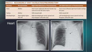
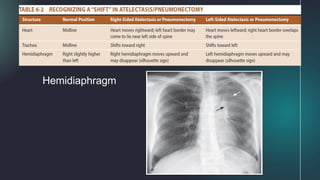
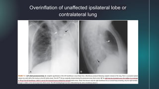
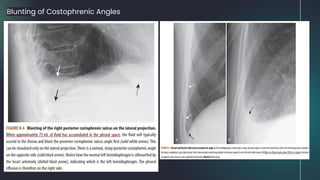
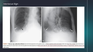
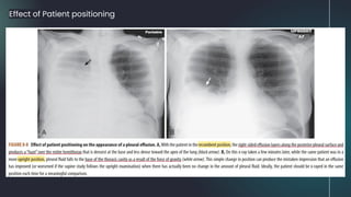
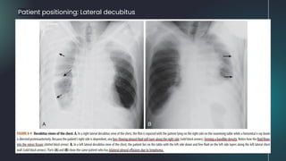
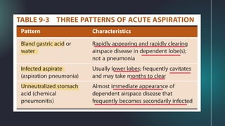
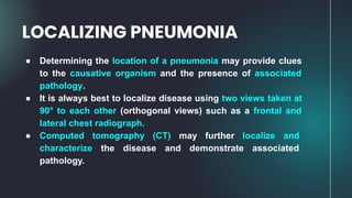
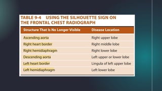
Recognizing Atelectasis,
Pleural Effusion and
Pneumonia
Recognizing
Atelectasis
01 02
03
Table of contents
2
Recognizing
Pleural Effusion
Recognizing
Penumonia
RECOGNIZING
PLEURAL
EFFUSION
01
Recognizing
Atelectasis
What is Atelectasis?
Loss of volume in some or all of the lung, usually leading to
increased density of the lung involved.
- part of the lung involved becomes whiter (denser or more
opaque)
- fluid or soft tissue density has substituted the air or the air in the
lung is resorbed (as it can be in atelectasis)
Signs of Atelectasis
*Toward the atelectasis
Increase in the density
of the affected lung
Displacement of the
interlobar fissures
Trachea
Heart
Hemidiaphragm
Overinflation of unaffected ipsilateral lobe or
contralateral lung
Types of Atelectasis
1. Subsegmental Atelectasis (Discoid or platelike)
2. Compressive Atelectasis (Passive)
a. Round Atelectasis
3. Obstructive Atelectasis
1. Subsegmental Atelectasis (Discoid or platelike)
● associated with deactivation of surfactant
○ leads to collapse of airspaces in a
nonsegmental or nonlobar distribution
(not due to bronchial obstruction)
● produces linear densities of varying thickness
usually parallel to the diaphragm
○ most commonly at the lung bases
● does not produce a sufficient amount of volume
loss to cause a shift of the mobile thoracic
structures
● looks identical to linear scarring
○ disappears within a matter of days with
resumption of normal, deep breathing,
whereas scarring remains
2. Compressive Atelectasis (Passive)
● due to passive compression of the lung can
be caused by:
○ poor inspiratory effort in which there is
passive atelectasis of the lung at the
bases (Fig. A)
○ large pleural effusion, large
pneumothorax, or a space-occupying
lesion (such as a large mass in the
lung) (Fig. B)
2. Compressive Atelectasis (Passive)
● when caused by a large effusion or
pneumothorax, the loss of volume associated
with compressive atelectasis may balance the
increased volume produced by either fluid (as in
pleural effusion) or air (as in pneumothorax)
● in an adult patient with an opacified hemithorax,
no air bronchograms, and little or no shift of the
mobile thoracic structures, it is important to
suspect an obstructing bronchogenic
carcinoma, perhaps with metastases to the
pleura
2. Compressive Atelectasis (Passive)
● Pitfall:
● Be suspicious of compressive atelectasis if the patient has taken less than an 8
posterior-rib breath.
○ Solution: Check the lateral projection for confirmation of the presence of real
airspace disease at the base
a. Round Atelectasis (Passive)
● usually seen at the periphery of the lung base
● develops from a combination of prior pleural
disease (such as from asbestos exposure or
tuberculosis) and formation of a pleural effusion
that produces adjacent compressive atelectasis
● underlying pleural disease leads to a portion of
the atelectatic lung becoming “trapped” when
pleural effusion recedes
○ produces a masslike lesion that can be
confused for a tumor
○ on chest CT scan, bronchovascular
markings produce a comet-tail
appearance
3. Obstructive Atelectasis
● associated with the resorption of air from the
alveoli, through the pulmonary capillary bed,
distal to an obstructing lesion of the
bronchial tree
Patterns of Collapse in Obstructive Atelectasis
1. Right Upper Lobe Atelectasis
A. Frontal radiograph:
● upward shift of the minor fissure
● rightward shift of the trachea
● combination of a hilar mass and the
upward shift of the minor fissure
produces S sign of Golden
B. Lateral radiograph:
● upward shift of the minor fissure
● forward shift of the major fissure
2. Left Upper Lobe Atelectasis
A. Frontal radiograph:
● increased density at left hilum
● leftward shift of the trachea
● may be an elevation with tenting or
peaking of left hemidiaphragm
● compensatory overinflation of lower lobe
B. Lateral radiograph:
● forward displacement of major fissure
● opacified upper lobe forms a band of
increased density parallel to the sternum
3. Lower Lobe Atelectasis
A. Frontal radiograph:
● both right and left lower lobes collapse to
form a triangular density (Fig. A)
● downward shift of major fissure (Fig. C)
● shift of the heart towards the atelectasis
● elevation of the hemidiaphragm on the
affected side
B. Lateral radiograph:
● both downward and posterior displacement
of the major fissure (Fig. B)
○ until the completely collapsed lower
lobe forms a small triangular density
at the posterior costophrenic angle
3. Lower Lobe Atelectasis
● In the critically ill patient, atelectasis occurs most frequently in the left lower lobe
○ Always check the left hemidiaphragm to be sure it is seen in its entire extent through the heart
shadow
■ left lower lobe atelectasis will manifest by disappearance (silhouetting) of all or part of the left
hemidiaphragm (Fig. C)
4. Right Middle Lobe Atelectasis
A. Frontal radiograph:
● triangular density with its base silhouetting
the right heart border and its apex pointing
the lateral chest wall
● downward shift of minor fissure
B. Lateral radiograph:
● triangular density with its base directed
anteriorly and its apex at the hilum
● shift of minor fissure inferiorly and major
fissure superiorly
5. Too Low Endotracheal Tube
6. Atelectasis of the Entire Lung
A. Frontal radiograph:
● opacification of the atelectatic lung
● shift of all mobile thoracic structures towards
the atelectasis
● hemidiaphragm on the side of atelectasis is
silhouetted by the above nonaerated lung
B. Lateral radiograph:
● hemidiaphragm on the side of atelectasis is
silhouetted by the above nonaerated lung
(only one hemidiaphragm is seen)
How Atelectasis resolves
Resolves within hours or lasts for many days once the
obstruction has been removed
- slowly resolving lobar or whole-lung atelectasis may manifest patchy areas
of airspace disease surrounded by progressively increasing zones of
aerated lung until the atelectasis has completely cleared.
RECOGNIZING
PLEURAL
EFFUSION
02
Recognizing
Pleural Effusion
What is Pleural Effusion?
Abnormal accumulation of fluid in the pleural cavity/space
between the visceral pleura and the parietal pleura
- normally 2-5 mL of pleural fluid in the pleural space
- first step in detecting pleural effusion is usually conventional
radiography
Types of Pleural Effusion
a. Transudate
- formed due to increased hydrostatic pressure or decreased
osmotic pressure
- Congestive heart failure: most common cause
b. Exudate
- formed due to inflammation
- Malignancy: most common cause
- empyema, hemothorax, chylothorax
Side-Specificity of Pleural Effusion
a. Diseases that produce bilateral effusions:
- CHF
- same amount of fluid in each hemithorax
- Parapneumonic effusion or malignancy
- different amount of fluid in each hemithorax
- SLE
b. Diseases that produce unilateral effusion (either side):
- TB and other diseases associated with infectious agents
- Pulmonary thromboembolic disease
- Trauma
c. Diseases that produce left-sided effusion:
- Pancreatitis, distal thoracic duct obstruction, Dressler syndrome
d. Diseases that produce right-sided effusion:
- Abdominal disease related to the liver or ovaries
- Meigs syndrome (ovarian tumor associated with right pleural effusion and
ascites)
- Proximal thoracic duct obstruction
- Rheumatoid arthritis
Dressler Syndrome
Appearances of Pleural
Effusion
a. Subpulmonic Effusion
Blunting of Costophrenic Angles
Meniscus Sign
Effect of Patient positioning
Patient positioning: Lateral decubitus
● When the hemithorax contains about 2 L of fluid, the entire
hemithorax will be opacified.
● As fluid fills the pleural space, the lung tends to undergo
passive collapse (atelectasis).
● Large effusions can act like a mass and displace the heart
and trachea away from the side of opacification.
Opacified Hemithorax
● Adhesions in the pleural space, caused most often by an
old infection or hemothorax, may limit the normal mobility of
a pleural effusion so that it remains in the same location no
matter what position the patient assumes.
● Suspected when an effusion has an unusual shape or
location in the thorax
Loculated Effusions
Loculated
Effusions
● Pseudotumors (also called vanishing tumors) are sharply
marginated collections of pleural fluid contained either
between the layers of an interlobar pulmonary fissure or in a
subpleural location just beneath the fissure.
● They are transudates that almost always occur in patients
with congestive heart failure.
● Imaging findings of a pseudotumor are characteristic and
should not be mistaken for an actual tumor.
Fissural Pseudotumors
● Is a form of pleural effusion in which the fluid assumes a
thin, bandlike density along the lateral chest wall, especially
near the costophrenic angle.
● The lateral costophrenic angle tends to maintain its acute
angle with a laminar effusion.
● They are usually not free-flowing.
● Can be recognized by the band of increased density that
separates the air-filled lung from the inside margin of the
ribs at the lung base on the frontal chest radiograph.
Laminar Effusions
Laminar
Effusions
● Presence of both air in the pleural space (pneumothorax)
and abnormal amounts of fluid in the pleural space (pleural
effusion or hydrothorax).
● Unlike pleural effusion alone, whose meniscoid shape is
governed by the elastic recoil of the lung, a
hydropneumothorax produces an air-fluid level in the
hemithorax marked by a straight edge and a sharp,
air-over-fluid interface when a horizontal x-ray beam is
exposed.
Hydropneumothorax
03
RECOGNIZING
PNEUMONIA
Recognizing
Pneumonia
Defined as consolidation of the lung produced by
inflammatory exudate, usually as a result of an infectious
agent.
- Most pneumonias produce airspace disease, either lobar
or segmental. Others demonstrate interstitial disease,
while some also present both.
What is Pneumonia?
● Appears denser (whiter) than surrounding normal lung.
● May contain Air Bronchograms
● If airspaces involved- appears fluffy and margins are
indistinct.
● For interstitial pneumonia- prominence of interstitial
markings noted.
● Usually homogenous in density
● In some types of pneumonia, atelectasis may be noted.
General Characteristics of
Pneumonia
● LOBAR
● SEGMENTAL
● INTERSTITIAL
● ROUND
● CAVITARY
Patterns of Pneumonia
LOBAR PNEUMONIA
● Prototypical Lobar pneumonia is caused by Streptococcus
pneumoniae.
● In its classic form, the disease fills most or all of a lobe of
the lung.
● If pneumonia is bound by a fissure, the margins may be
sharply distinct. If not bound by a fissure, it will have an
indistinct and irregular margin.
● Almost always produce a silhouette sign
● May contain air bronchograms if it involves the central
portions of the lung.
LOBAR
PNEUMONIA
● Prototypical Bronchopneumonia is caused by
Staphylococcus aureus and other Gram Negative Bacteria
such as Pseudomonas aeruginosa.
● Is spread centrifugally via the tracheobronchial tree to many
foci in the lung. Therefore, it frequently involves several
segments of the lung simultaenously.
● Margins tend to be fluffy and indistinct.
● Airbronchograms usually not present.
● Atelectasis may be noted.
SEGMENTAL PNEUMONIA
SEGMENTAL
PNEUMONIA
● Prototypes for Interstitial Pneumonia are Viral pnuemonia,
Mycoplasma pneumoniae and Pneumocystis pneumonia in
AIDS patients.
● Tends to involve the airway walls and alveolar septa and may
produce a fine, reticular pattern in the lungs.
● Most cases eventually spread to the adjacent alveoli and
produce patchy or confluent airspace disease
INTERSTITIAL PNEUMONIA
● PCP is the most common clinically recognized infection in
patients with AIDS.
● It classically presents as a perihilar, reticular interstitial
pneumonia or as airspace disease that may mimic the
central distribution pattern of pulmonary edema
● There are usually no pleural effusions and no hilar
adenopathy.
INTERSTITIAL PNEUMONIA
(Pneumocystis carinii pneumonia)
INTERSTITIAL
PNEUMONIA
● Some pneumonias may assume a spherical shape on chest
radiographs.
● Almost always posterior in the lungs, usually in the lower
lobes.
● Causative Agents: Haemophilus influenzae, Streptococcus
and Pneumococcus.
● May be confused with a tumor mass.
ROUND PNEUMONIA
ROUND
PNEUMONIA
● The prototypical organism produciing cavitary pneumonia is
Mycobacterium tuberculosis.
● Cavitation is common in postprimary tuberculosis (TB)
(reactivation tuberculosis) but rare in primary TB.
● The cavities are usually located in the upper lobes, are
bilateral and thin-walled, have a smooth inner margin, and
contain no air-fluid level.
● Transbronchial spread may be suggestive of infection with
Mycobacterium tuberculosis.
CAVITARY PNEUMONIA
CAVITARY
PNEUMONIA
● Almost always occurs in the most dependent portions of the
lung.
● In upright position, the most dependent portion of the lung is
the lower lobes.
● In recumbent position, aspiration usually occurs into the
superior segments of the lower lobes or the posterior segment
of the upper lobes.
● The right side is more often affected than the left because of the
straighter and wider nature of the right main bronchus.
● Acute aspiration will produce radiographic findings of airspace
disease.
ASPIRATION
● Determining the location of a pneumonia may provide clues
to the causative organism and the presence of associated
pathology.
● It is always best to localize disease using two views taken at
90° to each other (orthogonal views) such as a frontal and
lateral chest radiograph.
● Computed tomography (CT) may further localize and
characterize the disease and demonstrate associated
pathology.
LOCALIZING PNEUMONIA
● If two objects of the same radiographic density touch each
other, then the edge between them disappears
● The silhouette sign is valuable in localizing and identifying
tissue types throughout the body
SILHOUETTE SIGN
● On the lateral chest radiograph, the thoracic spine
normally appears to get darker from the shoulder girdle to
the diaphragm.
● When diseases of soft tissue or fluid density involves the
posterior portion of the lower lobe, more of the x-ray beam
will be absorbed by the new, added density, and the spine
will appear to become “whiter” just above the posterior
costophrenic sulcus.
SPINE SIGN
HOW PNEUMONIA RESOLVES
● Pneumonia can resolve in 2 to 3 days if the organism is
sensitive to the antibiotic administered.
● Most pneumonias typically resolve from within
(vacuolization), gradually disappearing in a patchy fashion
over days, or weeks.
THANK YOU
FOR LISTENING!

Recognizing Atelectasis, Pleural Effusion and Pneumonia

  • 1.
  • 2.
    Recognizing Atelectasis 01 02 03 Table ofcontents 2 Recognizing Pleural Effusion Recognizing Penumonia
  • 3.
  • 4.
    What is Atelectasis? Lossof volume in some or all of the lung, usually leading to increased density of the lung involved. - part of the lung involved becomes whiter (denser or more opaque) - fluid or soft tissue density has substituted the air or the air in the lung is resorbed (as it can be in atelectasis)
  • 6.
  • 7.
    Increase in thedensity of the affected lung Displacement of the interlobar fissures
  • 8.
  • 9.
  • 10.
  • 11.
    Overinflation of unaffectedipsilateral lobe or contralateral lung
  • 12.
    Types of Atelectasis 1.Subsegmental Atelectasis (Discoid or platelike) 2. Compressive Atelectasis (Passive) a. Round Atelectasis 3. Obstructive Atelectasis
  • 13.
    1. Subsegmental Atelectasis(Discoid or platelike) ● associated with deactivation of surfactant ○ leads to collapse of airspaces in a nonsegmental or nonlobar distribution (not due to bronchial obstruction) ● produces linear densities of varying thickness usually parallel to the diaphragm ○ most commonly at the lung bases ● does not produce a sufficient amount of volume loss to cause a shift of the mobile thoracic structures ● looks identical to linear scarring ○ disappears within a matter of days with resumption of normal, deep breathing, whereas scarring remains
  • 14.
    2. Compressive Atelectasis(Passive) ● due to passive compression of the lung can be caused by: ○ poor inspiratory effort in which there is passive atelectasis of the lung at the bases (Fig. A) ○ large pleural effusion, large pneumothorax, or a space-occupying lesion (such as a large mass in the lung) (Fig. B)
  • 15.
    2. Compressive Atelectasis(Passive) ● when caused by a large effusion or pneumothorax, the loss of volume associated with compressive atelectasis may balance the increased volume produced by either fluid (as in pleural effusion) or air (as in pneumothorax) ● in an adult patient with an opacified hemithorax, no air bronchograms, and little or no shift of the mobile thoracic structures, it is important to suspect an obstructing bronchogenic carcinoma, perhaps with metastases to the pleura
  • 16.
    2. Compressive Atelectasis(Passive) ● Pitfall: ● Be suspicious of compressive atelectasis if the patient has taken less than an 8 posterior-rib breath. ○ Solution: Check the lateral projection for confirmation of the presence of real airspace disease at the base
  • 17.
    a. Round Atelectasis(Passive) ● usually seen at the periphery of the lung base ● develops from a combination of prior pleural disease (such as from asbestos exposure or tuberculosis) and formation of a pleural effusion that produces adjacent compressive atelectasis ● underlying pleural disease leads to a portion of the atelectatic lung becoming “trapped” when pleural effusion recedes ○ produces a masslike lesion that can be confused for a tumor ○ on chest CT scan, bronchovascular markings produce a comet-tail appearance
  • 18.
    3. Obstructive Atelectasis ●associated with the resorption of air from the alveoli, through the pulmonary capillary bed, distal to an obstructing lesion of the bronchial tree
  • 20.
    Patterns of Collapsein Obstructive Atelectasis 1. Right Upper Lobe Atelectasis A. Frontal radiograph: ● upward shift of the minor fissure ● rightward shift of the trachea ● combination of a hilar mass and the upward shift of the minor fissure produces S sign of Golden B. Lateral radiograph: ● upward shift of the minor fissure ● forward shift of the major fissure
  • 21.
    2. Left UpperLobe Atelectasis A. Frontal radiograph: ● increased density at left hilum ● leftward shift of the trachea ● may be an elevation with tenting or peaking of left hemidiaphragm ● compensatory overinflation of lower lobe B. Lateral radiograph: ● forward displacement of major fissure ● opacified upper lobe forms a band of increased density parallel to the sternum
  • 22.
    3. Lower LobeAtelectasis A. Frontal radiograph: ● both right and left lower lobes collapse to form a triangular density (Fig. A) ● downward shift of major fissure (Fig. C) ● shift of the heart towards the atelectasis ● elevation of the hemidiaphragm on the affected side B. Lateral radiograph: ● both downward and posterior displacement of the major fissure (Fig. B) ○ until the completely collapsed lower lobe forms a small triangular density at the posterior costophrenic angle
  • 23.
    3. Lower LobeAtelectasis ● In the critically ill patient, atelectasis occurs most frequently in the left lower lobe ○ Always check the left hemidiaphragm to be sure it is seen in its entire extent through the heart shadow ■ left lower lobe atelectasis will manifest by disappearance (silhouetting) of all or part of the left hemidiaphragm (Fig. C)
  • 24.
    4. Right MiddleLobe Atelectasis A. Frontal radiograph: ● triangular density with its base silhouetting the right heart border and its apex pointing the lateral chest wall ● downward shift of minor fissure B. Lateral radiograph: ● triangular density with its base directed anteriorly and its apex at the hilum ● shift of minor fissure inferiorly and major fissure superiorly
  • 25.
    5. Too LowEndotracheal Tube
  • 26.
    6. Atelectasis ofthe Entire Lung A. Frontal radiograph: ● opacification of the atelectatic lung ● shift of all mobile thoracic structures towards the atelectasis ● hemidiaphragm on the side of atelectasis is silhouetted by the above nonaerated lung B. Lateral radiograph: ● hemidiaphragm on the side of atelectasis is silhouetted by the above nonaerated lung (only one hemidiaphragm is seen)
  • 27.
    How Atelectasis resolves Resolveswithin hours or lasts for many days once the obstruction has been removed - slowly resolving lobar or whole-lung atelectasis may manifest patchy areas of airspace disease surrounded by progressively increasing zones of aerated lung until the atelectasis has completely cleared.
  • 28.
  • 29.
    What is PleuralEffusion? Abnormal accumulation of fluid in the pleural cavity/space between the visceral pleura and the parietal pleura - normally 2-5 mL of pleural fluid in the pleural space - first step in detecting pleural effusion is usually conventional radiography
  • 31.
    Types of PleuralEffusion a. Transudate - formed due to increased hydrostatic pressure or decreased osmotic pressure - Congestive heart failure: most common cause b. Exudate - formed due to inflammation - Malignancy: most common cause - empyema, hemothorax, chylothorax
  • 32.
    Side-Specificity of PleuralEffusion a. Diseases that produce bilateral effusions: - CHF - same amount of fluid in each hemithorax - Parapneumonic effusion or malignancy - different amount of fluid in each hemithorax - SLE b. Diseases that produce unilateral effusion (either side): - TB and other diseases associated with infectious agents - Pulmonary thromboembolic disease - Trauma c. Diseases that produce left-sided effusion: - Pancreatitis, distal thoracic duct obstruction, Dressler syndrome d. Diseases that produce right-sided effusion: - Abdominal disease related to the liver or ovaries - Meigs syndrome (ovarian tumor associated with right pleural effusion and ascites) - Proximal thoracic duct obstruction - Rheumatoid arthritis
  • 33.
  • 34.
  • 35.
  • 36.
  • 37.
  • 38.
  • 39.
    Effect of Patientpositioning
  • 40.
  • 41.
    ● When thehemithorax contains about 2 L of fluid, the entire hemithorax will be opacified. ● As fluid fills the pleural space, the lung tends to undergo passive collapse (atelectasis). ● Large effusions can act like a mass and displace the heart and trachea away from the side of opacification. Opacified Hemithorax
  • 43.
    ● Adhesions inthe pleural space, caused most often by an old infection or hemothorax, may limit the normal mobility of a pleural effusion so that it remains in the same location no matter what position the patient assumes. ● Suspected when an effusion has an unusual shape or location in the thorax Loculated Effusions
  • 44.
  • 45.
    ● Pseudotumors (alsocalled vanishing tumors) are sharply marginated collections of pleural fluid contained either between the layers of an interlobar pulmonary fissure or in a subpleural location just beneath the fissure. ● They are transudates that almost always occur in patients with congestive heart failure. ● Imaging findings of a pseudotumor are characteristic and should not be mistaken for an actual tumor. Fissural Pseudotumors
  • 47.
    ● Is aform of pleural effusion in which the fluid assumes a thin, bandlike density along the lateral chest wall, especially near the costophrenic angle. ● The lateral costophrenic angle tends to maintain its acute angle with a laminar effusion. ● They are usually not free-flowing. ● Can be recognized by the band of increased density that separates the air-filled lung from the inside margin of the ribs at the lung base on the frontal chest radiograph. Laminar Effusions
  • 48.
  • 49.
    ● Presence ofboth air in the pleural space (pneumothorax) and abnormal amounts of fluid in the pleural space (pleural effusion or hydrothorax). ● Unlike pleural effusion alone, whose meniscoid shape is governed by the elastic recoil of the lung, a hydropneumothorax produces an air-fluid level in the hemithorax marked by a straight edge and a sharp, air-over-fluid interface when a horizontal x-ray beam is exposed. Hydropneumothorax
  • 52.
  • 53.
    Defined as consolidationof the lung produced by inflammatory exudate, usually as a result of an infectious agent. - Most pneumonias produce airspace disease, either lobar or segmental. Others demonstrate interstitial disease, while some also present both. What is Pneumonia?
  • 55.
    ● Appears denser(whiter) than surrounding normal lung. ● May contain Air Bronchograms ● If airspaces involved- appears fluffy and margins are indistinct. ● For interstitial pneumonia- prominence of interstitial markings noted. ● Usually homogenous in density ● In some types of pneumonia, atelectasis may be noted. General Characteristics of Pneumonia
  • 58.
    ● LOBAR ● SEGMENTAL ●INTERSTITIAL ● ROUND ● CAVITARY Patterns of Pneumonia
  • 59.
    LOBAR PNEUMONIA ● PrototypicalLobar pneumonia is caused by Streptococcus pneumoniae. ● In its classic form, the disease fills most or all of a lobe of the lung. ● If pneumonia is bound by a fissure, the margins may be sharply distinct. If not bound by a fissure, it will have an indistinct and irregular margin. ● Almost always produce a silhouette sign ● May contain air bronchograms if it involves the central portions of the lung.
  • 60.
  • 61.
    ● Prototypical Bronchopneumoniais caused by Staphylococcus aureus and other Gram Negative Bacteria such as Pseudomonas aeruginosa. ● Is spread centrifugally via the tracheobronchial tree to many foci in the lung. Therefore, it frequently involves several segments of the lung simultaenously. ● Margins tend to be fluffy and indistinct. ● Airbronchograms usually not present. ● Atelectasis may be noted. SEGMENTAL PNEUMONIA
  • 62.
  • 63.
    ● Prototypes forInterstitial Pneumonia are Viral pnuemonia, Mycoplasma pneumoniae and Pneumocystis pneumonia in AIDS patients. ● Tends to involve the airway walls and alveolar septa and may produce a fine, reticular pattern in the lungs. ● Most cases eventually spread to the adjacent alveoli and produce patchy or confluent airspace disease INTERSTITIAL PNEUMONIA
  • 64.
    ● PCP isthe most common clinically recognized infection in patients with AIDS. ● It classically presents as a perihilar, reticular interstitial pneumonia or as airspace disease that may mimic the central distribution pattern of pulmonary edema ● There are usually no pleural effusions and no hilar adenopathy. INTERSTITIAL PNEUMONIA (Pneumocystis carinii pneumonia)
  • 65.
  • 66.
    ● Some pneumoniasmay assume a spherical shape on chest radiographs. ● Almost always posterior in the lungs, usually in the lower lobes. ● Causative Agents: Haemophilus influenzae, Streptococcus and Pneumococcus. ● May be confused with a tumor mass. ROUND PNEUMONIA
  • 67.
  • 68.
    ● The prototypicalorganism produciing cavitary pneumonia is Mycobacterium tuberculosis. ● Cavitation is common in postprimary tuberculosis (TB) (reactivation tuberculosis) but rare in primary TB. ● The cavities are usually located in the upper lobes, are bilateral and thin-walled, have a smooth inner margin, and contain no air-fluid level. ● Transbronchial spread may be suggestive of infection with Mycobacterium tuberculosis. CAVITARY PNEUMONIA
  • 69.
  • 71.
    ● Almost alwaysoccurs in the most dependent portions of the lung. ● In upright position, the most dependent portion of the lung is the lower lobes. ● In recumbent position, aspiration usually occurs into the superior segments of the lower lobes or the posterior segment of the upper lobes. ● The right side is more often affected than the left because of the straighter and wider nature of the right main bronchus. ● Acute aspiration will produce radiographic findings of airspace disease. ASPIRATION
  • 73.
    ● Determining thelocation of a pneumonia may provide clues to the causative organism and the presence of associated pathology. ● It is always best to localize disease using two views taken at 90° to each other (orthogonal views) such as a frontal and lateral chest radiograph. ● Computed tomography (CT) may further localize and characterize the disease and demonstrate associated pathology. LOCALIZING PNEUMONIA
  • 74.
    ● If twoobjects of the same radiographic density touch each other, then the edge between them disappears ● The silhouette sign is valuable in localizing and identifying tissue types throughout the body SILHOUETTE SIGN
  • 79.
    ● On thelateral chest radiograph, the thoracic spine normally appears to get darker from the shoulder girdle to the diaphragm. ● When diseases of soft tissue or fluid density involves the posterior portion of the lower lobe, more of the x-ray beam will be absorbed by the new, added density, and the spine will appear to become “whiter” just above the posterior costophrenic sulcus. SPINE SIGN
  • 81.
    HOW PNEUMONIA RESOLVES ●Pneumonia can resolve in 2 to 3 days if the organism is sensitive to the antibiotic administered. ● Most pneumonias typically resolve from within (vacuolization), gradually disappearing in a patchy fashion over days, or weeks.
  • 84.