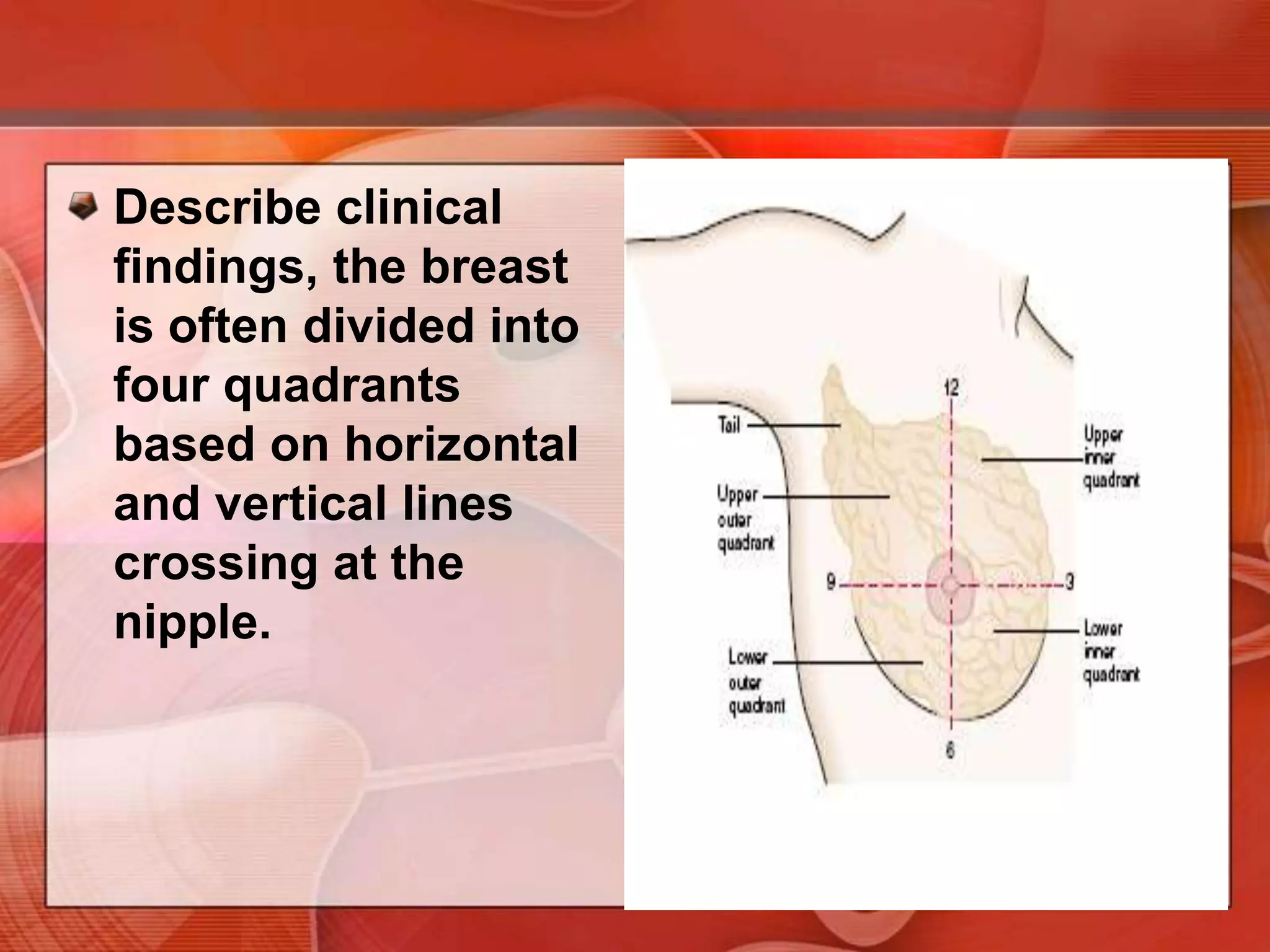
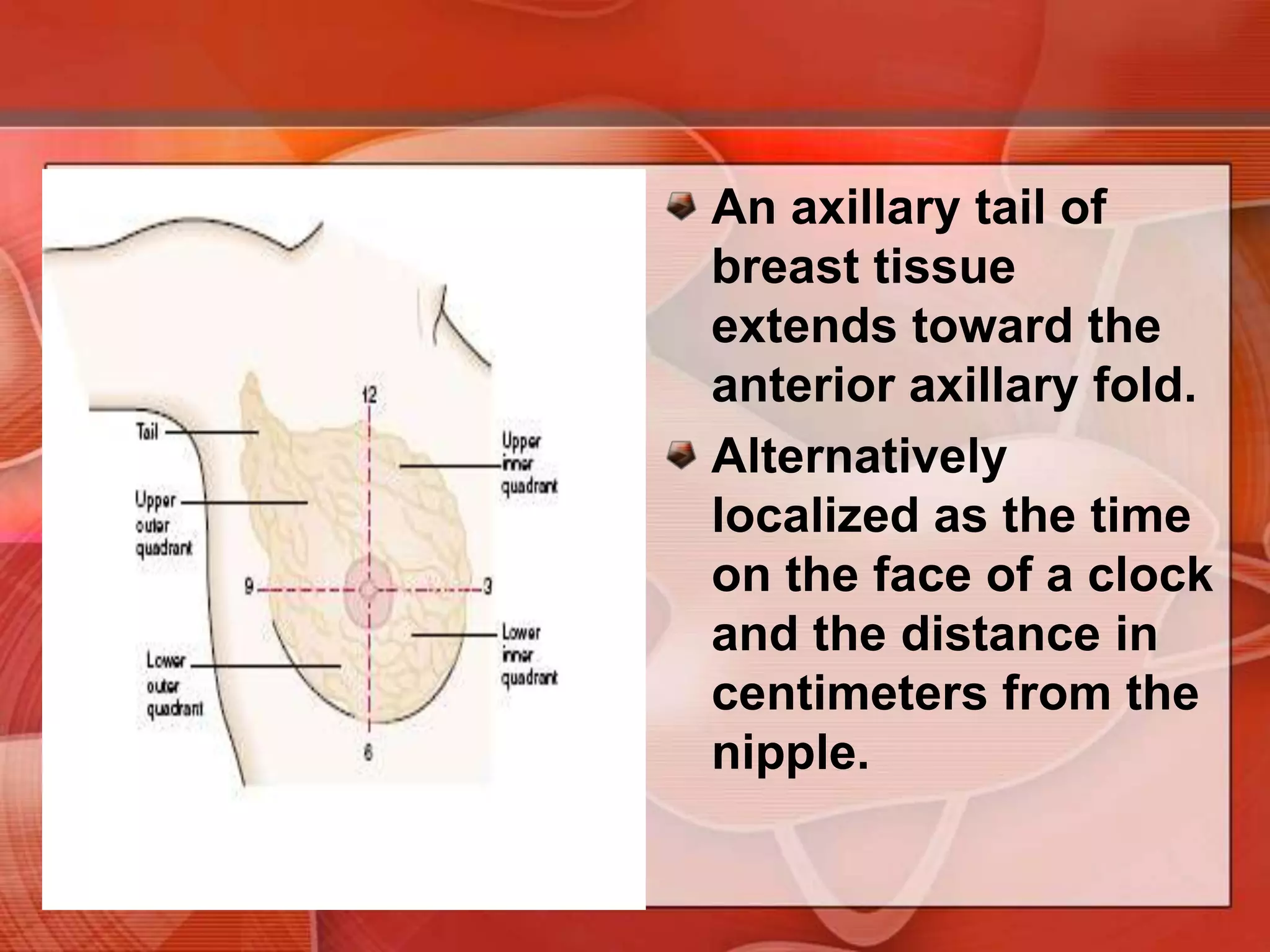
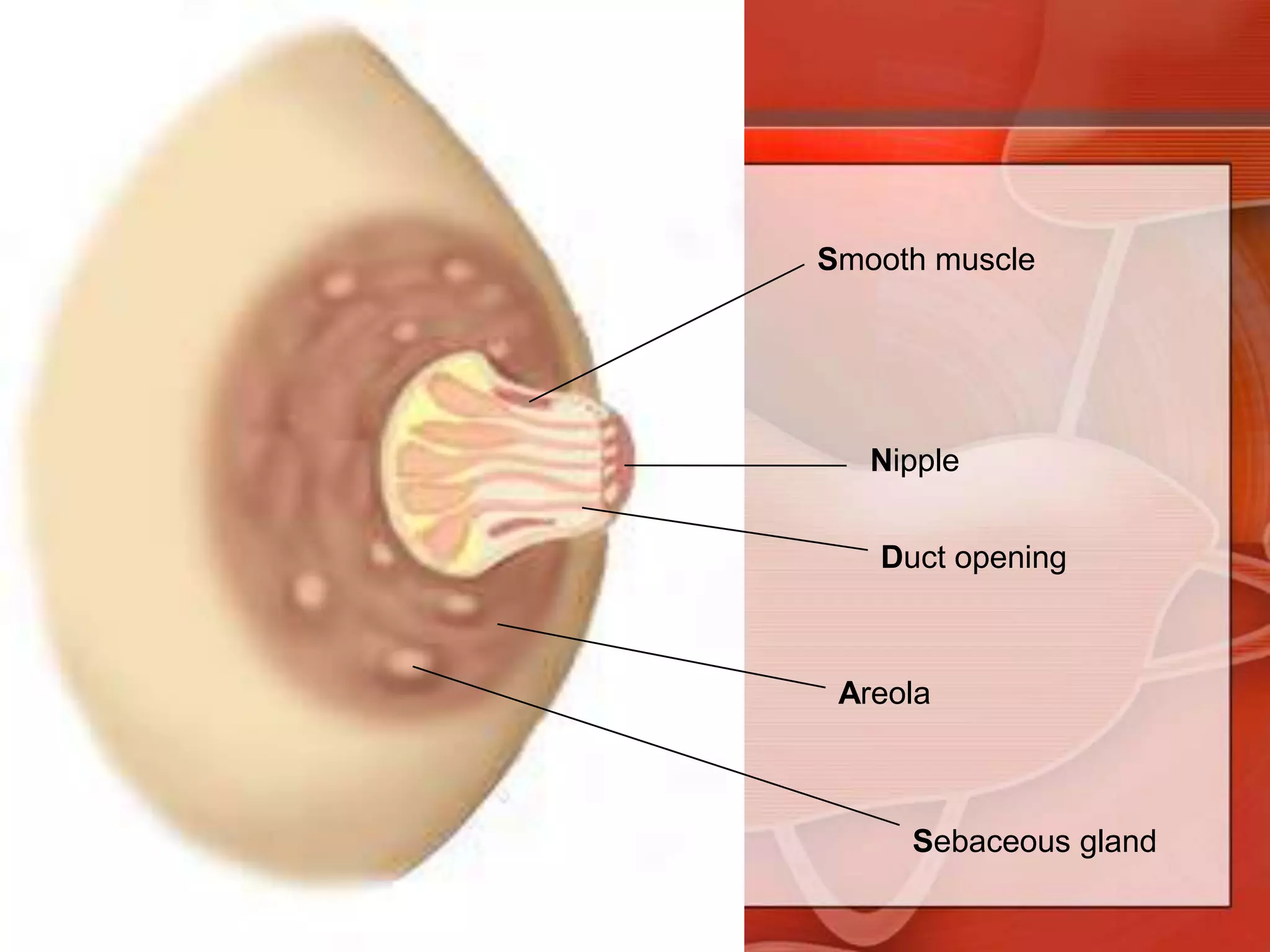
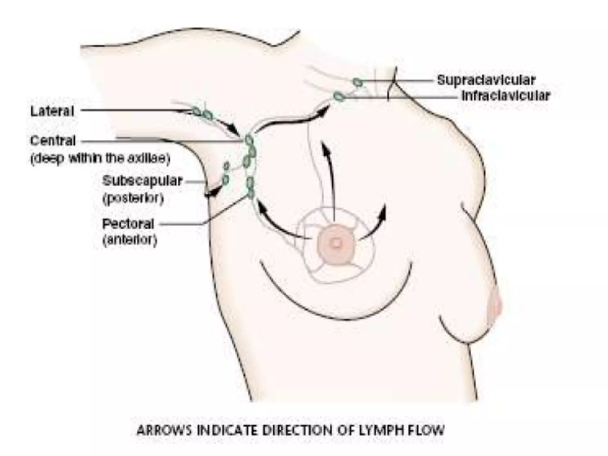
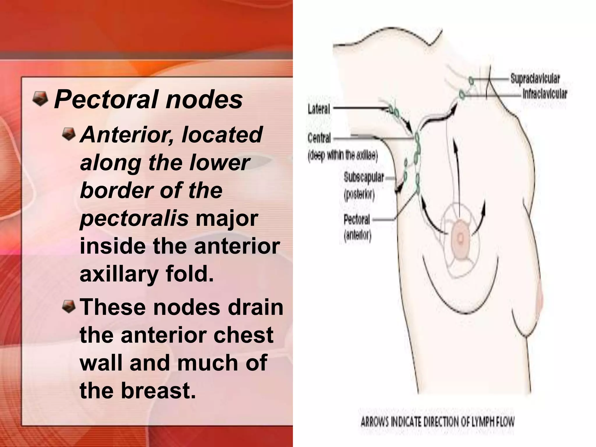
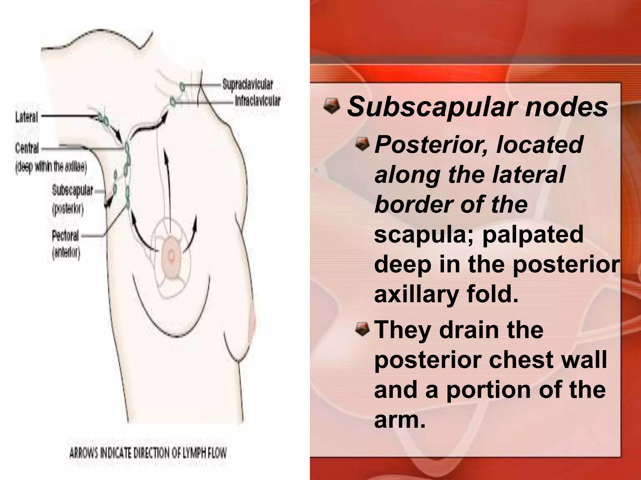
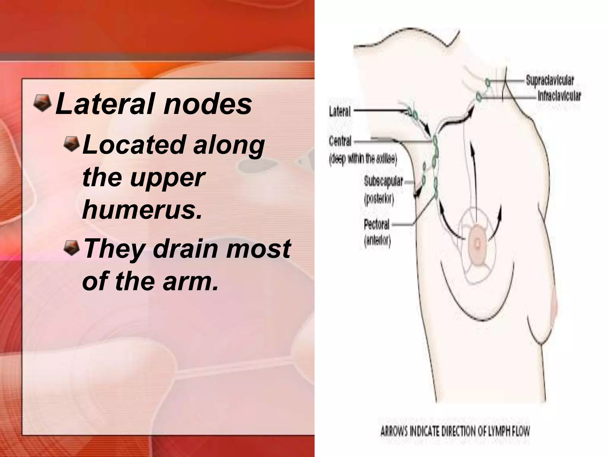
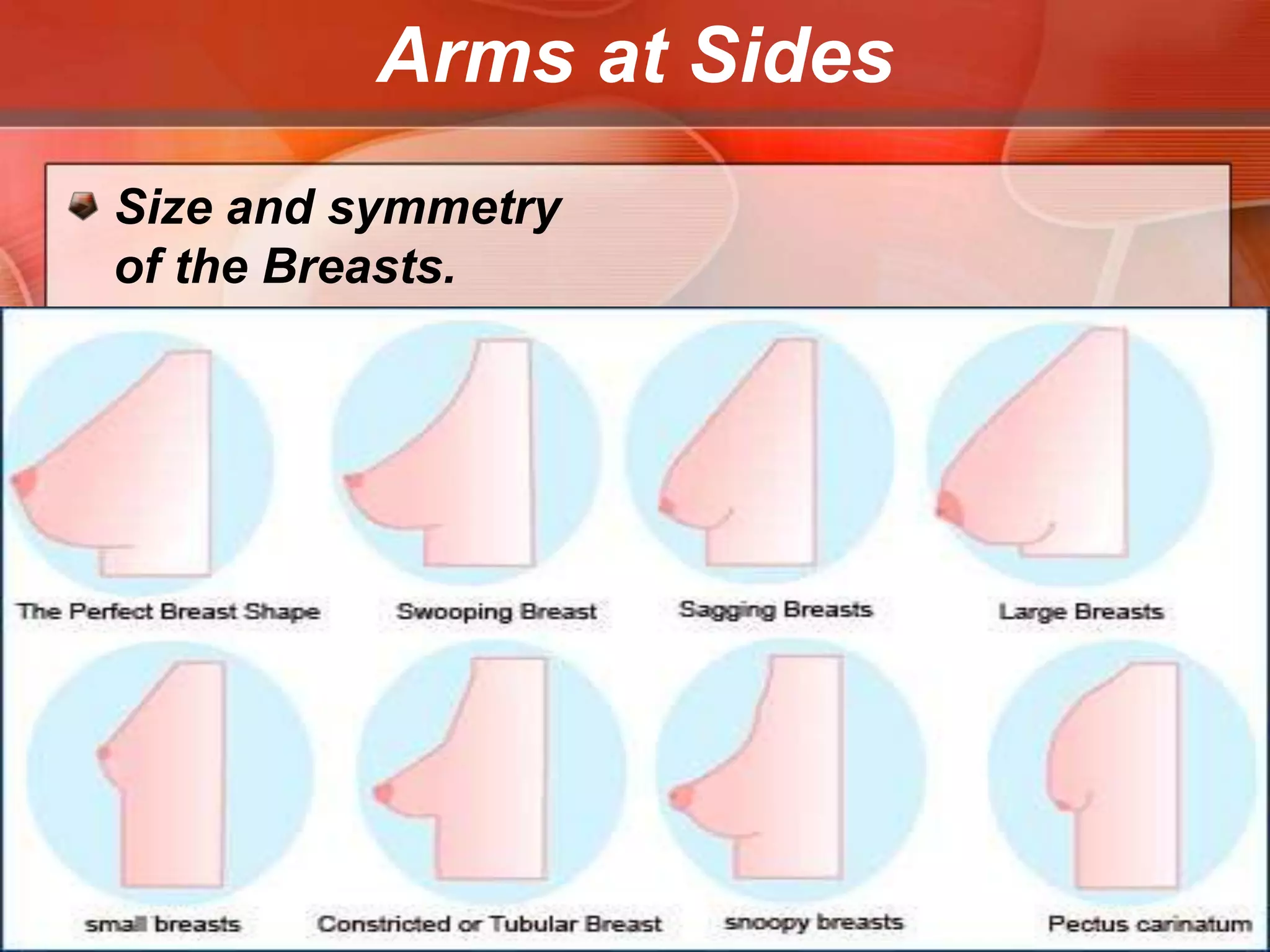
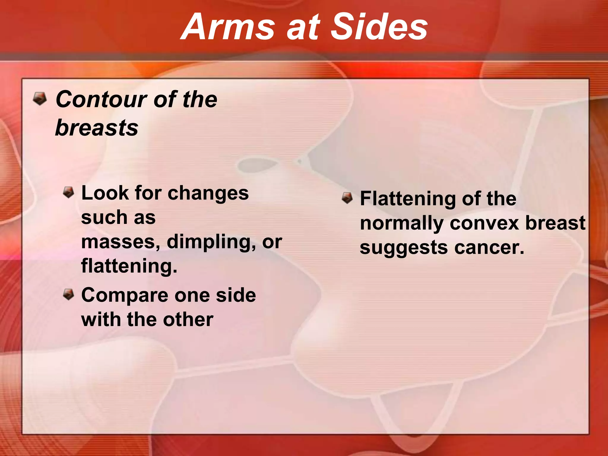
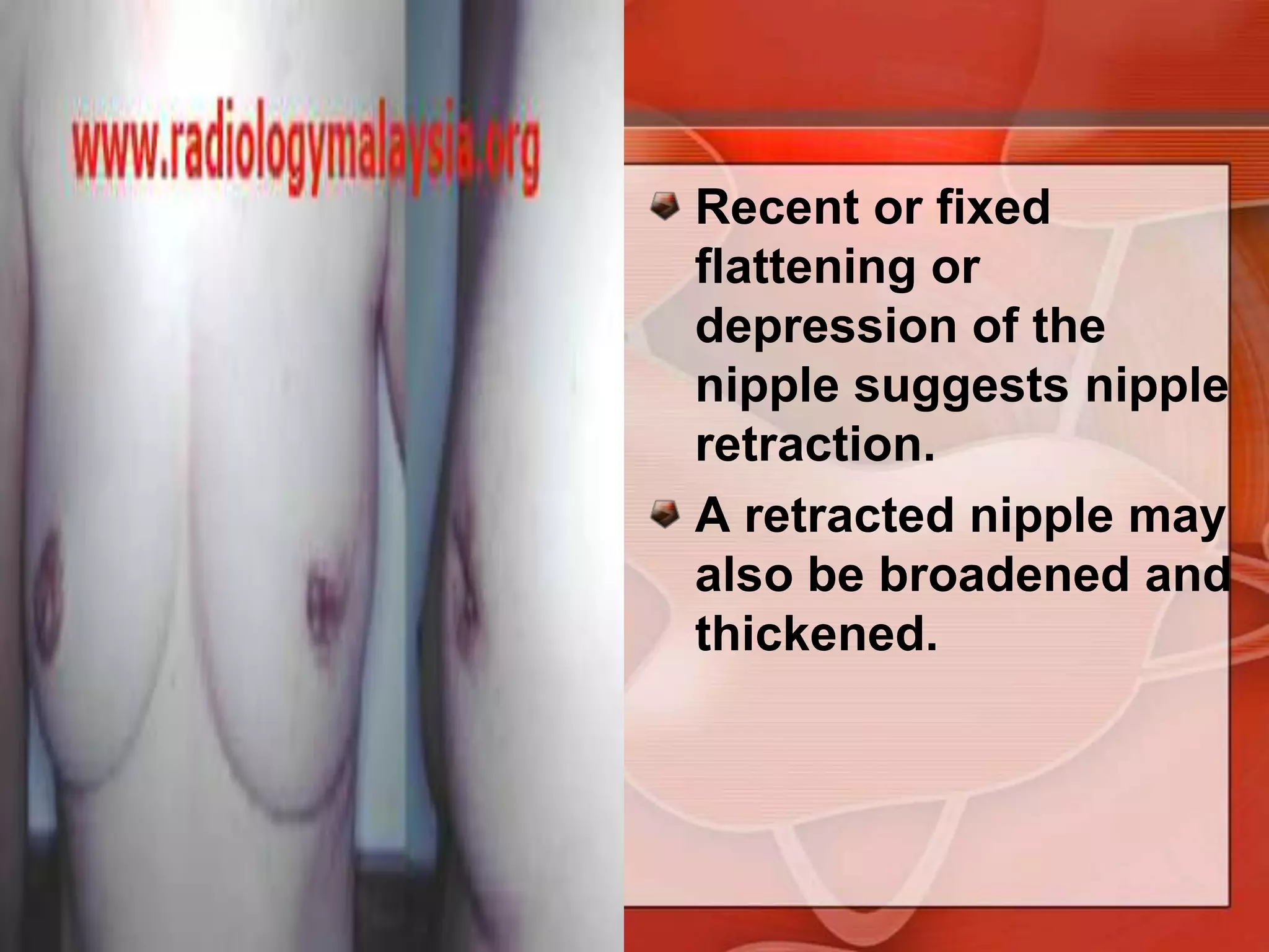
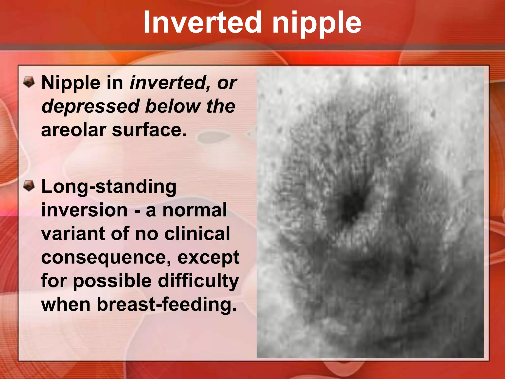
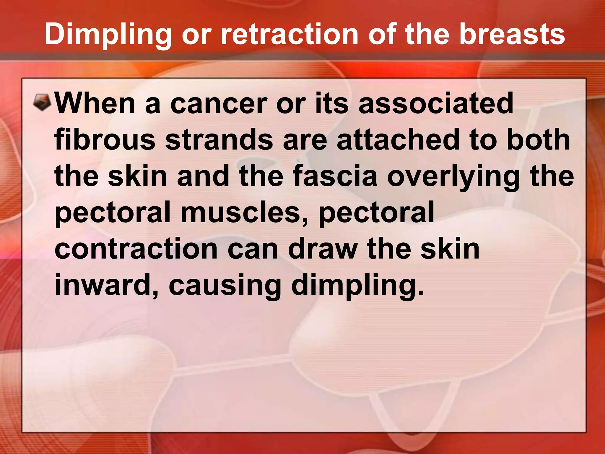
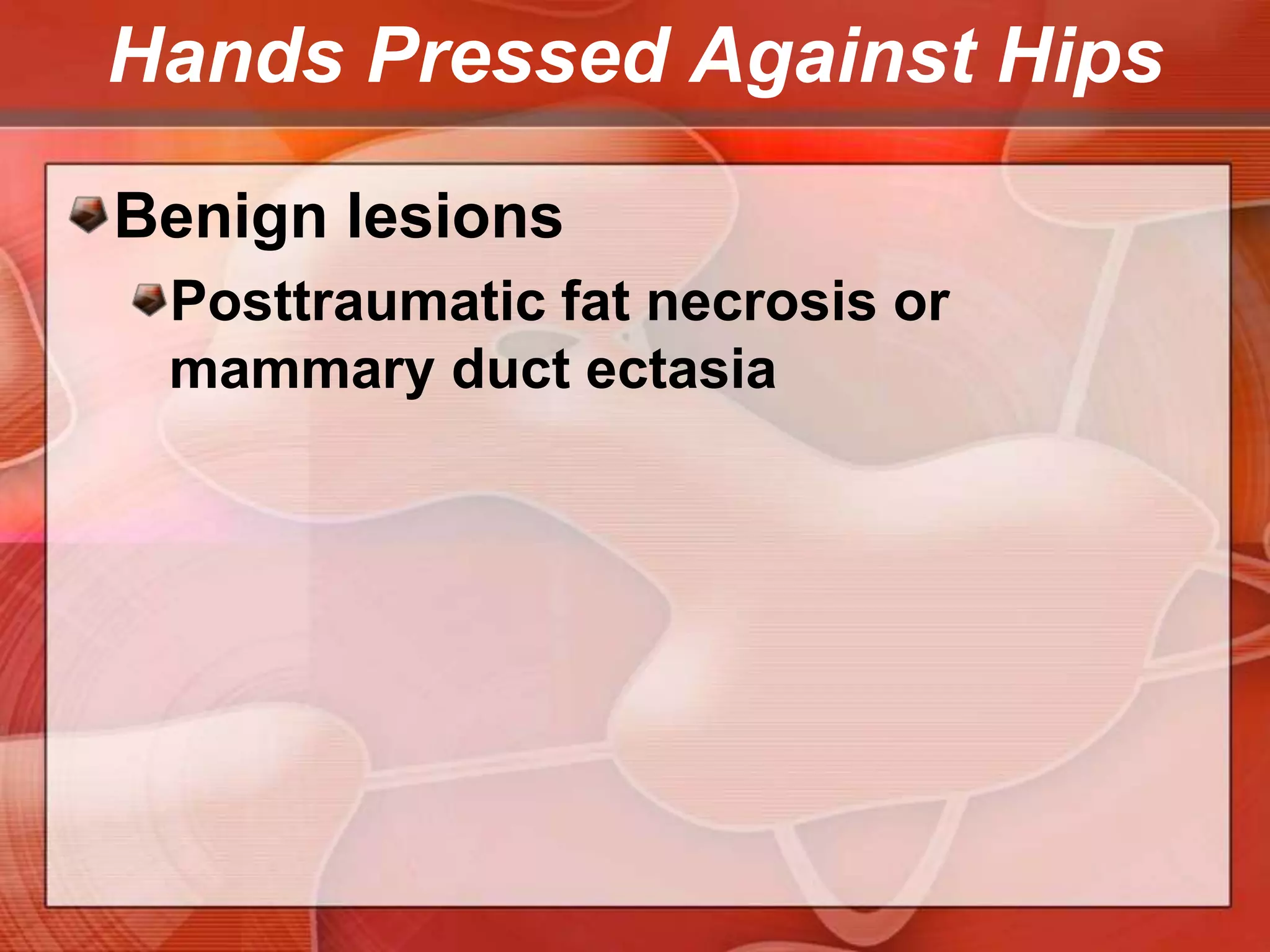
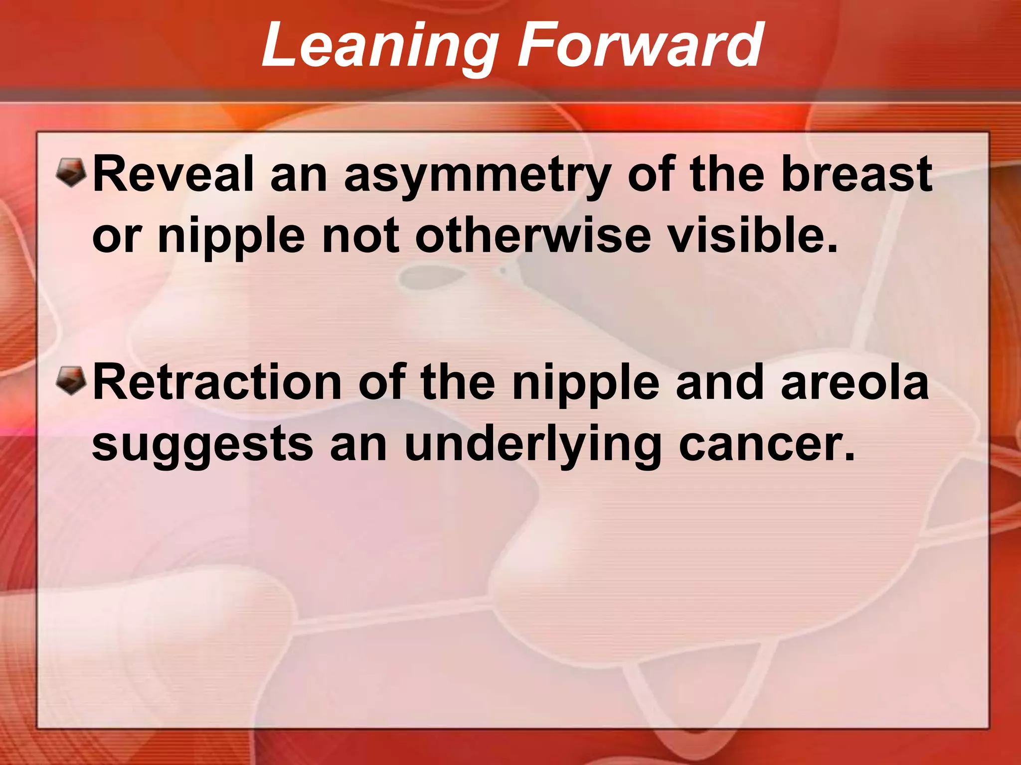
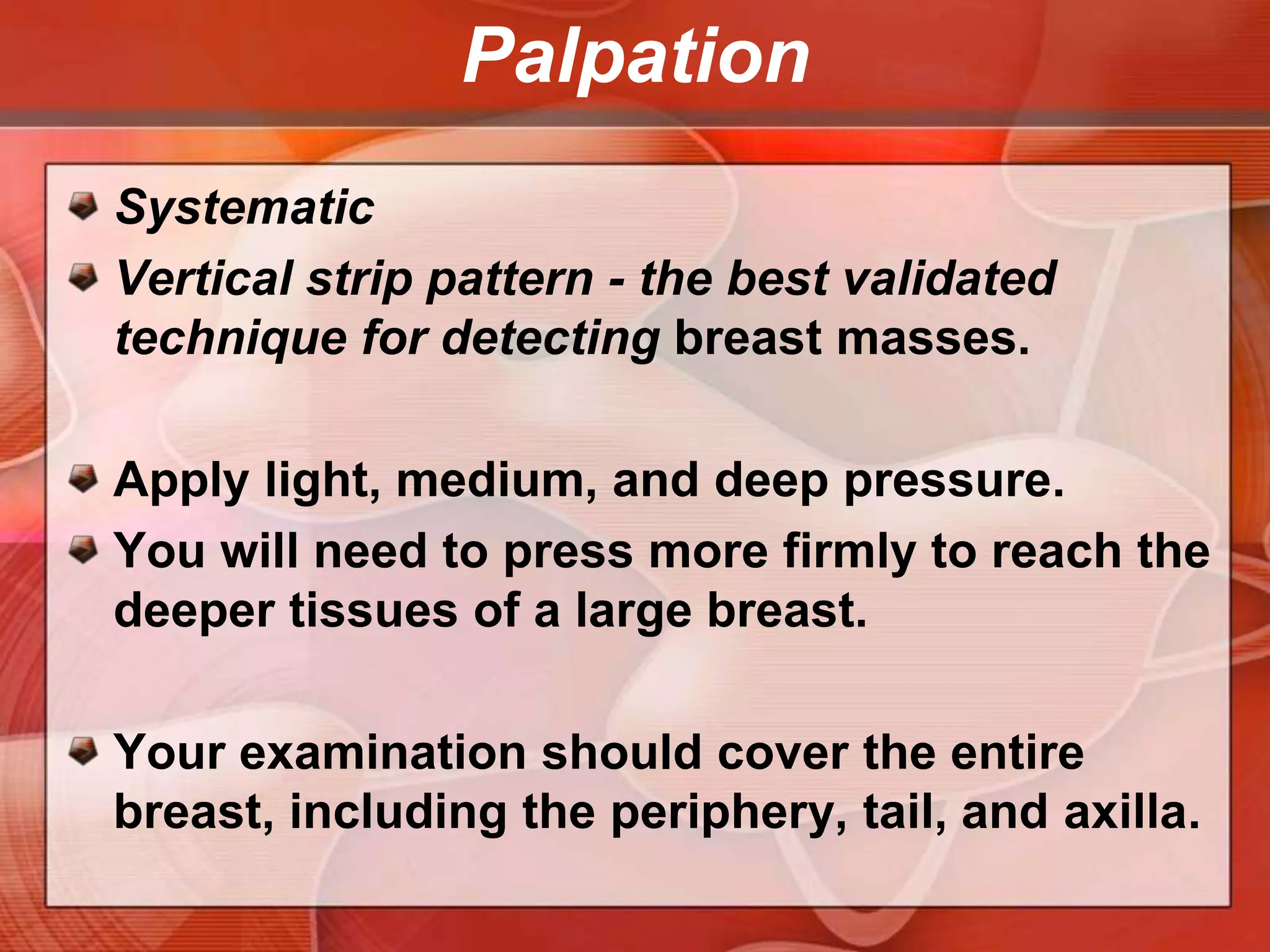
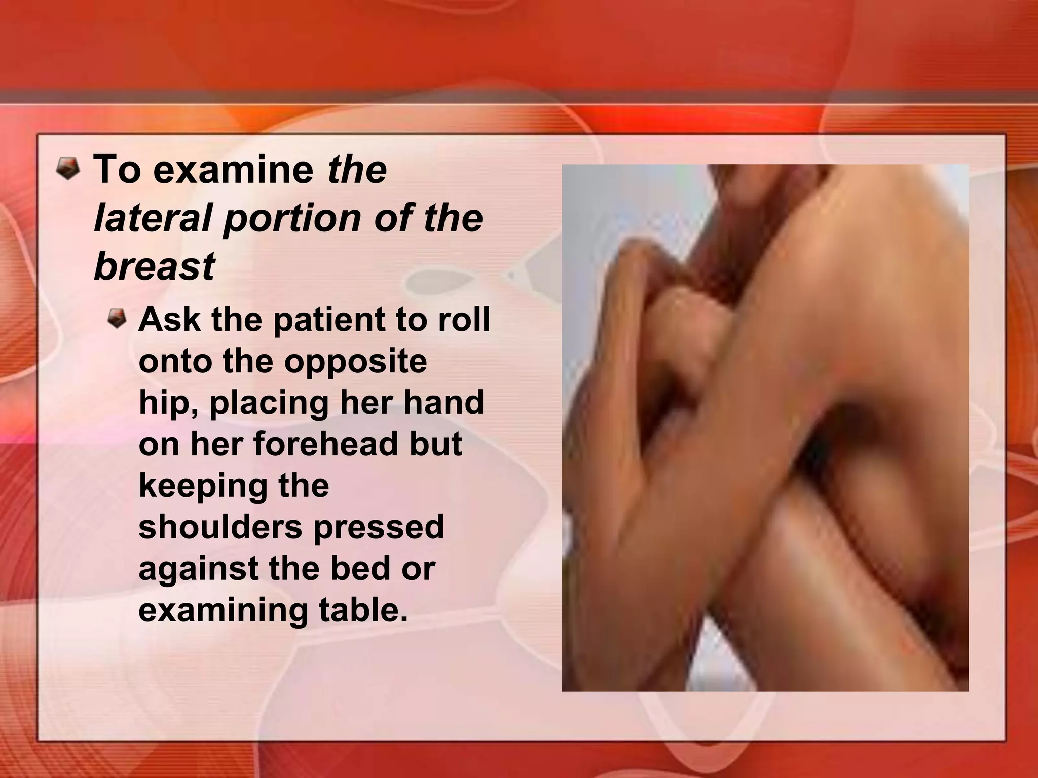
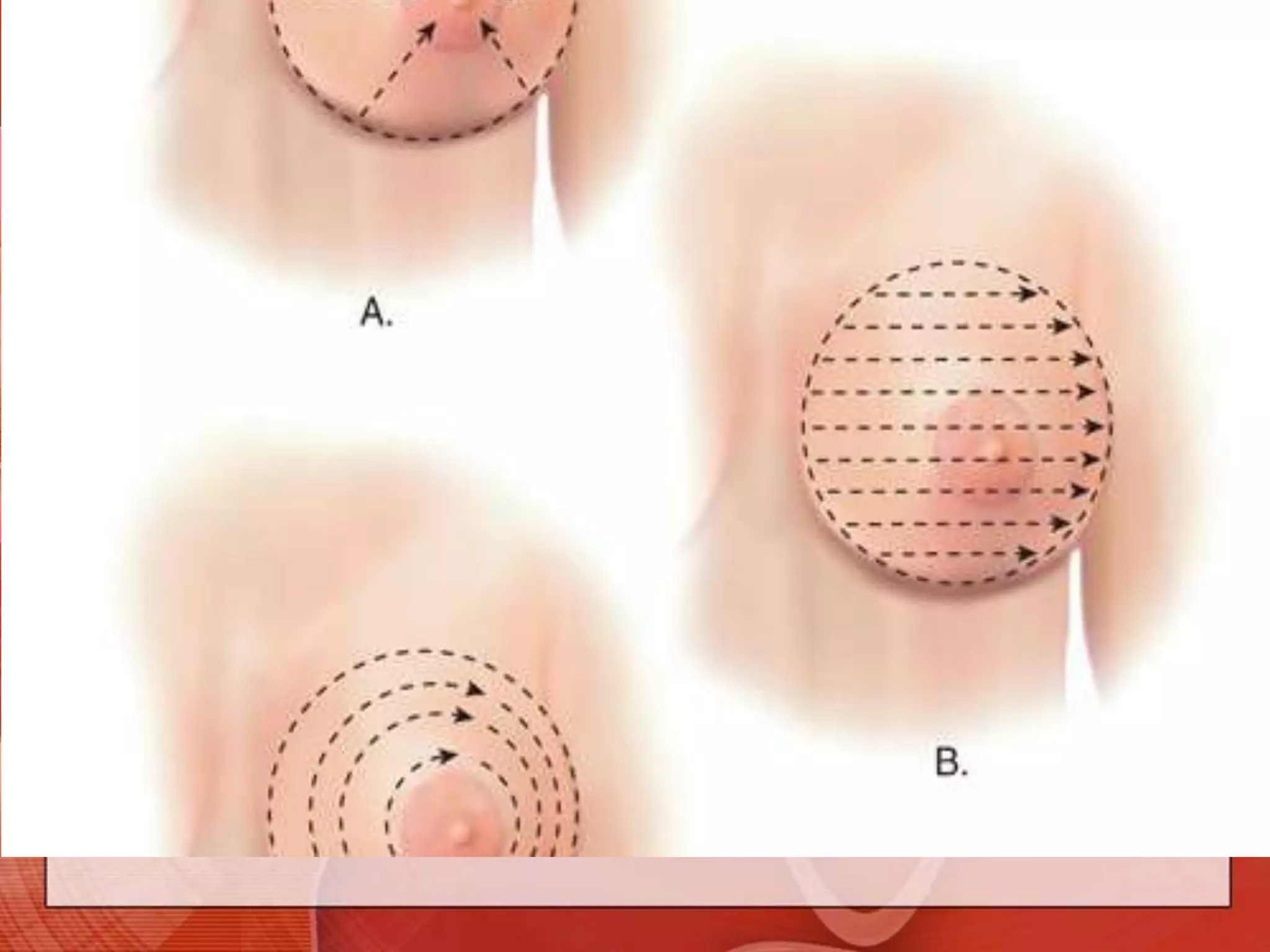
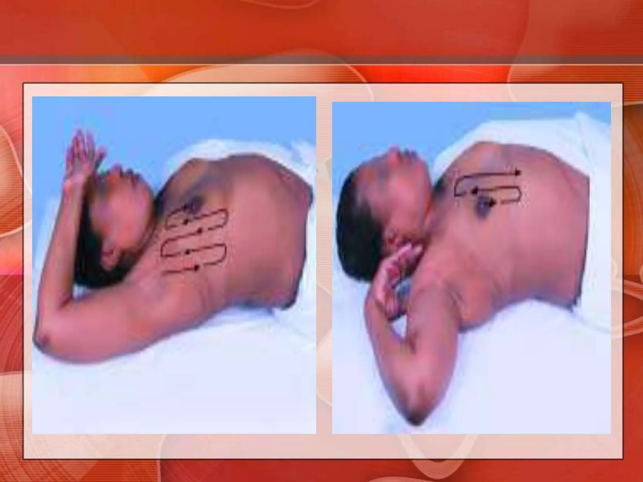
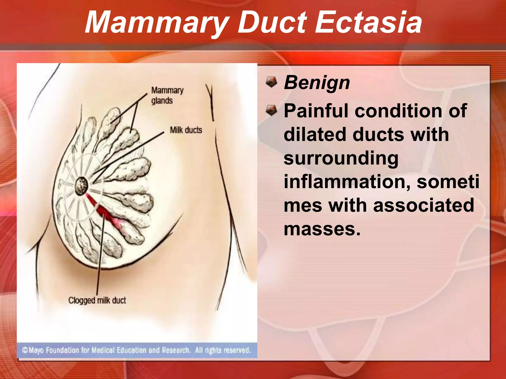
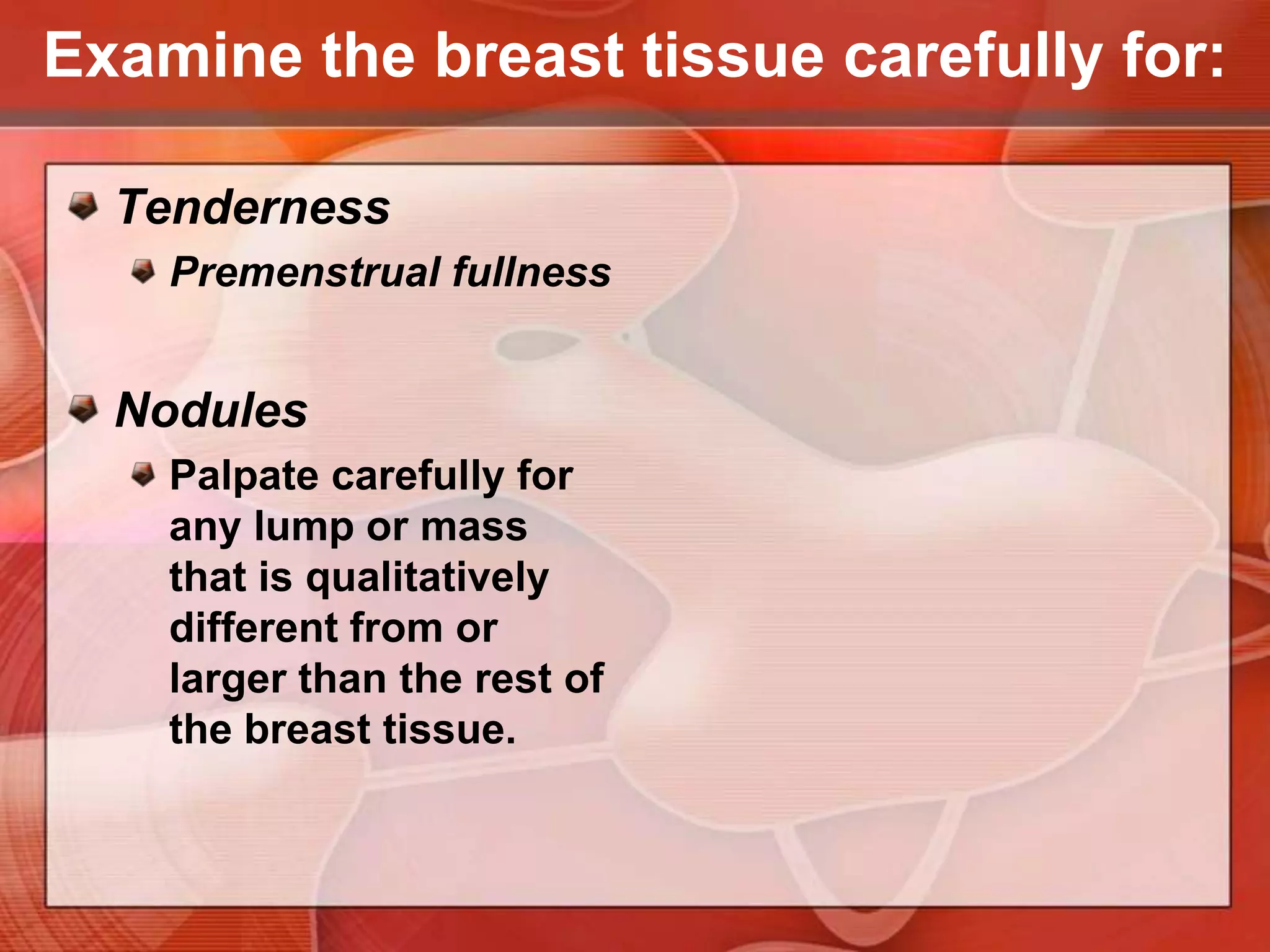
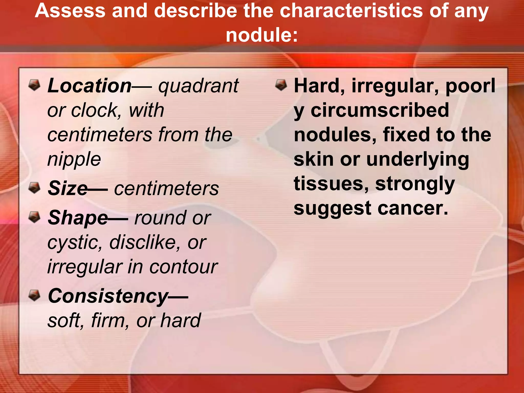
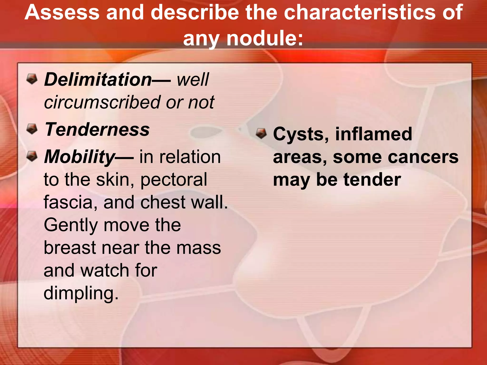
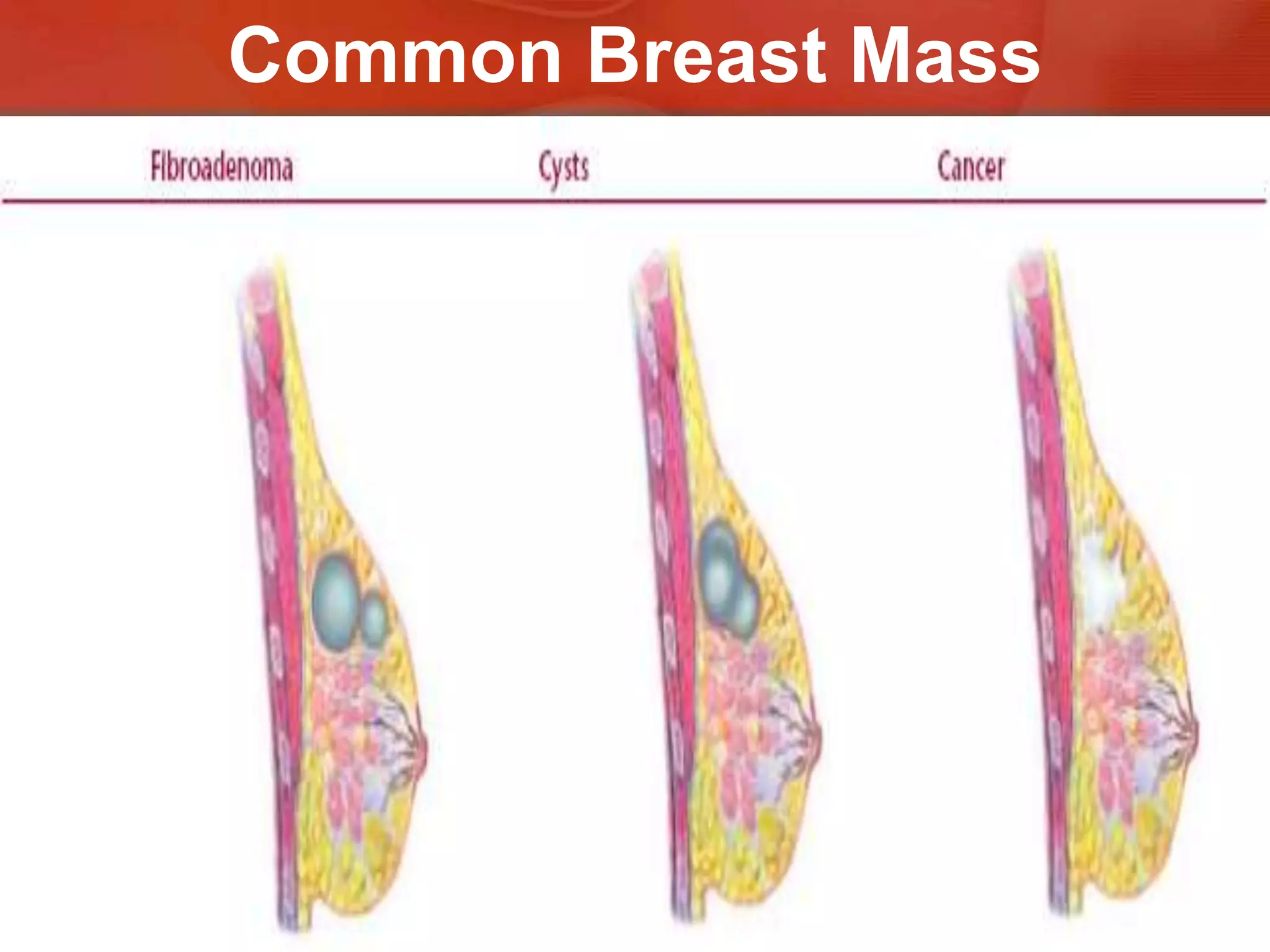
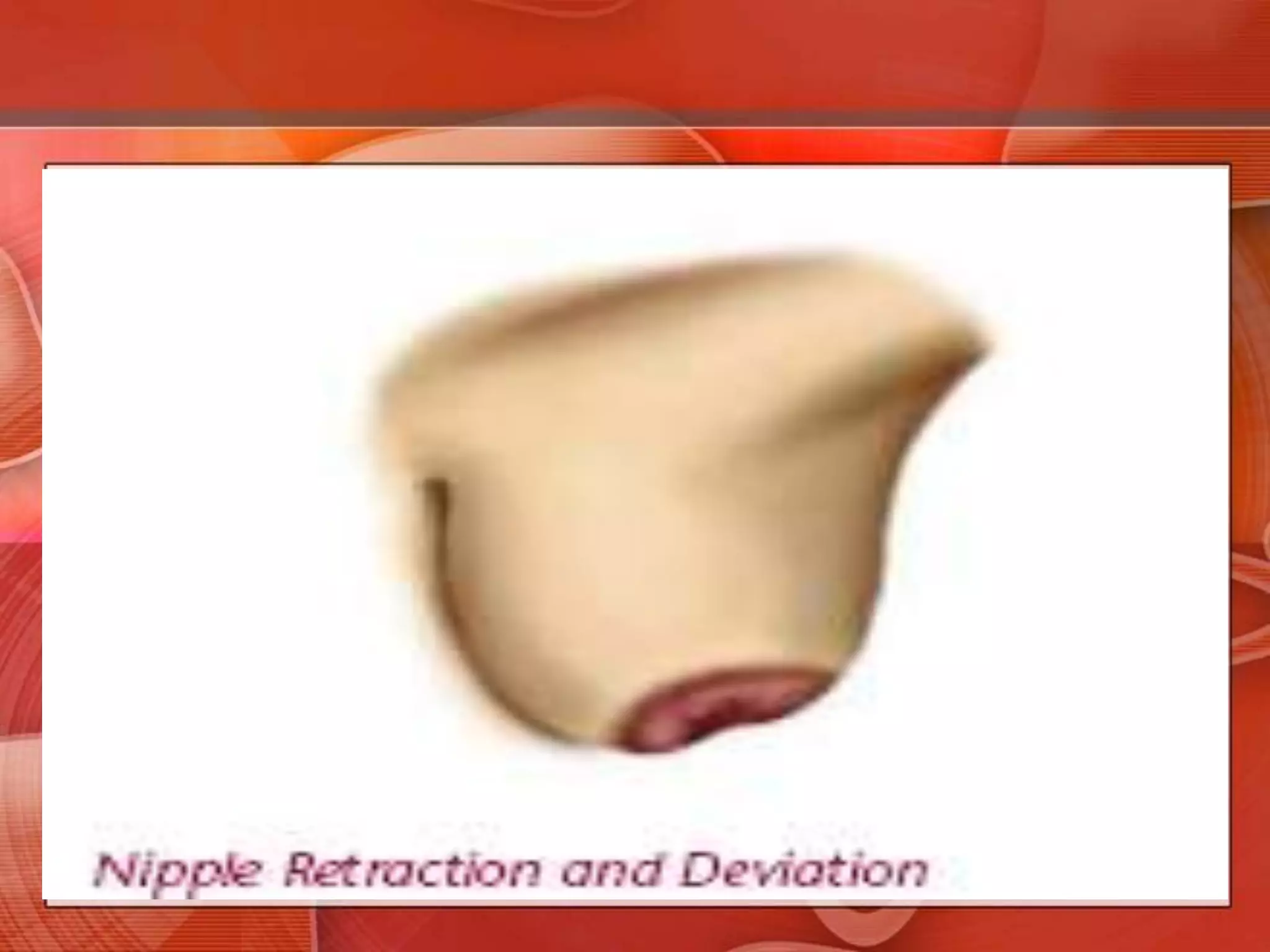
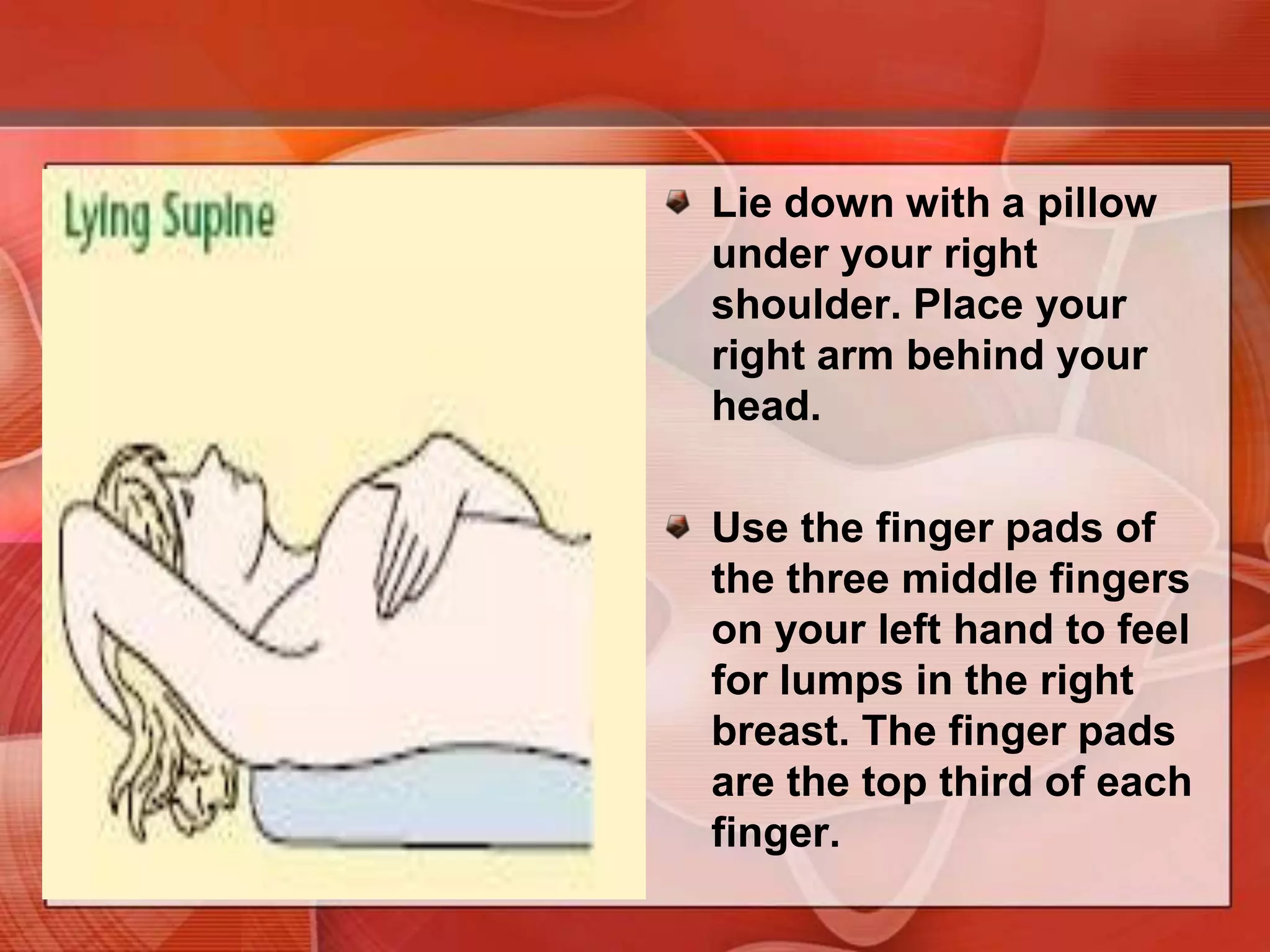
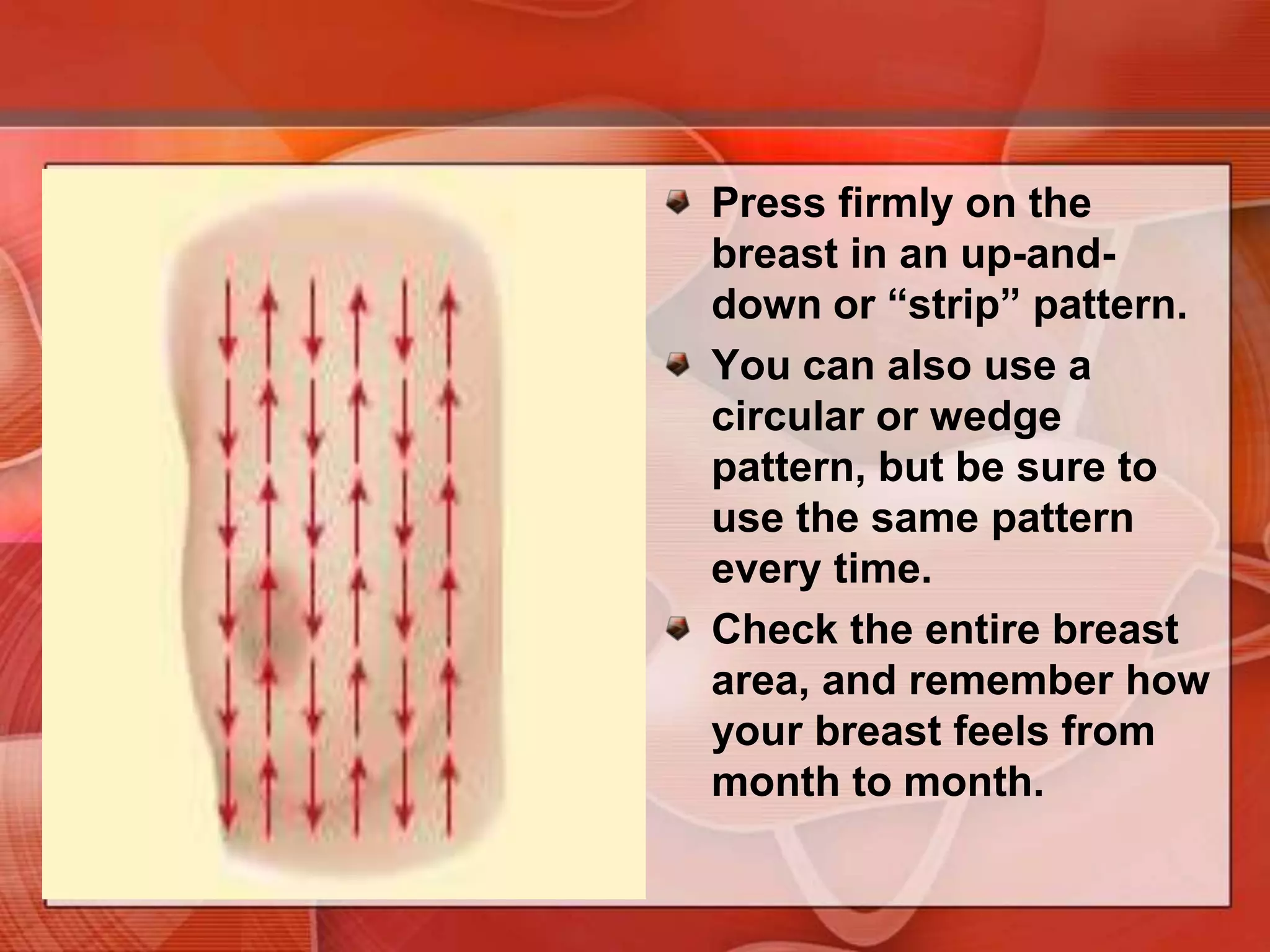
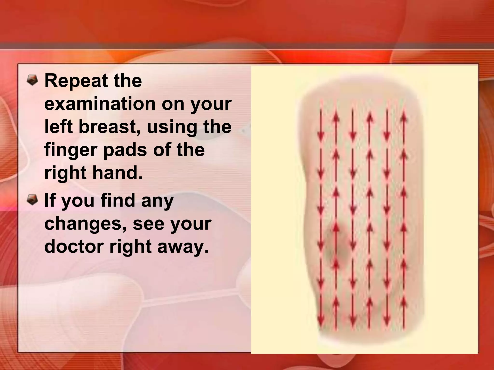
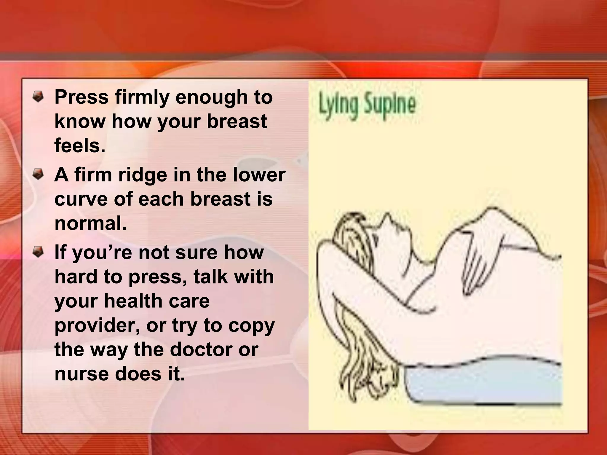
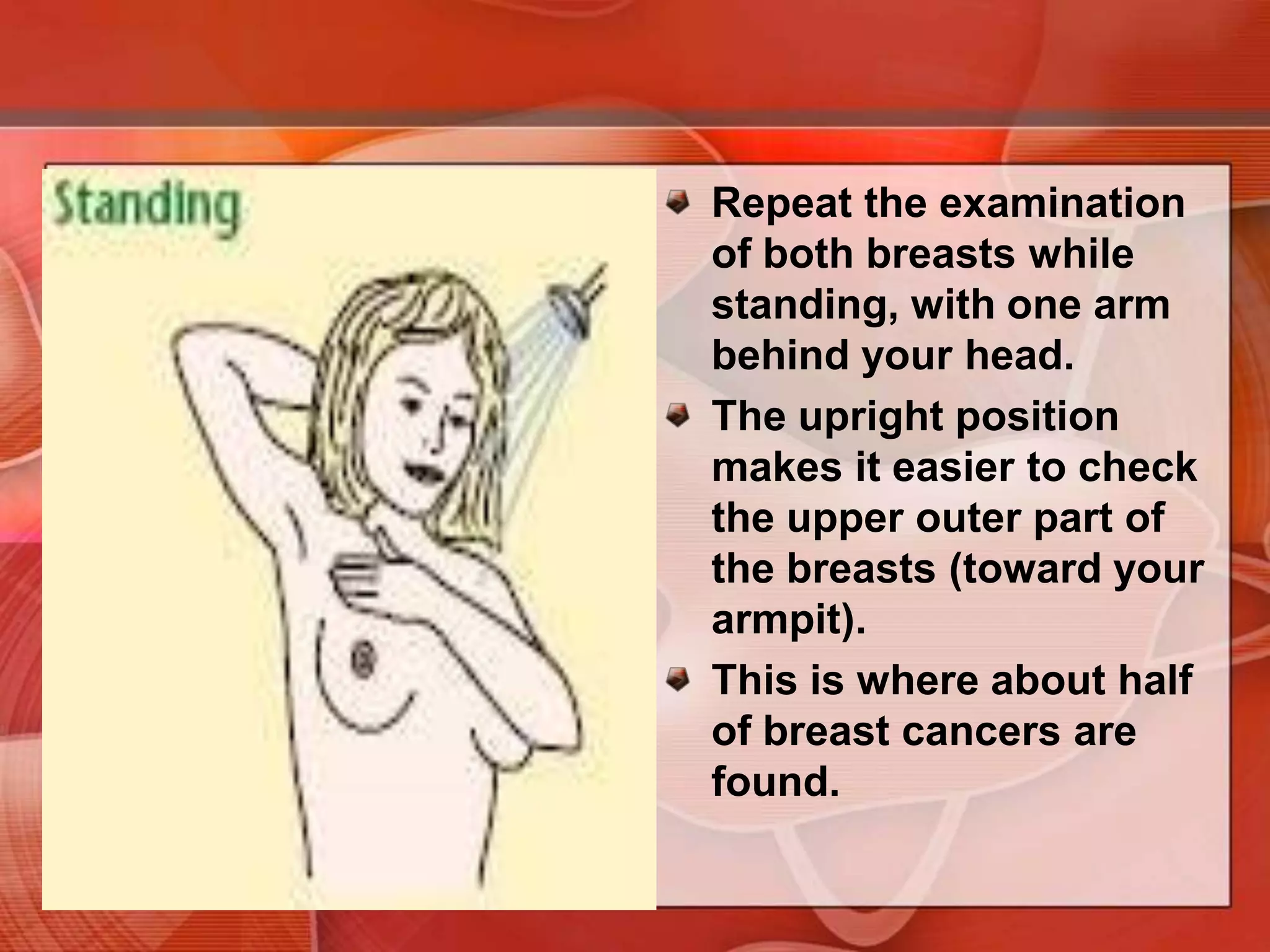
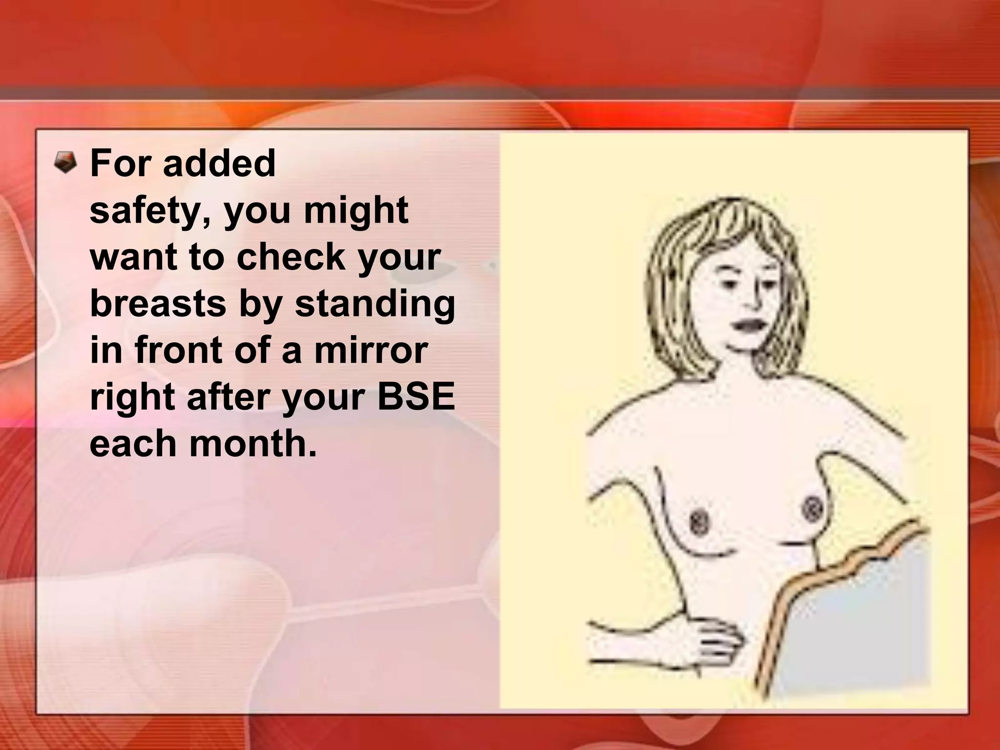
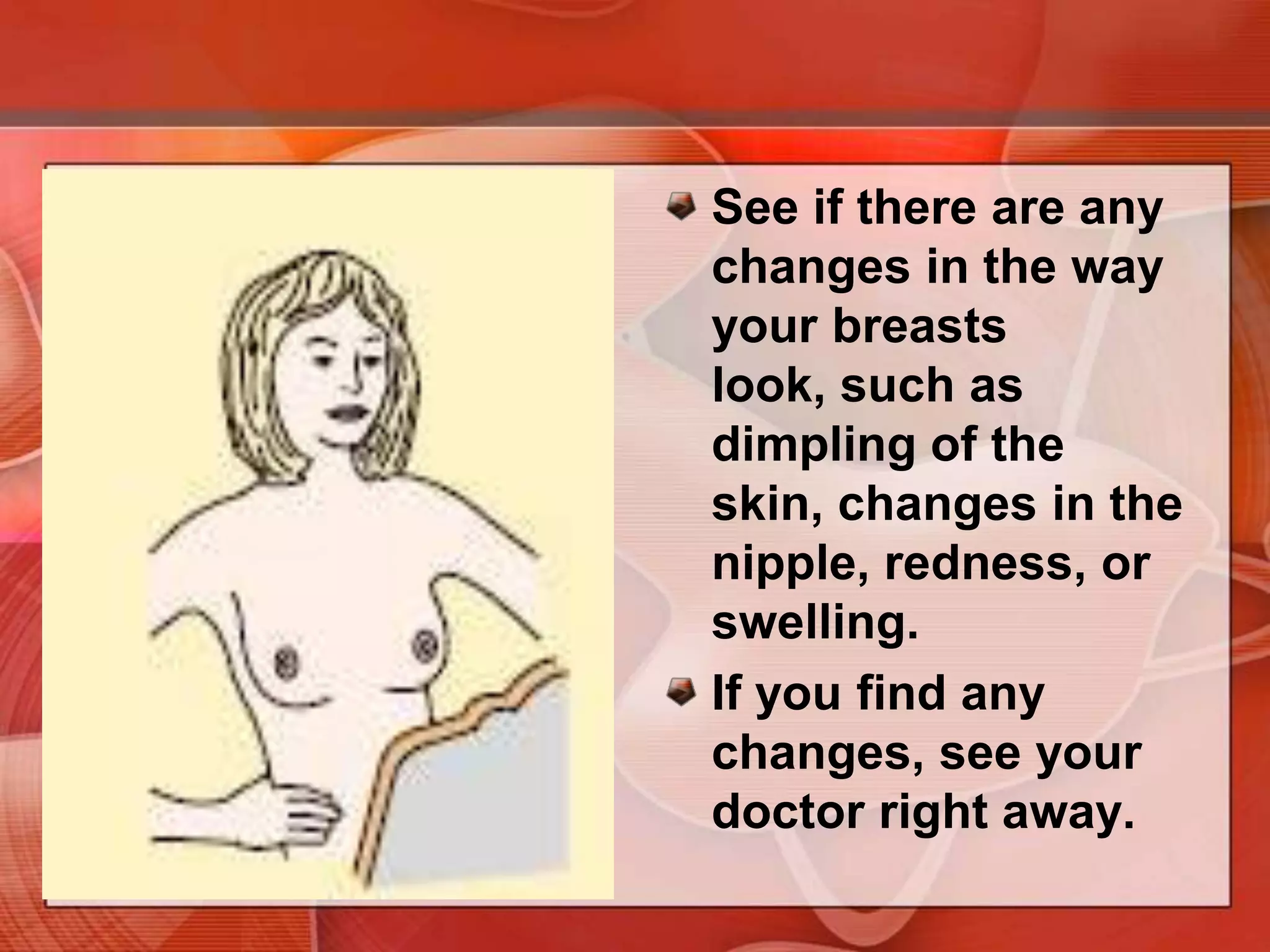
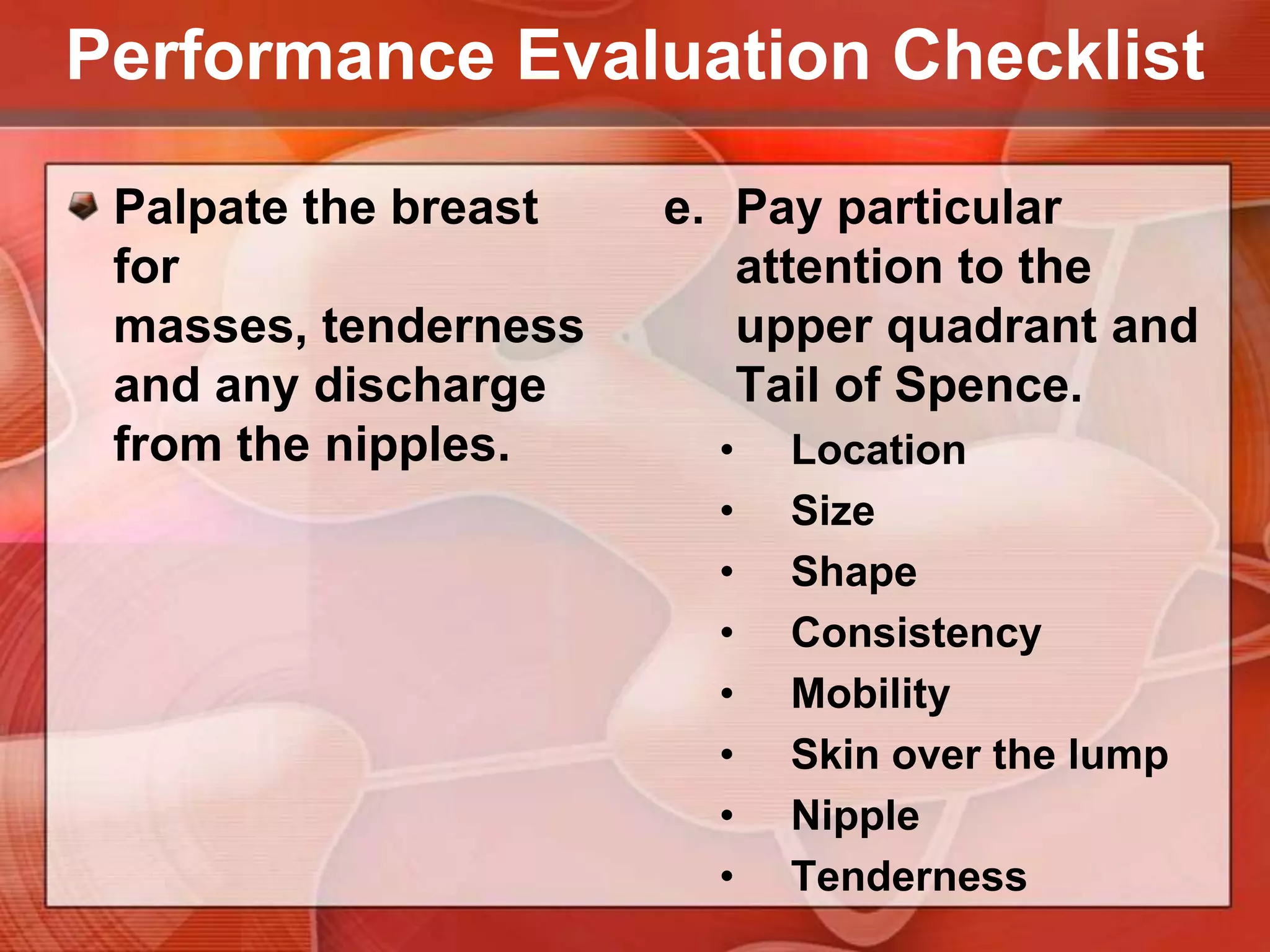
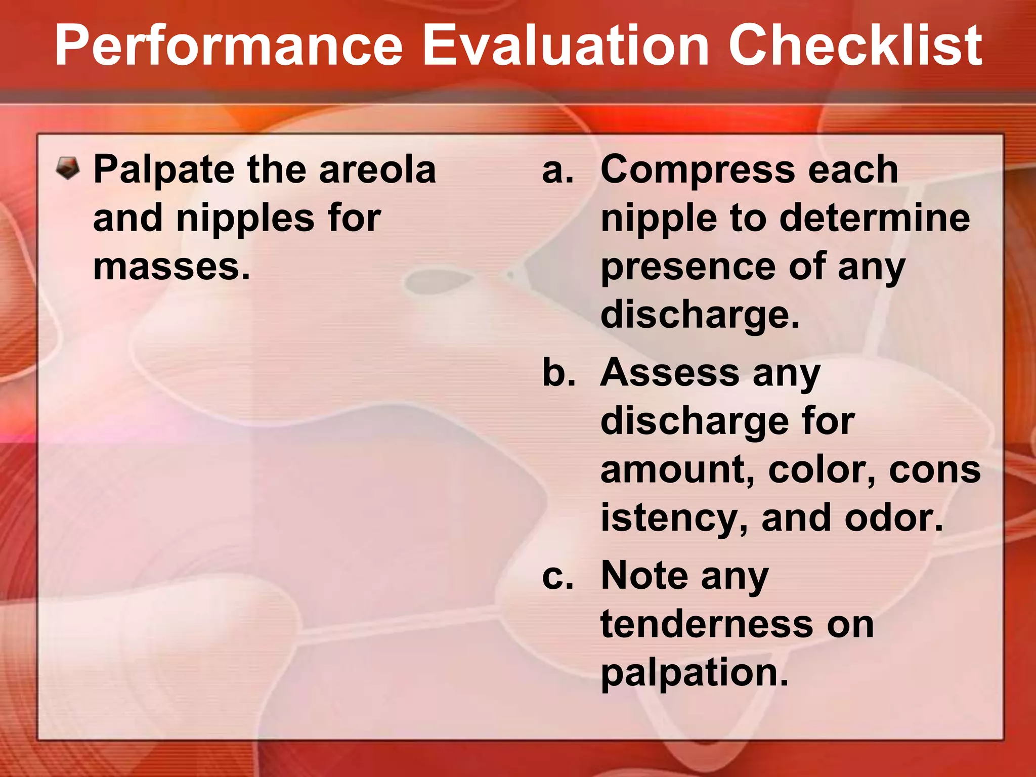
Procedure on Assessing the Breasts and Axillae
OBJECTIVESReview the anatomy of the breasts & axillae.
Discuss and demonstrate the procedure on assessing the breasts & axillae examination.
Return – demonstrate the assessing the breasts & axillae examination properly.Anatomy of the breasts & axillae
The female breast lies against the anterior thoracic wall, extending from the clavicle and 2nd rib down to the 6th rib, and from the sternum across to the midaxillary line. Its surface area is generally rectangular rather than round.The breast overlies the pectoralis major and at its inferior margin, the serratus anterior.
Describe clinical findings, the breast is often divided into four quadrants based on horizontal and vertical lines crossing at the nipple.
An axillary tail of breast tissue extends toward the anterior axillary fold. Alternatively localized as the time on the face of a clock and the distance in centimeters from the nipple.
Breast structures
Smooth muscleNippleDuct openingAreolaSebaceous gland
Lymph nodes
Lymphatics from most of the breast drain toward the axilla. Central nodes are palpable most frequently. They lie along the chest wall, usually high in the axilla and midway between the anterior and posterior axillary folds.
Pectoral nodesAnterior, located along the lower border of the pectoralismajor inside the anterior axillary fold. These nodes drain the anterior chest wall and much of the breast.
SubscapularnodesPosterior, located along the lateral border of the scapula; palpated deep in the posterior axillary fold. They drain the posterior chest wall and a portion of the arm.
Lateral nodesLocated along the upper humerus. They drain most of the arm.
Health history
Common or Concerning SymptomsBreast lump or massLumps may be physiologic or pathologic, ranging from cysts and fibroadenomas to breast cancer.Breast pain or discomfortNipple dischargeMilky bilateral discharge, or galactorrhea, may reflect pregnancy or prolactin or other hormonal imbalance. Non milky unilateral discharge suggests local breast disease.
Risk Factors for Breast CancerBreast cancer - most common cause of cancer in women worldwide, accounting for 18% of all female malignancies.>12% - lifetime risk of developing breast cancer Approximately 22% - risk of dying from the disease70% - no known predisposing factors definite risk factors are well-established
Risk Factors for Breast CancerAgeFamily HistoryRelative risk of breast cancer menstrual historypregnancybreast conditions and diseasesRisk from familial breast cancer family history of breast cancer genetic predisposition
Risk Factors for Breast CancerMenstrual History and PregnancyEarly menarcheDelayed menopauseFirst live birth after age 35 or no pregnancy all raise the risk of breast cancer two- to three-fold.
Risk Factors for Breast CancerBreast Conditions and DiseasesBenign breast disease with biopsy findings Atypical hyperplasia Lobular carcinomaBreast Cancer ScreeningBreast self-examination method
Recording the Physical Examination —Breasts and Axillae
Recording the Physical Examination Breasts and Axillae“Breasts symmetric and without masses. Nipples without discharge.”
Recording the Physical Examination Breasts and Axillae“Breasts pendulous with diffuse fibrocystic changes. Single firm 1 × 1 cm mass, mobile and nontender, with overlying peaud’orange appearance in right breast, upper outer quadrant at 11 o’clock.”
procedure on assessing the breasts & axillae examination
The Female Breast
INSPECTIONASSESSMENT TECHNIQUES
Inspect the breasts and nipples with the patient in the sitting position and disrobed to the waist.
Arms at SidesAppearance of the skinColorThickening of the skin and unusually prominent pores, which may accompany lymphatic obstructionRedness from local infection or inflammatory carcinomaThickening and prominent pores suggest a breast cancer.
Arms at Sides
Arms at Sides
Arms at SidesSize and symmetry of the Breasts.
Arms at SidesContour of the breastsLook for changes such as masses, dimpling, or flattening. Compare one side with the otherFlattening of the normally convex breast suggests cancer.
Arms at SidesCharacteristics of the nipples, size and shape, direction in which they point, any rashes or ulceration, or any dischargeAsymmetry of directions in which nipples point suggests an underlying cancer. Rash or ulceration in Paget’s disease of the breast
Paget’s Disease
Recent or fixed flattening or depression of the nipple suggests nipple retraction. A retracted nipple may also be broadened and thickened.
Inverted nippleNipple in inverted, or depressed below the areolar surface. Long-standing inversion - a normal variant of no clinical consequence, except for possible difficulty when breast-feeding.
Retracted nipple
Arms Over HeadHands Pressed Against Hips Leaning Forward
To bring out dimpling or retraction that may otherwise be invisibleAsk the patient to raise her arms over her head, then press her hands against her hips to contract the pectoral muscles. Inspect the breast contours carefully in each position. If the breasts are large or pendulous, it may be useful to have the patient stand and lean forward, supported by the back of the chair or the examiner’s hands.
Arms Over Head
Dimpling or retraction of the breastsWhen a cancer or its associated fibrous strands are attached to both the skin and the fascia overlying the pectoral muscles, pectoral contraction can draw the skin inward, causing dimpling.
Dimpling breast
Hands Pressed Against Hips
Hands Pressed Against HipsBenign lesions Posttraumatic fat necrosis or mammary duct ectasia
Leaning Forward
Leaning ForwardReveal an asymmetry of the breast or nipple not otherwise visible. Retraction of the nipple and areola suggests an underlying cancer.
palpationASSESSMENT TECHNIQUES
BS3 MNEUMONICBest performed when the breast tissue is flattened.Supine position3 minutes thorough examination for each breast. Use the fingerpadsof the 2nd, 3rd, and 4th fingers, keeping the fingers slightly flexed.
PalpationSystematicVertical strip pattern - the best validated technique for detecting breast masses.Apply light, medium, and deep pressure. You will need to press more firmly to reach the deeper tissues of a large breast. Your examination should cover the entire breast, including the periphery, tail, and axilla.
To examine the lateral portion of the breastAsk the patient to roll onto the opposite hip, placing her hand on her forehead but keeping the shoulders pressed against the bed or examining table.
Begin palpation in the axilla, moving in a straight line down to the bra line, then move the fingers medially and palpate in a vertical strip up the chest to the clavicle. Continue in vertical overlapping strips until you reach the nipple, then reposition the patient to flatten the medial portion of the breast.Nodules in the tail of the breast are sometimes mistaken for enlarged axillary lymph nodes (and vice versa).
To examine the medial portion of the breast,Ask the patient to lie with her shoulders flat against the bed or examining table, placing her hand at her neck and lifting up her elbow until it is even with her shoulder. Palpate in a straight line down from the nipple to the bra line, then back to the clavicle, continuing in vertical overlapping strips to the midsternum.
Examine the breast tissue carefully for:Consistency of the tissues.Normal consistency varies widely, depending in part on the relative proportions of firmer glandular tissue and soft fat.Physiologic nodularity may be present, increasing before menses.
Examine the breast tissue carefully for:There may be a firm transverse ridge of compressed tissue along the lower margin of the breast, especially in large breasts. This is the normal inframammary ridge, not a tumor.Tender cords suggest mammary duct ectasia, a benign but sometimespainful condition of dilated ducts with surrounding inflammation, sometimes with associatedmasses.
Mammary Duct EctasiaBenign Painful condition of dilated ducts with surrounding inflammation, sometimes with associated masses.
Examine the breast tissue carefully for:Tenderness Premenstrual fullnessNodulesPalpate carefully for any lump or mass that is qualitatively different from or larger than the rest of the breast tissue.
Hard, irregular, poorly circumscribed nodules, fixed to the skin or underlying tissues, strongly suggest cancer.Location— quadrant or clock, with centimeters from the nippleSize— centimetersShape— round or cystic, disclike, or irregular in contourConsistency— soft, firm, or hardAssess and describe the characteristics of any nodule:
Assess and describe the characteristics of any nodule:Delimitation— well circumscribed or notTendernessMobility— in relation to the skin, pectoral fascia, and chest wall. Gently move the breast near the mass and watch for dimpling.Cysts, inflamed areas, some cancers may be tender
The NipplePalpate each nipple, noting its elasticity.Thickening of the nipple and loss of elasticity suggest an underlying cancer.
The Male Breast
The Male BreastInspect the nipple and areola for nodules, swelling, or ulceration.Palpate the areola and breast tissue for nodules. If the breast appears enlarged, distinguish between the soft fatty enlargement of obesity and the firm disc of glandular enlargement, called gynecomastia.Gynecomastia - imbalance of estrogens & androgens, sometimes drug-related. Breast cancer - A hard,  irregular, eccentric, or ulcerating nodule.
The Axillae
INSPECTIONInspect the skin of each axillaRashInfectionUnusual pigmentationDeodorant and other rashesSweat gland infection (hidradenitissuppurativa)Deeply pigmented, velvety axillary skin suggests acanthosisnigricans,one form of which is associated with internal malignancy.
PALPATIONTo examine the left axillaAsk the patient to relax with the left arm down. Help by supporting the left wrist or hand with your left hand.Cup together the fingers of your right hand and reach as high as you can toward the apex of the axilla. Warn the patient that this may feel uncomfortable.
PALPATIONYour fingers should lie directly behind the pectoral muscles, pointing toward the midclavicle. Now press your fingers in toward the chest wall and slide them downward, trying to feel the central nodes against the chest wall. Of the axillary nodes, these are the most often palpable. One or more soft, small (<1 cm), nontendernodesare frequently felt.
PALPATION: ABNORMALITIESEnlarged axillary nodes from infection of the hand or arm, recent immunizations or skin tests in the arm, or part of a generalized lymphadenopathy. Check the epitrochlear nodes and other groups of lymph nodes.Nodes that are large (≥1 cm) andfirm or hard, matted together, or fixed to the skin or to underlying tissues suggest malignant involvement.
Use your left hand to examine the right axilla.
Groups of axillary lymph nodes
Assessment of Spontaneous Nipple DischargeSPECIAL TECHNIQUES
History of spontaneous nipple dischargeDetermine its origin by compressing the areola with your index finger, placed in radial positions around the nipple.Watch for discharge appearing through one of the duct openings on the nipple’s surface. Note the color, consistency, and quantity of any discharge and the exact location where it appears.
PapillomaNonmilky unilateral discharge - local breast disease. Causative lesion – benignMalignant - elderly womenNote the drop of blood exuding from a duct opening.
Examination of The Mastectomy Patient
Examination of The Mastectomy PatientWarrants special care on examination. Inspect the mastectomy scar and axilla carefully for any masses or unusual nodularity. Note any change in color or signs of inflammation.Palpate gently along the scar—these tissues may be unusually sensitive. Use a circular motion with two or three fingers.
Examination of The Mastectomy PatientUpper outer quadrant and axilla. Note any enlargement of the lymph nodes or signs of inflammation or infection.Lymphedemamay be present in the axilla and upper arm from impaired lymph drainage after surgery.
Examination of The Mastectomy PatientMassesNodularityChange in color or inflammation
Common Breast Mass
Visible Signs of Cancer
Instructions for The Breast Self-Examination
Teach the patient how to perform the breast self-examination (BSE).A high proportion of breast masses are detected by women examining their own breasts.Inexpensive and may promote stronger health awareness and more active self-care.
Early detection of breast cancerBSE is most useful when coupled with regular breast examination by an experienced clinician and mammography. BSE is best timed just after menses, when hormonal stimulation of breast tissue is low.
Lie down with a pillow under your right shoulder. Place your right arm behind your head.Use the finger pads of the three middle fingers on your left hand to feel for lumps in the right breast. The finger pads are the top third of each finger.
Press firmly on the breast in an up-and-down or “strip” pattern.You can also use a circular or wedge pattern, but be sure to use the same pattern every time.Check the entire breast area, and remember how your breast feels from month to month.
Repeat the  examination on your left breast, using the finger pads of the right hand.If you find any changes, see your doctor right away.
Press firmly enough to know how your breast feels. A firm ridge in the lower curve of each breast is normal. If you’re not sure how hard to press, talk with your health care provider, or try to copy the way the doctor or nurse does it.
Repeat the examination of both breasts while standing, with one arm behind your head. The upright position makes it easier to check the upper outer part of the breasts (toward your armpit). This is where about half of breast cancers are found.
You may want to do the upright part of the BSE while you are in the shower. Your soapy hands will make it easy to check how your breasts feel as they glide over the wet skin.
For added safety, you might want to check your breasts by standing in front of a mirror right after your BSE each month.
See if there are any changes in the way your breasts look, such as dimpling of the skin, changes in the nipple, redness, or swelling. If you find any changes, see your doctor right away.
Performance Evaluation Checklist
Performance Evaluation ChecklistInspect breasts for size, symmetry & contour or shape.Using a good source of light, carefully inspect the breasts while the client is in sitting position.
Performance Evaluation ChecklistInspect the skin of the breast for localized discolorations or hyperpigmentation, retraction or dimpling, localized hypervascular areas, swelling or edema.Emphasize any retraction by having the client:Raise the arms above the head Push the hands together, with elbows flexed.
Performance Evaluation ChecklistInspect the areola area for size, shape, symmetry, color, surface characteristics & any masses or lesions.
Performance Evaluation ChecklistInspect the nipples for size, shape, position, color discharges & lesions.
Performance Evaluation ChecklistPalpate the axillary, subclavicular lymph nodes.The client is seated with the arms abducted and supported on the nurse’s forearm.Use the flat surface of all fingertips to palpate these areas.
Performance Evaluation ChecklistPalpate the breast for masses, tenderness and any discharge from the nipples.Enhance flattening of the breast, instruct client to abduct arm & place her hand behind her head. Then, place a small pillow or rolled towel under the client’s shoulder.
Performance Evaluation ChecklistPalpate the breast for masses, tenderness and any discharge from the nipples.Use palmar surface of the middle 3 fingertips held together & make gentle circular motion on the breast.
Performance Evaluation ChecklistPalpate the breast for masses, tenderness and any discharge from the nipples.Choose 1 of 3 patterns for palpation.Hand of clock or spokes on a wheel
Concentric circles
Vertical strips patternPerformance Evaluation ChecklistPalpate the breast for masses, tenderness and any discharge from the nipples.Start at one point for palpation, & more symmetrically to the end point to ensure that all breast surfaces are assessed.
Performance Evaluation ChecklistPalpate the breast for masses, tenderness and any discharge from the nipples.Pay particular attention to the upper quadrant and Tail of Spence.Location

Assessing the breasts and axillae

  • 1.
    Procedure on Assessingthe Breasts and Axillae
  • 2.
    OBJECTIVESReview the anatomyof the breasts & axillae.
  • 3.
    Discuss and demonstratethe procedure on assessing the breasts & axillae examination.
  • 4.
    Return – demonstratethe assessing the breasts & axillae examination properly.Anatomy of the breasts & axillae
  • 5.
    The female breastlies against the anterior thoracic wall, extending from the clavicle and 2nd rib down to the 6th rib, and from the sternum across to the midaxillary line. Its surface area is generally rectangular rather than round.The breast overlies the pectoralis major and at its inferior margin, the serratus anterior.
  • 7.
    Describe clinical findings,the breast is often divided into four quadrants based on horizontal and vertical lines crossing at the nipple.
  • 8.
    An axillary tailof breast tissue extends toward the anterior axillary fold. Alternatively localized as the time on the face of a clock and the distance in centimeters from the nipple.
  • 9.
  • 10.
  • 11.
  • 12.
    Lymphatics from mostof the breast drain toward the axilla. Central nodes are palpable most frequently. They lie along the chest wall, usually high in the axilla and midway between the anterior and posterior axillary folds.
  • 14.
    Pectoral nodesAnterior, locatedalong the lower border of the pectoralismajor inside the anterior axillary fold. These nodes drain the anterior chest wall and much of the breast.
  • 15.
    SubscapularnodesPosterior, located alongthe lateral border of the scapula; palpated deep in the posterior axillary fold. They drain the posterior chest wall and a portion of the arm.
  • 16.
    Lateral nodesLocated alongthe upper humerus. They drain most of the arm.
  • 17.
  • 18.
    Common or ConcerningSymptomsBreast lump or massLumps may be physiologic or pathologic, ranging from cysts and fibroadenomas to breast cancer.Breast pain or discomfortNipple dischargeMilky bilateral discharge, or galactorrhea, may reflect pregnancy or prolactin or other hormonal imbalance. Non milky unilateral discharge suggests local breast disease.
  • 20.
    Risk Factors forBreast CancerBreast cancer - most common cause of cancer in women worldwide, accounting for 18% of all female malignancies.>12% - lifetime risk of developing breast cancer Approximately 22% - risk of dying from the disease70% - no known predisposing factors definite risk factors are well-established
  • 21.
    Risk Factors forBreast CancerAgeFamily HistoryRelative risk of breast cancer menstrual historypregnancybreast conditions and diseasesRisk from familial breast cancer family history of breast cancer genetic predisposition
  • 22.
    Risk Factors forBreast CancerMenstrual History and PregnancyEarly menarcheDelayed menopauseFirst live birth after age 35 or no pregnancy all raise the risk of breast cancer two- to three-fold.
  • 23.
    Risk Factors forBreast CancerBreast Conditions and DiseasesBenign breast disease with biopsy findings Atypical hyperplasia Lobular carcinomaBreast Cancer ScreeningBreast self-examination method
  • 24.
    Recording the PhysicalExamination —Breasts and Axillae
  • 25.
    Recording the PhysicalExamination Breasts and Axillae“Breasts symmetric and without masses. Nipples without discharge.”
  • 26.
    Recording the PhysicalExamination Breasts and Axillae“Breasts pendulous with diffuse fibrocystic changes. Single firm 1 × 1 cm mass, mobile and nontender, with overlying peaud’orange appearance in right breast, upper outer quadrant at 11 o’clock.”
  • 27.
    procedure on assessingthe breasts & axillae examination
  • 28.
  • 29.
  • 30.
    Inspect the breastsand nipples with the patient in the sitting position and disrobed to the waist.
  • 31.
    Arms at SidesAppearanceof the skinColorThickening of the skin and unusually prominent pores, which may accompany lymphatic obstructionRedness from local infection or inflammatory carcinomaThickening and prominent pores suggest a breast cancer.
  • 32.
  • 33.
  • 34.
    Arms at SidesSizeand symmetry of the Breasts.
  • 35.
    Arms at SidesContourof the breastsLook for changes such as masses, dimpling, or flattening. Compare one side with the otherFlattening of the normally convex breast suggests cancer.
  • 36.
    Arms at SidesCharacteristicsof the nipples, size and shape, direction in which they point, any rashes or ulceration, or any dischargeAsymmetry of directions in which nipples point suggests an underlying cancer. Rash or ulceration in Paget’s disease of the breast
  • 37.
  • 38.
    Recent or fixedflattening or depression of the nipple suggests nipple retraction. A retracted nipple may also be broadened and thickened.
  • 39.
    Inverted nippleNipple ininverted, or depressed below the areolar surface. Long-standing inversion - a normal variant of no clinical consequence, except for possible difficulty when breast-feeding.
  • 40.
  • 41.
    Arms Over HeadHandsPressed Against Hips Leaning Forward
  • 42.
    To bring outdimpling or retraction that may otherwise be invisibleAsk the patient to raise her arms over her head, then press her hands against her hips to contract the pectoral muscles. Inspect the breast contours carefully in each position. If the breasts are large or pendulous, it may be useful to have the patient stand and lean forward, supported by the back of the chair or the examiner’s hands.
  • 43.
  • 44.
    Dimpling or retractionof the breastsWhen a cancer or its associated fibrous strands are attached to both the skin and the fascia overlying the pectoral muscles, pectoral contraction can draw the skin inward, causing dimpling.
  • 45.
  • 46.
  • 47.
    Hands Pressed AgainstHipsBenign lesions Posttraumatic fat necrosis or mammary duct ectasia
  • 48.
  • 49.
    Leaning ForwardReveal anasymmetry of the breast or nipple not otherwise visible. Retraction of the nipple and areola suggests an underlying cancer.
  • 50.
  • 51.
    BS3 MNEUMONICBest performedwhen the breast tissue is flattened.Supine position3 minutes thorough examination for each breast. Use the fingerpadsof the 2nd, 3rd, and 4th fingers, keeping the fingers slightly flexed.
  • 52.
    PalpationSystematicVertical strip pattern- the best validated technique for detecting breast masses.Apply light, medium, and deep pressure. You will need to press more firmly to reach the deeper tissues of a large breast. Your examination should cover the entire breast, including the periphery, tail, and axilla.
  • 53.
    To examine thelateral portion of the breastAsk the patient to roll onto the opposite hip, placing her hand on her forehead but keeping the shoulders pressed against the bed or examining table.
  • 55.
    Begin palpation inthe axilla, moving in a straight line down to the bra line, then move the fingers medially and palpate in a vertical strip up the chest to the clavicle. Continue in vertical overlapping strips until you reach the nipple, then reposition the patient to flatten the medial portion of the breast.Nodules in the tail of the breast are sometimes mistaken for enlarged axillary lymph nodes (and vice versa).
  • 57.
    To examine themedial portion of the breast,Ask the patient to lie with her shoulders flat against the bed or examining table, placing her hand at her neck and lifting up her elbow until it is even with her shoulder. Palpate in a straight line down from the nipple to the bra line, then back to the clavicle, continuing in vertical overlapping strips to the midsternum.
  • 58.
    Examine the breasttissue carefully for:Consistency of the tissues.Normal consistency varies widely, depending in part on the relative proportions of firmer glandular tissue and soft fat.Physiologic nodularity may be present, increasing before menses.
  • 59.
    Examine the breasttissue carefully for:There may be a firm transverse ridge of compressed tissue along the lower margin of the breast, especially in large breasts. This is the normal inframammary ridge, not a tumor.Tender cords suggest mammary duct ectasia, a benign but sometimespainful condition of dilated ducts with surrounding inflammation, sometimes with associatedmasses.
  • 60.
    Mammary Duct EctasiaBenignPainful condition of dilated ducts with surrounding inflammation, sometimes with associated masses.
  • 61.
    Examine the breasttissue carefully for:Tenderness Premenstrual fullnessNodulesPalpate carefully for any lump or mass that is qualitatively different from or larger than the rest of the breast tissue.
  • 62.
    Hard, irregular, poorlycircumscribed nodules, fixed to the skin or underlying tissues, strongly suggest cancer.Location— quadrant or clock, with centimeters from the nippleSize— centimetersShape— round or cystic, disclike, or irregular in contourConsistency— soft, firm, or hardAssess and describe the characteristics of any nodule:
  • 63.
    Assess and describethe characteristics of any nodule:Delimitation— well circumscribed or notTendernessMobility— in relation to the skin, pectoral fascia, and chest wall. Gently move the breast near the mass and watch for dimpling.Cysts, inflamed areas, some cancers may be tender
  • 64.
    The NipplePalpate eachnipple, noting its elasticity.Thickening of the nipple and loss of elasticity suggest an underlying cancer.
  • 65.
  • 66.
    The Male BreastInspectthe nipple and areola for nodules, swelling, or ulceration.Palpate the areola and breast tissue for nodules. If the breast appears enlarged, distinguish between the soft fatty enlargement of obesity and the firm disc of glandular enlargement, called gynecomastia.Gynecomastia - imbalance of estrogens & androgens, sometimes drug-related. Breast cancer - A hard, irregular, eccentric, or ulcerating nodule.
  • 67.
  • 69.
    INSPECTIONInspect the skinof each axillaRashInfectionUnusual pigmentationDeodorant and other rashesSweat gland infection (hidradenitissuppurativa)Deeply pigmented, velvety axillary skin suggests acanthosisnigricans,one form of which is associated with internal malignancy.
  • 70.
    PALPATIONTo examine theleft axillaAsk the patient to relax with the left arm down. Help by supporting the left wrist or hand with your left hand.Cup together the fingers of your right hand and reach as high as you can toward the apex of the axilla. Warn the patient that this may feel uncomfortable.
  • 71.
    PALPATIONYour fingers shouldlie directly behind the pectoral muscles, pointing toward the midclavicle. Now press your fingers in toward the chest wall and slide them downward, trying to feel the central nodes against the chest wall. Of the axillary nodes, these are the most often palpable. One or more soft, small (<1 cm), nontendernodesare frequently felt.
  • 72.
    PALPATION: ABNORMALITIESEnlarged axillarynodes from infection of the hand or arm, recent immunizations or skin tests in the arm, or part of a generalized lymphadenopathy. Check the epitrochlear nodes and other groups of lymph nodes.Nodes that are large (≥1 cm) andfirm or hard, matted together, or fixed to the skin or to underlying tissues suggest malignant involvement.
  • 73.
    Use your lefthand to examine the right axilla.
  • 74.
  • 76.
  • 77.
    Assessment of SpontaneousNipple DischargeSPECIAL TECHNIQUES
  • 78.
    History of spontaneousnipple dischargeDetermine its origin by compressing the areola with your index finger, placed in radial positions around the nipple.Watch for discharge appearing through one of the duct openings on the nipple’s surface. Note the color, consistency, and quantity of any discharge and the exact location where it appears.
  • 80.
    PapillomaNonmilky unilateral discharge- local breast disease. Causative lesion – benignMalignant - elderly womenNote the drop of blood exuding from a duct opening.
  • 81.
    Examination of TheMastectomy Patient
  • 82.
    Examination of TheMastectomy PatientWarrants special care on examination. Inspect the mastectomy scar and axilla carefully for any masses or unusual nodularity. Note any change in color or signs of inflammation.Palpate gently along the scar—these tissues may be unusually sensitive. Use a circular motion with two or three fingers.
  • 83.
    Examination of TheMastectomy PatientUpper outer quadrant and axilla. Note any enlargement of the lymph nodes or signs of inflammation or infection.Lymphedemamay be present in the axilla and upper arm from impaired lymph drainage after surgery.
  • 84.
    Examination of TheMastectomy PatientMassesNodularityChange in color or inflammation
  • 85.
  • 86.
  • 92.
    Instructions for TheBreast Self-Examination
  • 93.
    Teach the patienthow to perform the breast self-examination (BSE).A high proportion of breast masses are detected by women examining their own breasts.Inexpensive and may promote stronger health awareness and more active self-care.
  • 94.
    Early detection ofbreast cancerBSE is most useful when coupled with regular breast examination by an experienced clinician and mammography. BSE is best timed just after menses, when hormonal stimulation of breast tissue is low.
  • 95.
    Lie down witha pillow under your right shoulder. Place your right arm behind your head.Use the finger pads of the three middle fingers on your left hand to feel for lumps in the right breast. The finger pads are the top third of each finger.
  • 96.
    Press firmly onthe breast in an up-and-down or “strip” pattern.You can also use a circular or wedge pattern, but be sure to use the same pattern every time.Check the entire breast area, and remember how your breast feels from month to month.
  • 97.
    Repeat the examination on your left breast, using the finger pads of the right hand.If you find any changes, see your doctor right away.
  • 98.
    Press firmly enoughto know how your breast feels. A firm ridge in the lower curve of each breast is normal. If you’re not sure how hard to press, talk with your health care provider, or try to copy the way the doctor or nurse does it.
  • 99.
    Repeat the examinationof both breasts while standing, with one arm behind your head. The upright position makes it easier to check the upper outer part of the breasts (toward your armpit). This is where about half of breast cancers are found.
  • 100.
    You may wantto do the upright part of the BSE while you are in the shower. Your soapy hands will make it easy to check how your breasts feel as they glide over the wet skin.
  • 101.
    For added safety,you might want to check your breasts by standing in front of a mirror right after your BSE each month.
  • 102.
    See if thereare any changes in the way your breasts look, such as dimpling of the skin, changes in the nipple, redness, or swelling. If you find any changes, see your doctor right away.
  • 103.
  • 104.
    Performance Evaluation ChecklistInspectbreasts for size, symmetry & contour or shape.Using a good source of light, carefully inspect the breasts while the client is in sitting position.
  • 105.
    Performance Evaluation ChecklistInspectthe skin of the breast for localized discolorations or hyperpigmentation, retraction or dimpling, localized hypervascular areas, swelling or edema.Emphasize any retraction by having the client:Raise the arms above the head Push the hands together, with elbows flexed.
  • 106.
    Performance Evaluation ChecklistInspectthe areola area for size, shape, symmetry, color, surface characteristics & any masses or lesions.
  • 107.
    Performance Evaluation ChecklistInspectthe nipples for size, shape, position, color discharges & lesions.
  • 108.
    Performance Evaluation ChecklistPalpatethe axillary, subclavicular lymph nodes.The client is seated with the arms abducted and supported on the nurse’s forearm.Use the flat surface of all fingertips to palpate these areas.
  • 109.
    Performance Evaluation ChecklistPalpatethe breast for masses, tenderness and any discharge from the nipples.Enhance flattening of the breast, instruct client to abduct arm & place her hand behind her head. Then, place a small pillow or rolled towel under the client’s shoulder.
  • 110.
    Performance Evaluation ChecklistPalpatethe breast for masses, tenderness and any discharge from the nipples.Use palmar surface of the middle 3 fingertips held together & make gentle circular motion on the breast.
  • 111.
    Performance Evaluation ChecklistPalpatethe breast for masses, tenderness and any discharge from the nipples.Choose 1 of 3 patterns for palpation.Hand of clock or spokes on a wheel
  • 112.
  • 113.
    Vertical strips patternPerformanceEvaluation ChecklistPalpate the breast for masses, tenderness and any discharge from the nipples.Start at one point for palpation, & more symmetrically to the end point to ensure that all breast surfaces are assessed.
  • 114.
    Performance Evaluation ChecklistPalpatethe breast for masses, tenderness and any discharge from the nipples.Pay particular attention to the upper quadrant and Tail of Spence.Location
  • 115.
  • 116.
  • 117.
  • 118.
  • 119.
  • 120.
  • 121.
    Tenderness Performance EvaluationChecklistPalpate the areola and nipples for masses.Compress each nipple to determine presence of any discharge.Assess any discharge for amount, color, consistency, and odor.Note any tenderness on palpation.
  • 122.

Editor's Notes

  • #13 Pectoral nodes—anterior, located along the lower border of the pectoralismajor inside the anterior axillary fold. These nodes drain the anterior chest wall and much of the breast.Subscapular nodes—posterior, located along the lateral border of thescapula; palpated deep in the posterior axillary fold. They drain the posterior chest wall and a portion of the arm.Lateral nodes—located along the upper humerus. They drain most of the arm.Lymph drains from the central axillary nodes to the infraclavicular andsupraclavicular nodes.Not all the lymphatics of the breast drain into the axilla. Malignant cells from a breast cancer may spread directly to the infraclavicular nodes or into deep channels within the chest.
  • #21 The relative risk (or risk relative to an individual without a given risk factor)First-degree relatives, namely a mother or sister with breast cancer, establish a “positive family history.” Within this group, menopausal status and extent of disease play a key role. Having firstdegreerelatives with breast cancer who are premenopausal with bilateral diseaseconfers the highest risk. Even when a mother and a sister have bilateralbreast cancer, however, the probability of breast cancer is only 25%.-
  • #25 Note that initially you may use sentences to describe your findings; later you will use phrases. The style below contains phrases appropriate for most write-ups. Unfamiliar terms are explained in the next section, Techniques of Examination.“Breasts symmetric and without masses. Nipples without discharge.” (Axillaryadenopathy usually included after Neck in section on LymphNodes,
  • #34 Some difference in the size of the breasts,including the areolae, is common and is usually normal
  • #37 Uncommon type of breast cancer Mammary Paget’s Disease
  • #38 A retracted nipple may also be broadened and thickened – underlying CANCER
  • #47 Occasionally, these signs may be associated with benign lesionssuch as posttraumatic fat necrosis or mammary duct ectasia, but theymust always be evaluated with great care.
  • #52 When pressing deeply on thebreast, you may mistake a normalrib for a hard breast mass.
  • #53 This flattens the lateral breast tissue.
  • #61 NODULES: This is sometimes called a dominant mass and may reflect a pathologic change that requires evaluation by mammogram, aspiration, or biopsy.
  • #73 If the central nodes feel large, hard, or tender, or if there is a suspicious lesionin the drainage areas for the axillary nodes, feel for the other groups of axillarylymph nodes:Pectoral nodes—grasp the anterior axillary fold between your thumb andfingers, and with your fingers palpate inside the border of the pectoralmuscle.Lateral nodes—from high in the axilla, feel along the upper humerus.Subscapular nodes—step behind the patient and with your fingers feel insidethe muscle of the posterior axillary fold.Also, feel for infraclavicular nodes and reexamine the supraclavicular nodes.
  • #80 Benign intraductalPapilloma - subareolar location.
  • #84 MassesNodularityChange in color or inflammation, especially in the incision line, suggest recurrence ofbreast cancer.