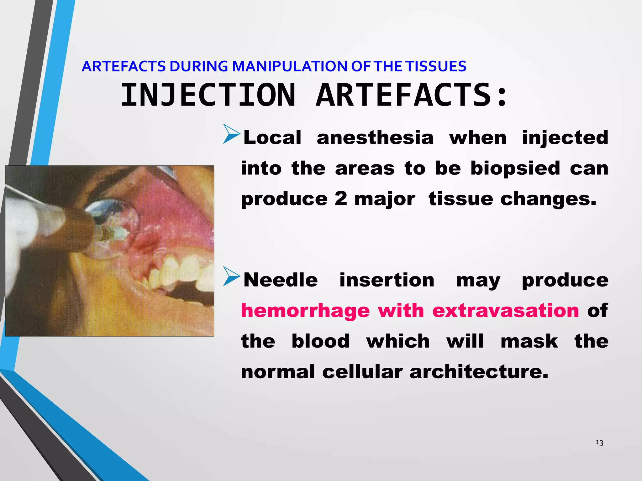
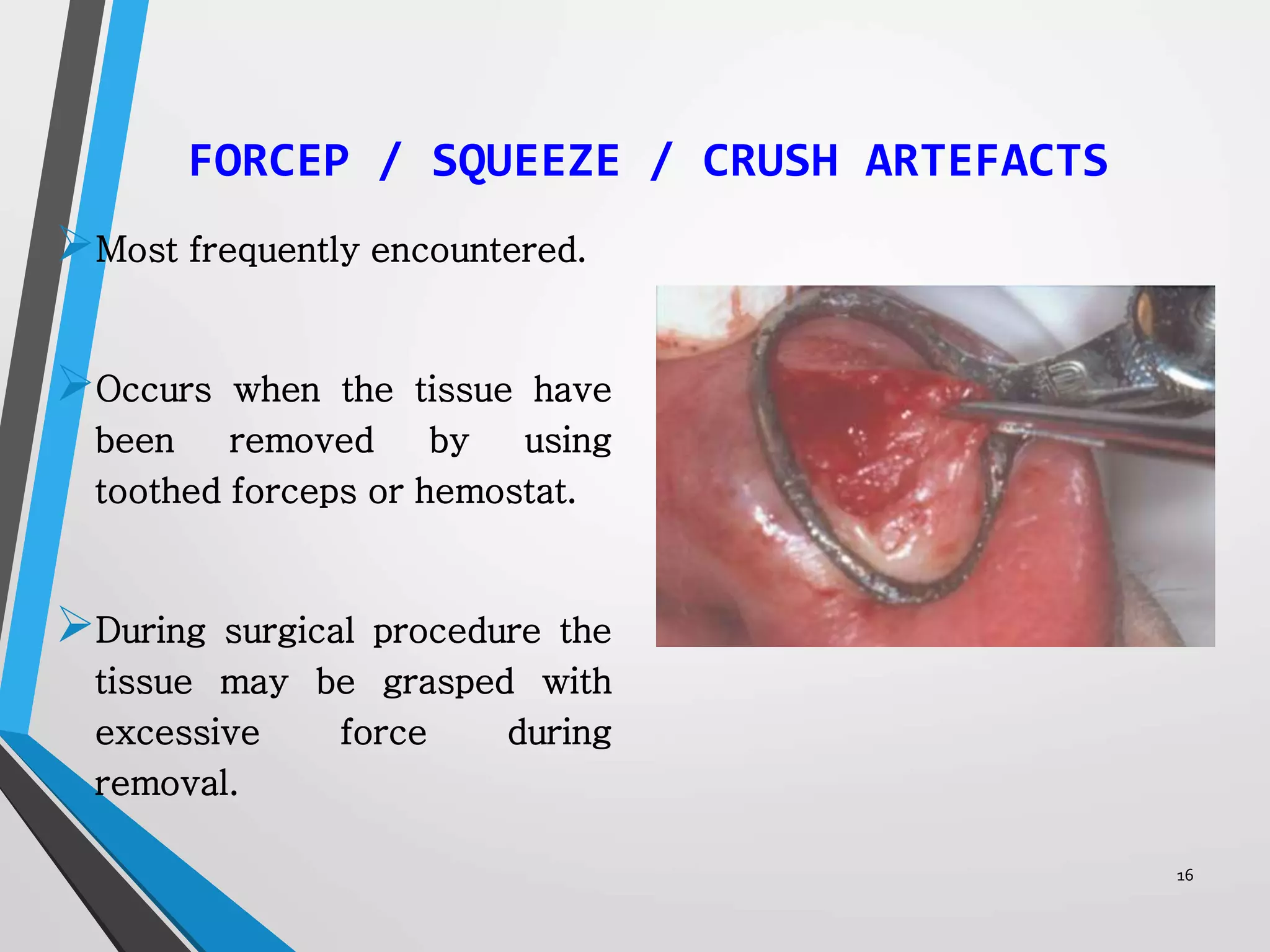
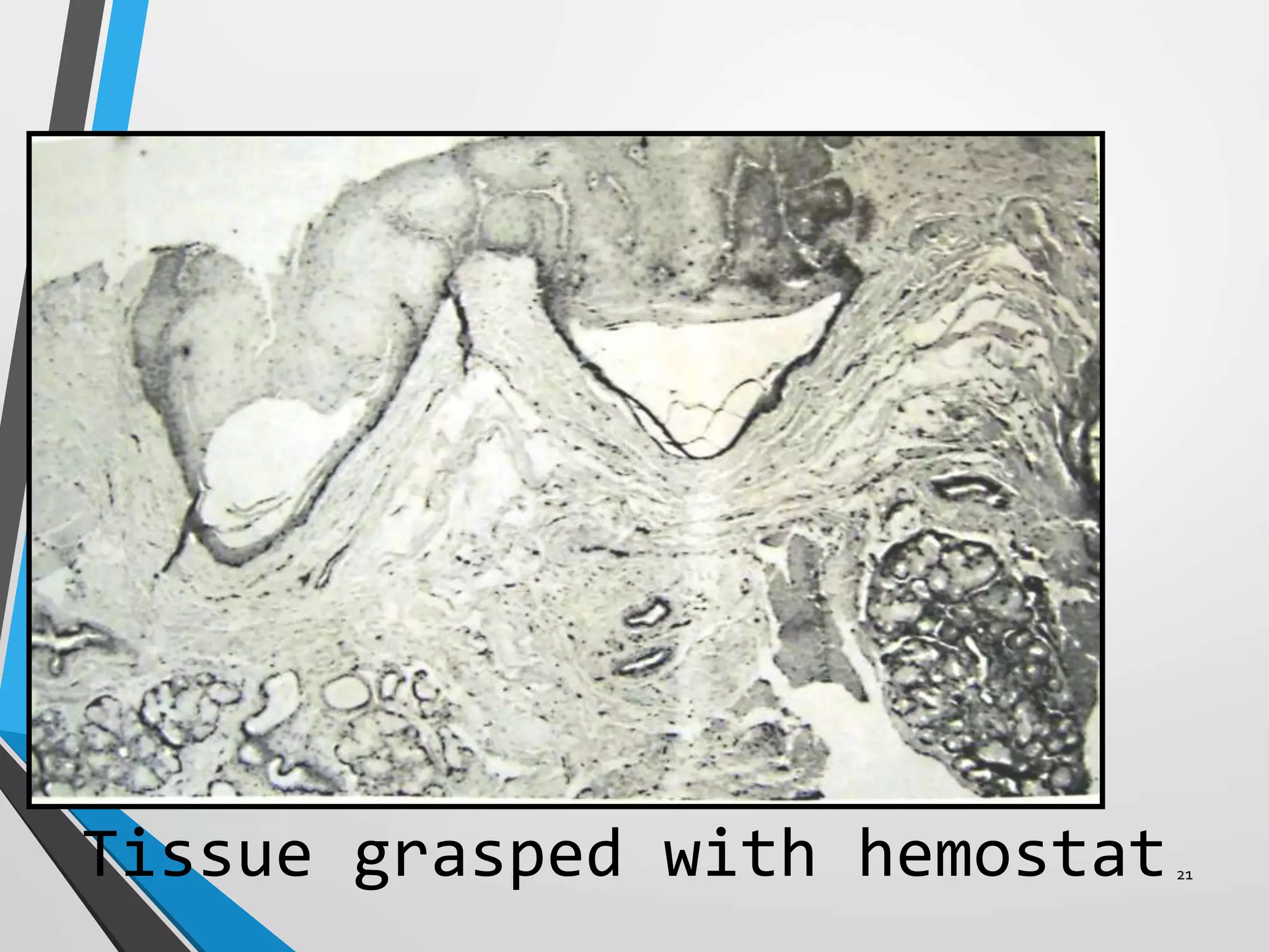
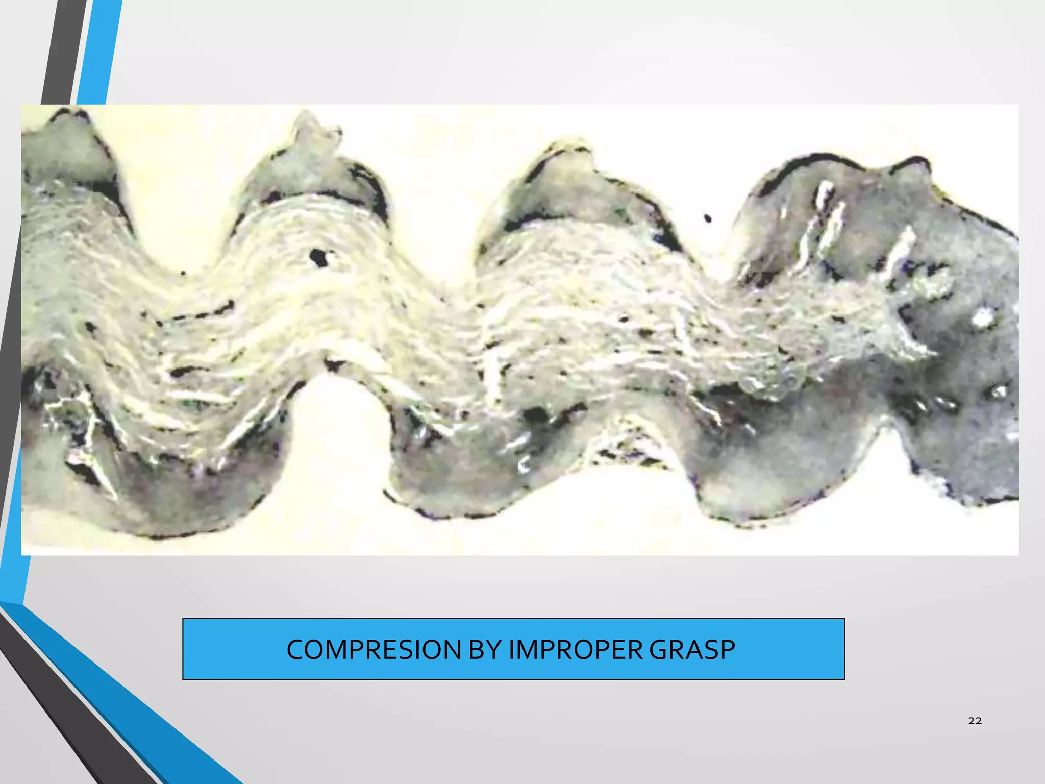
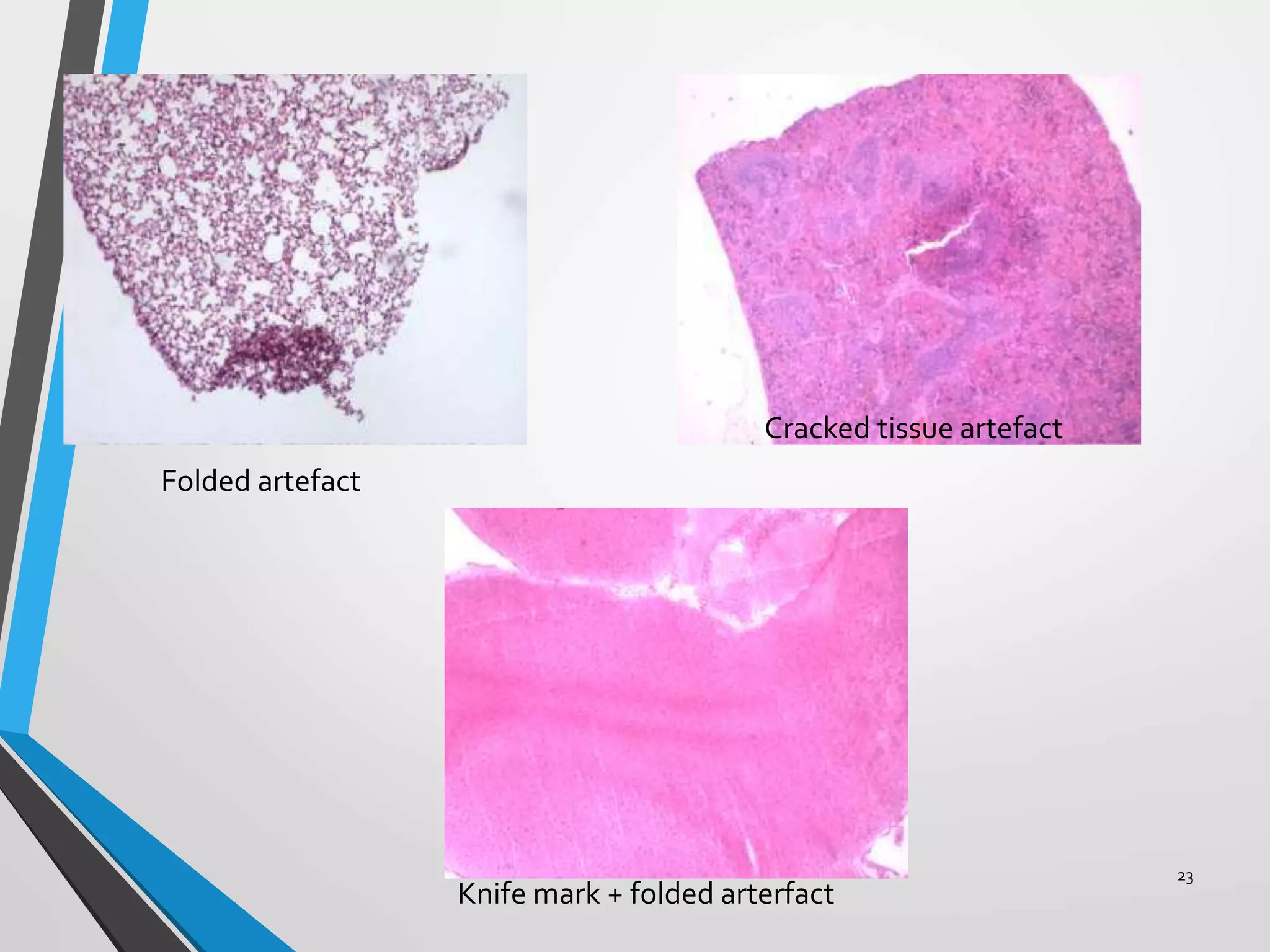
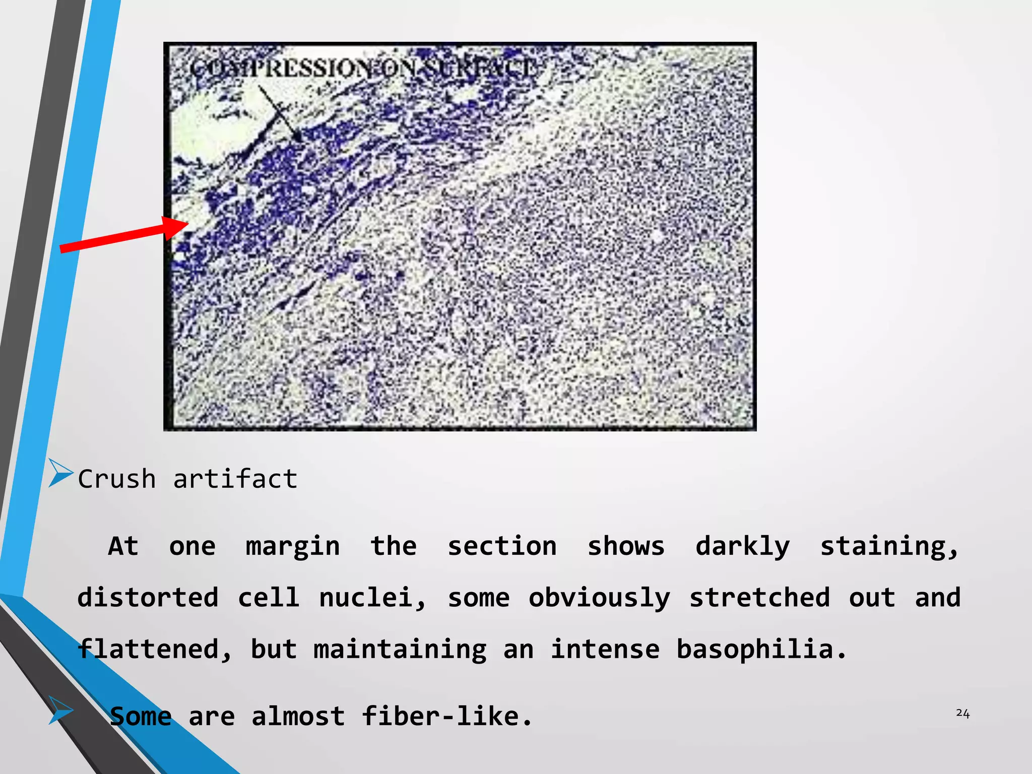
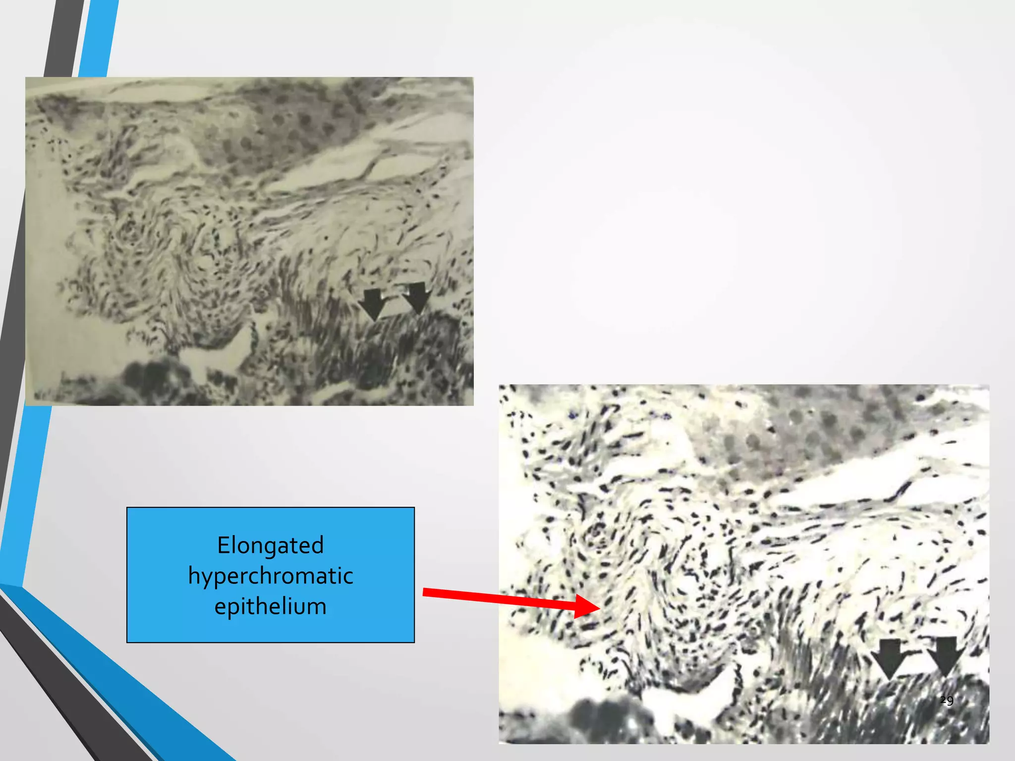
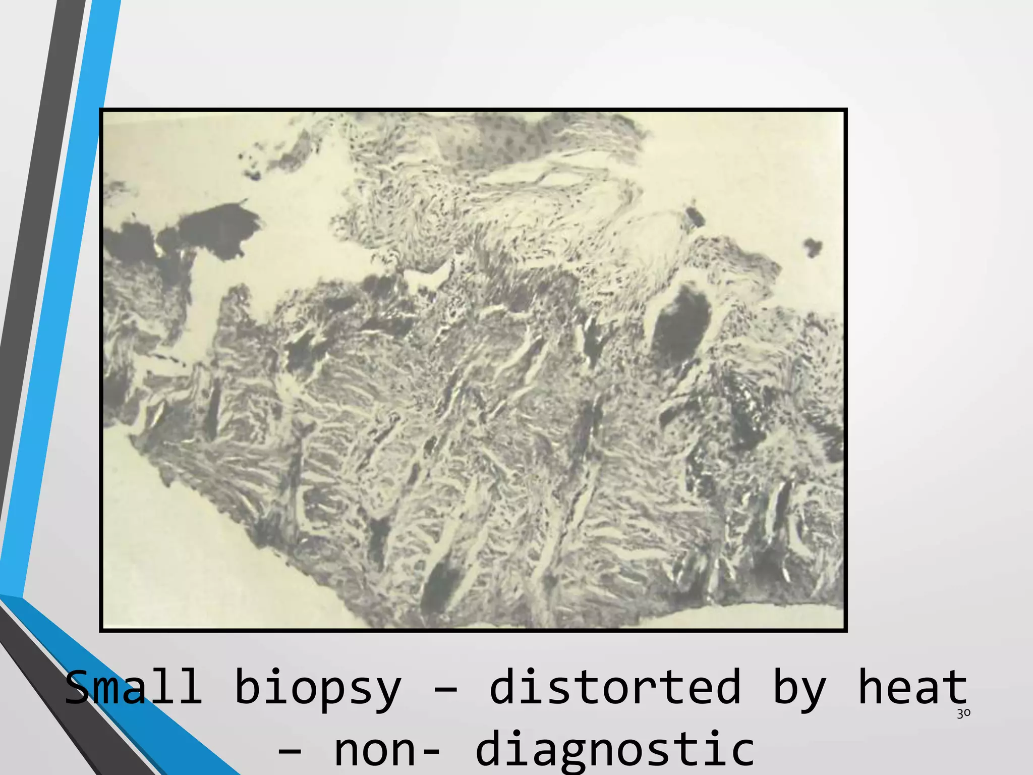
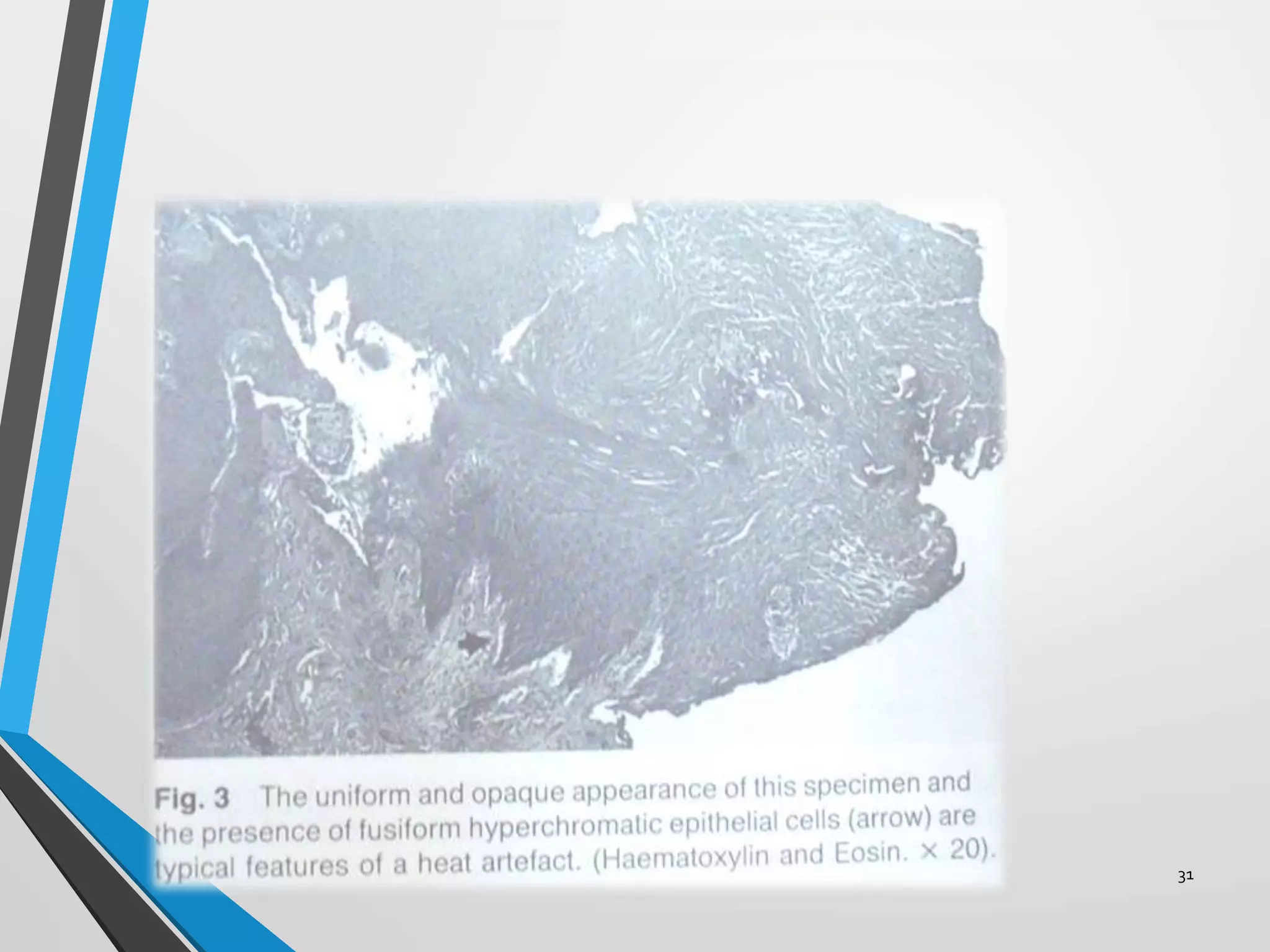
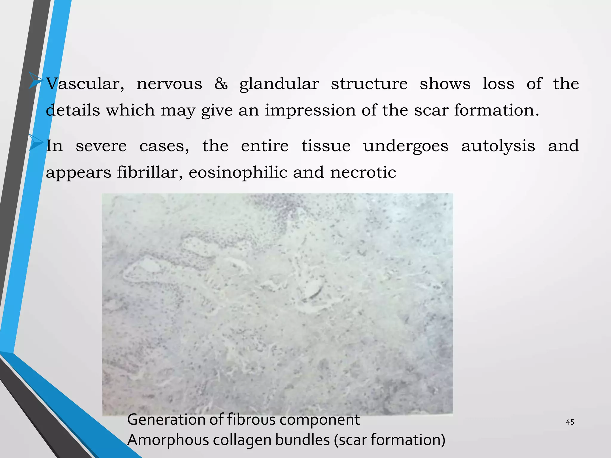
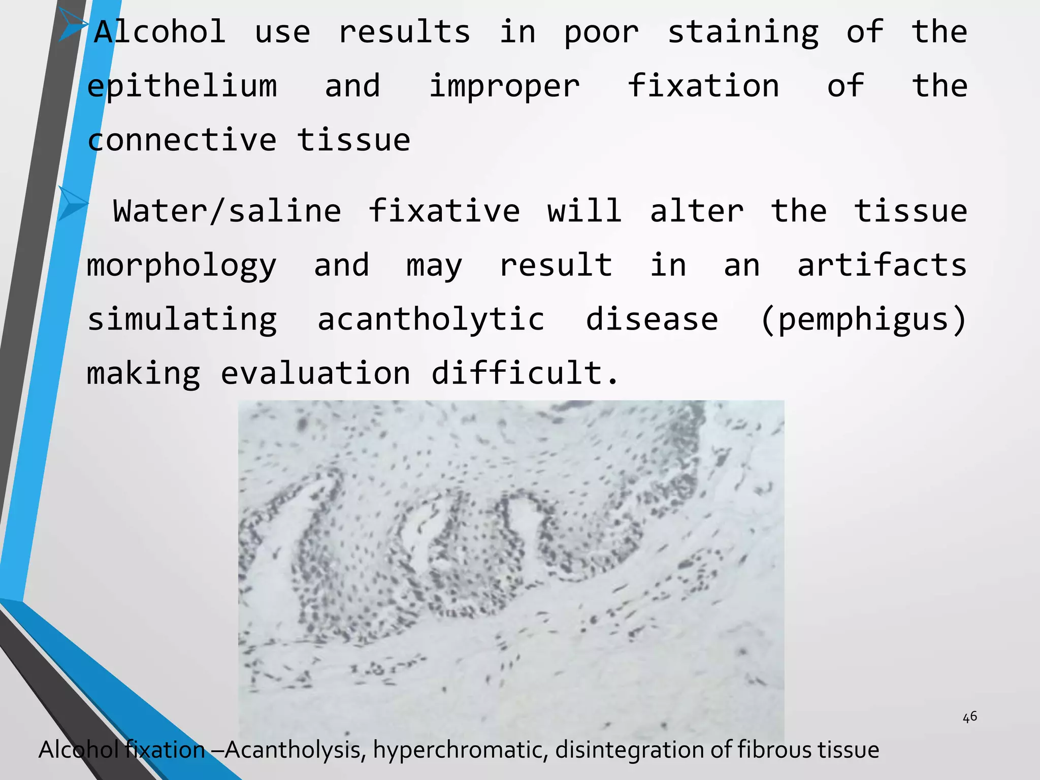
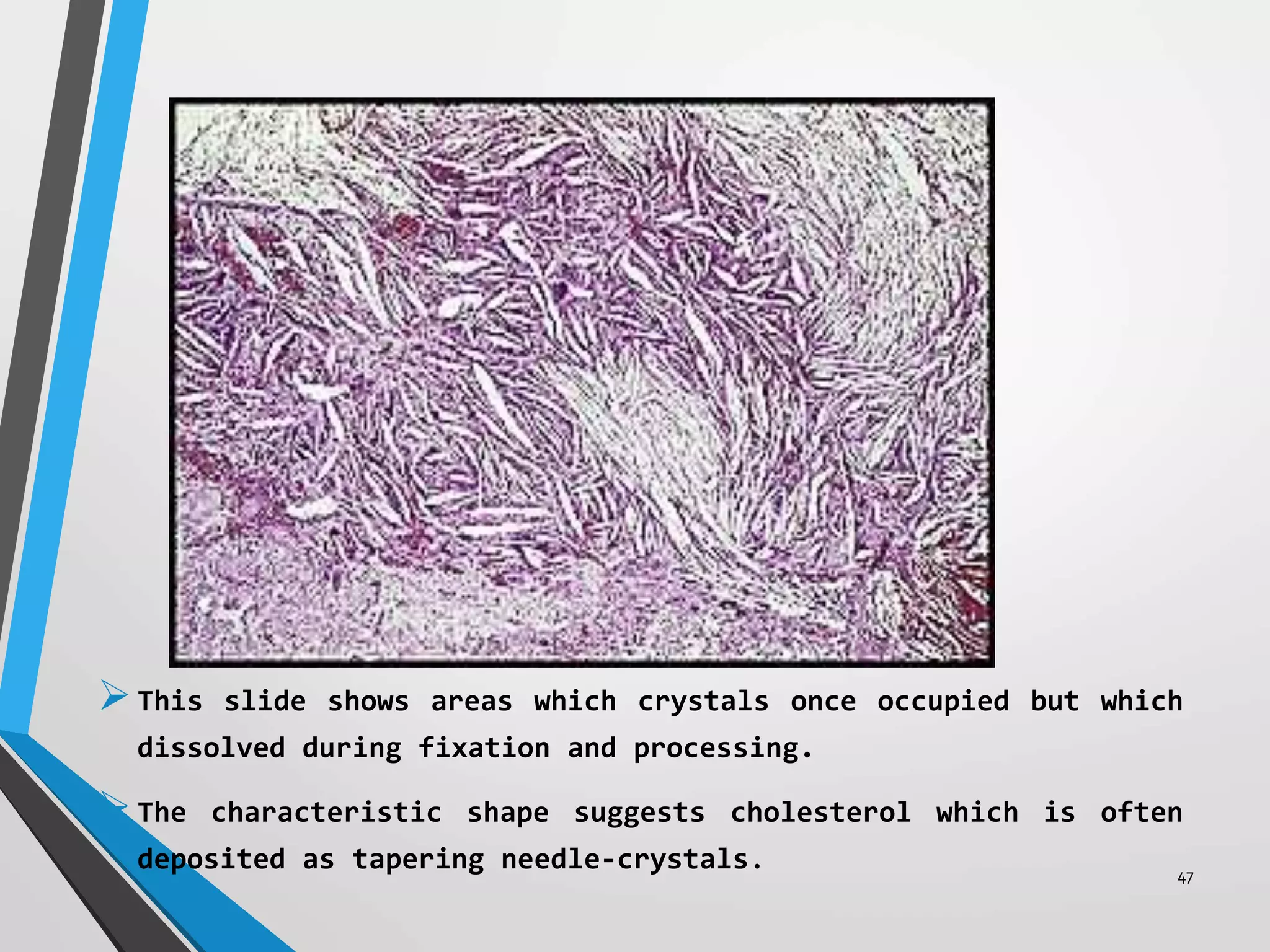
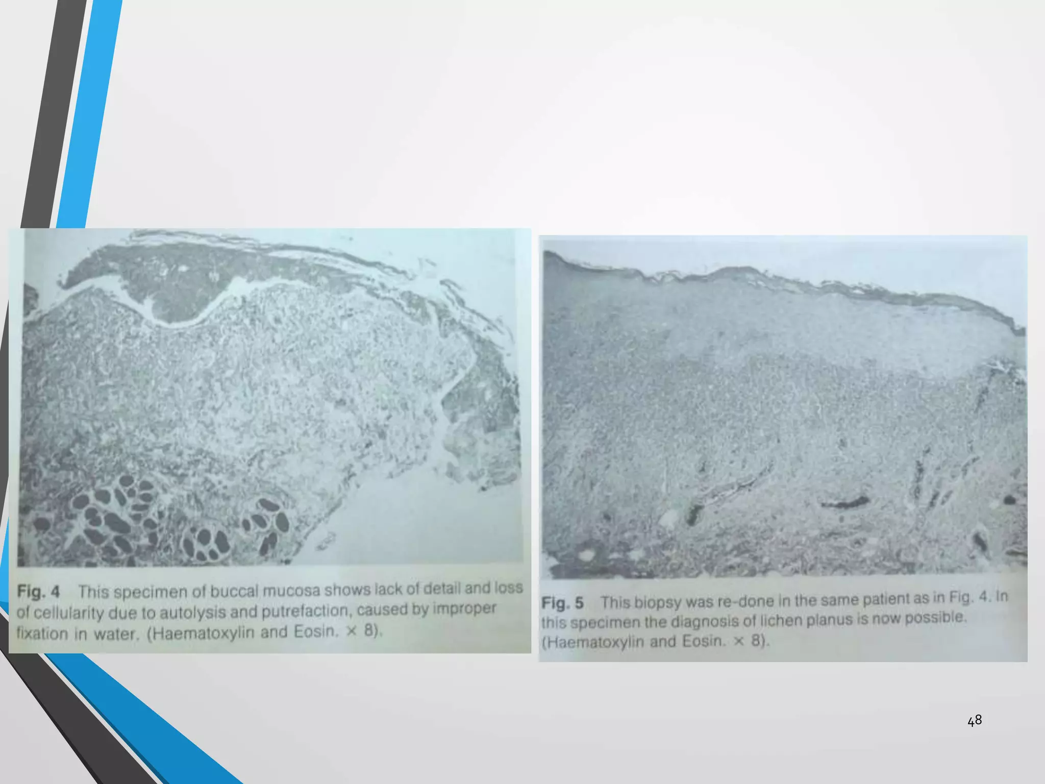
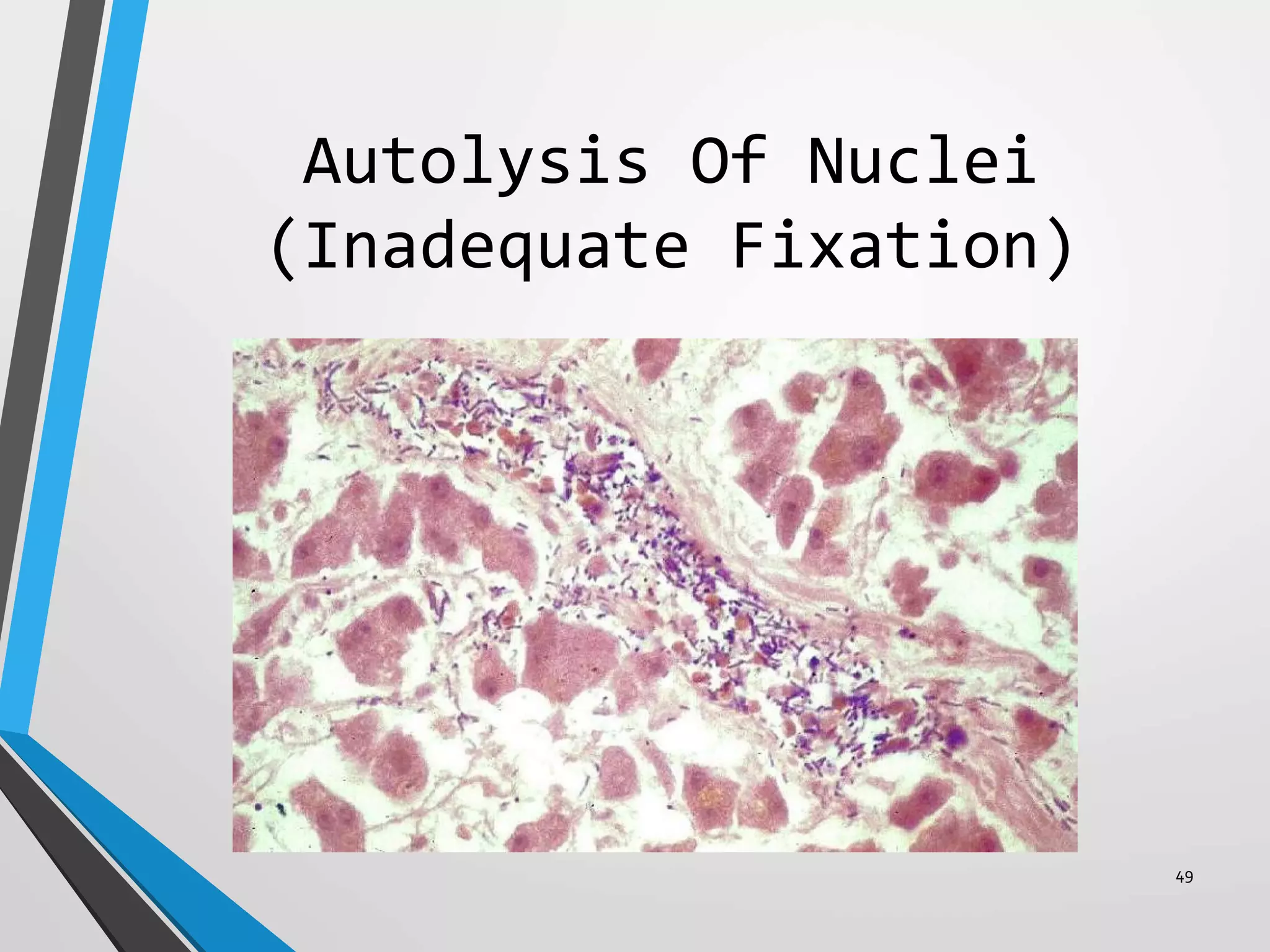
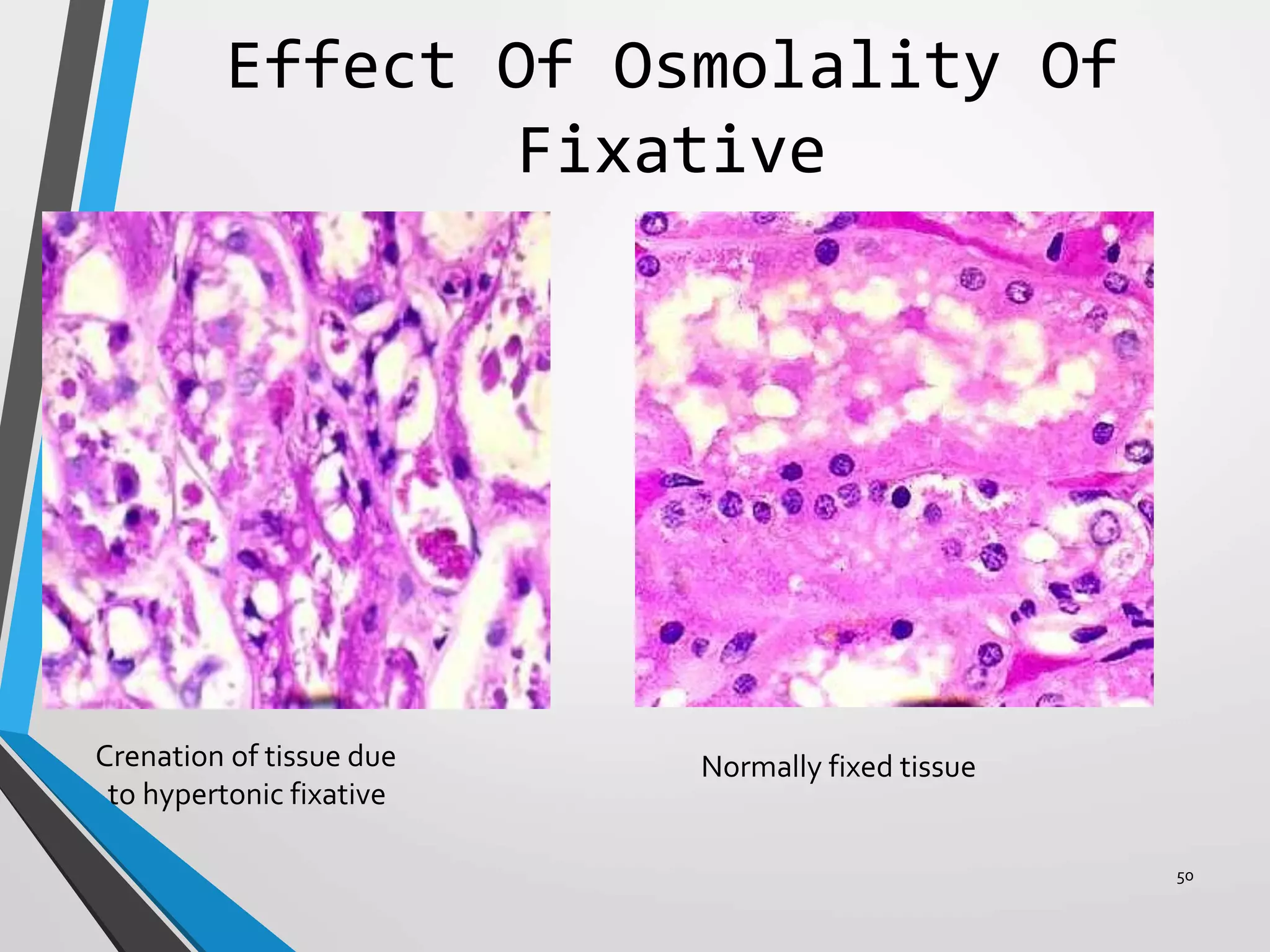
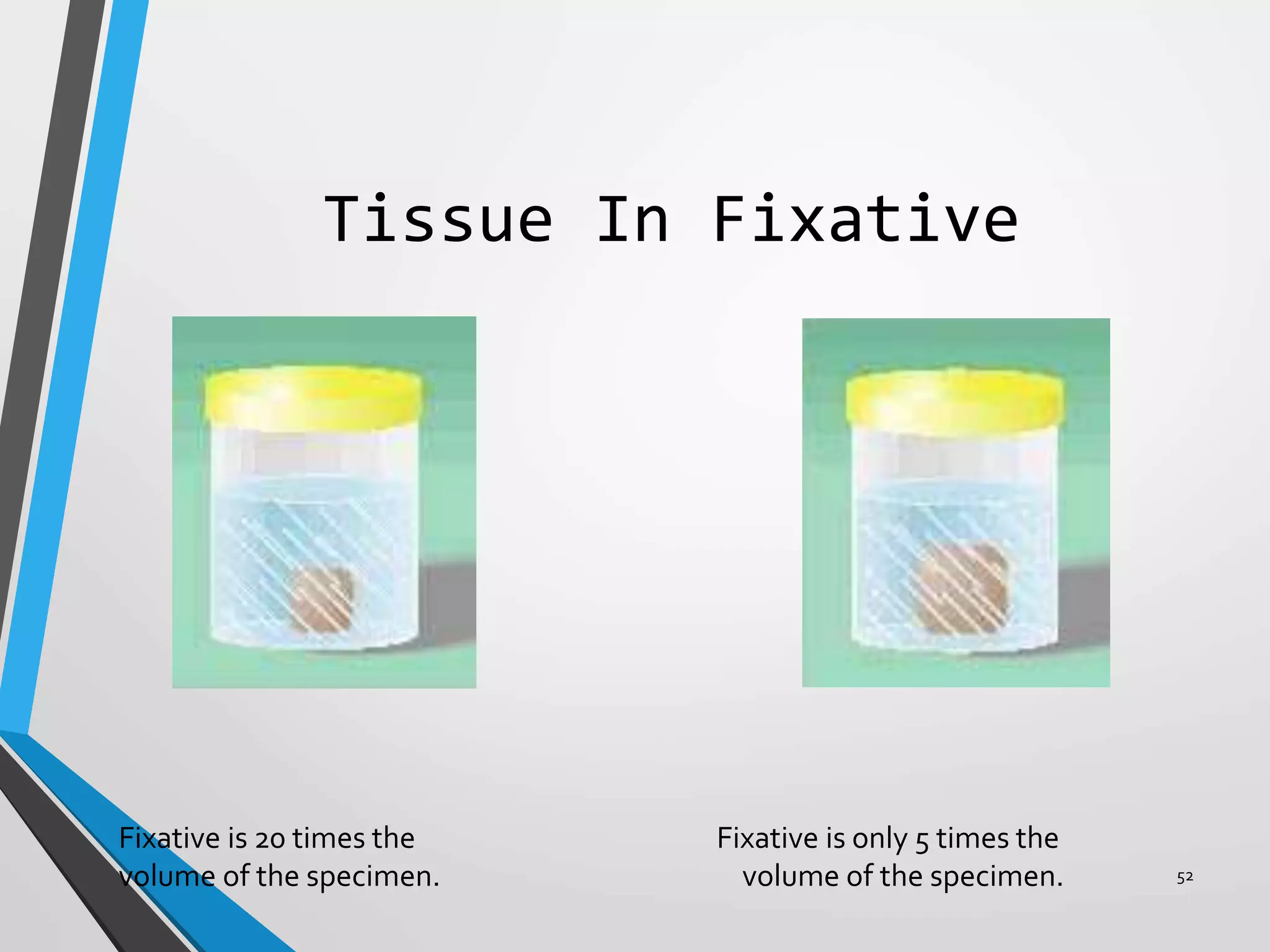
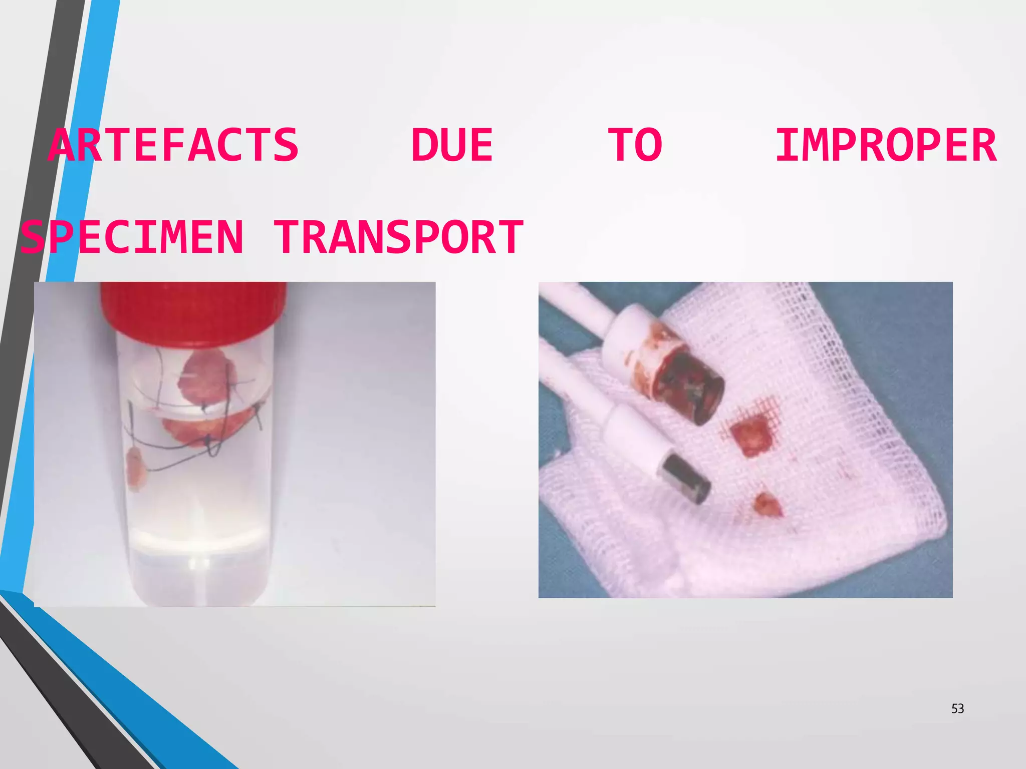
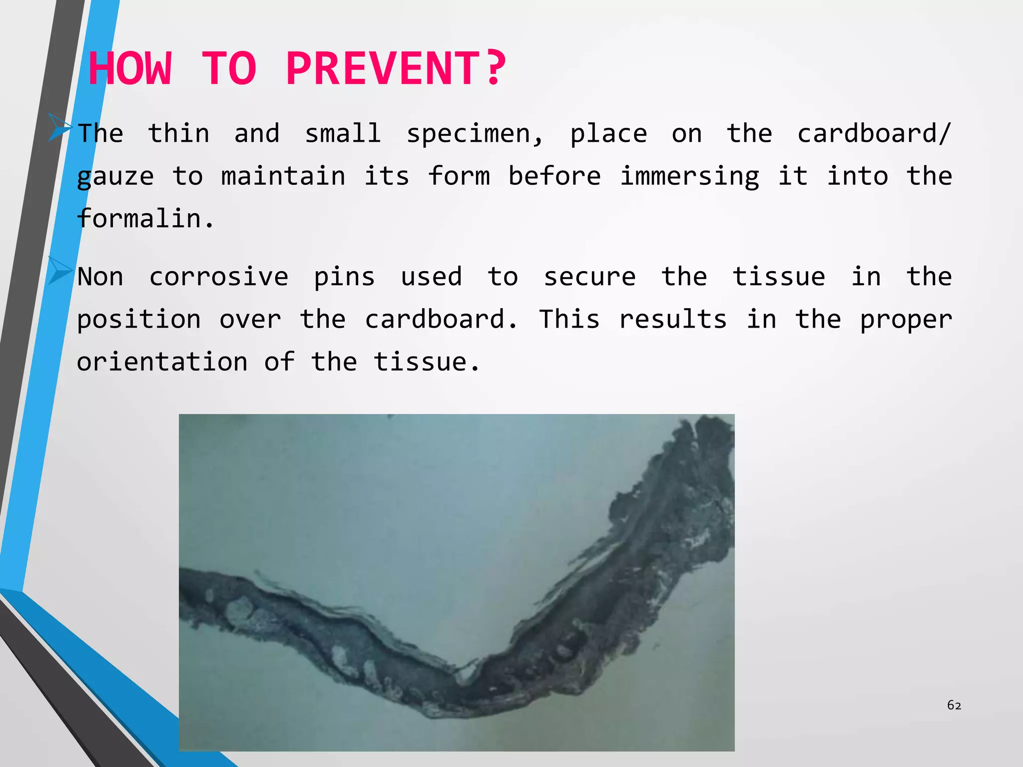
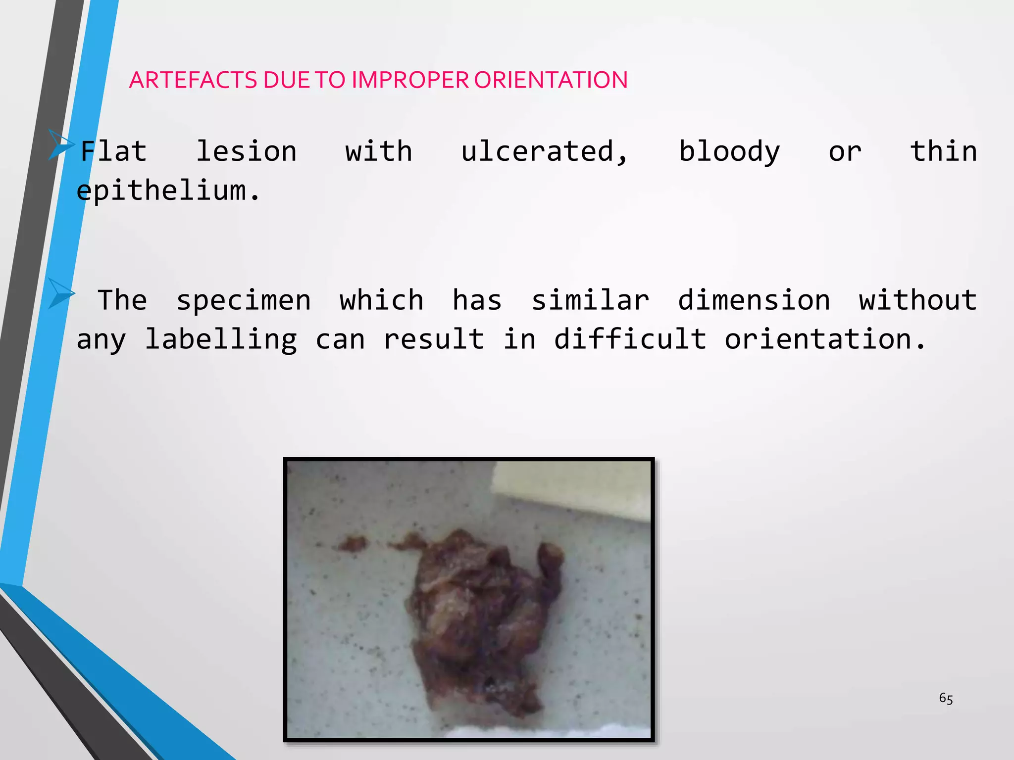
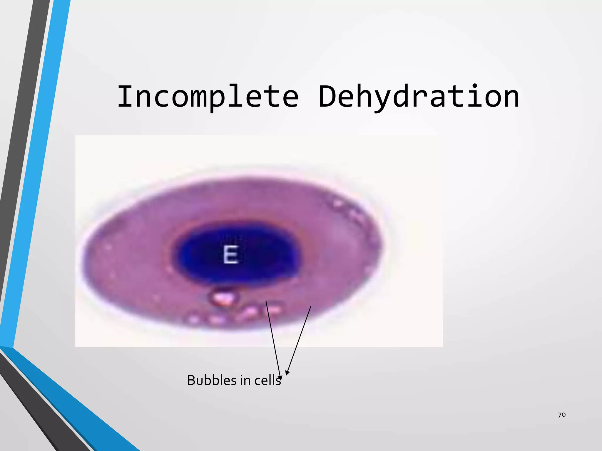
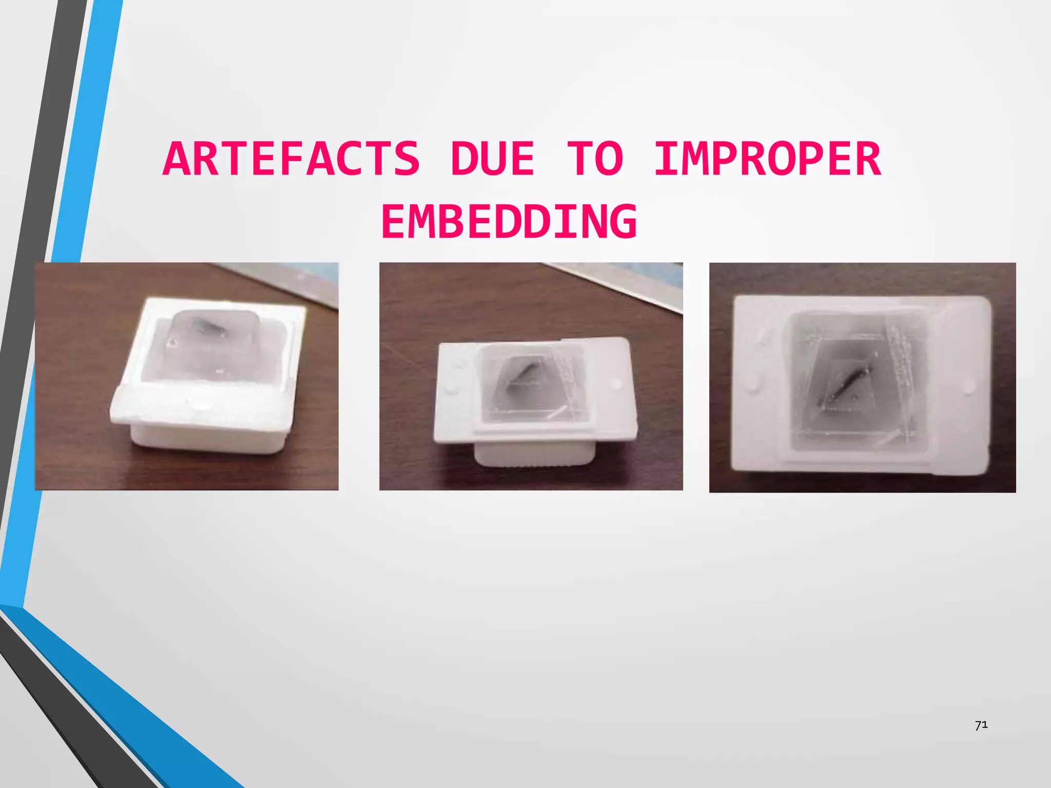
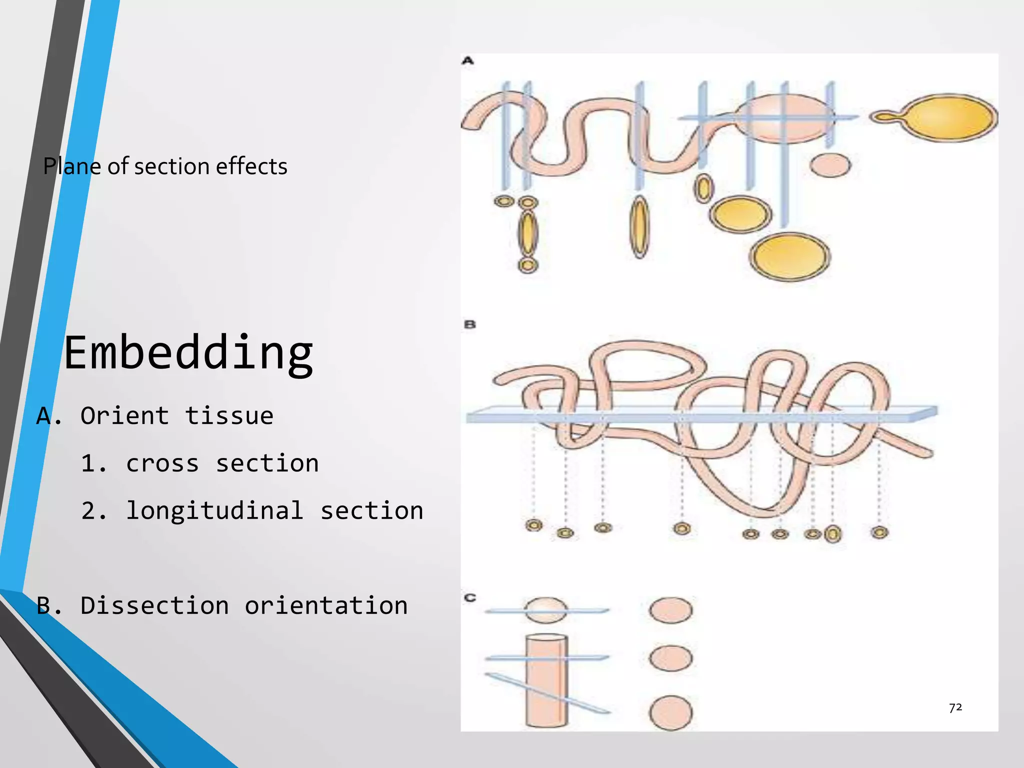
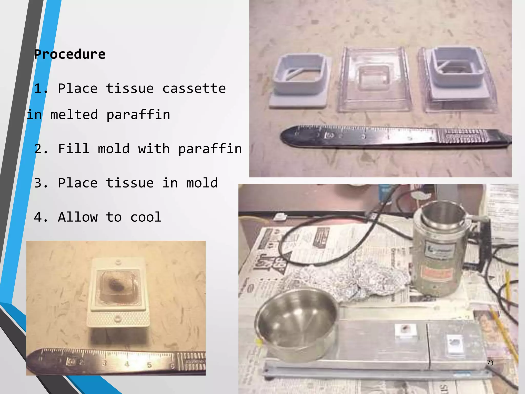
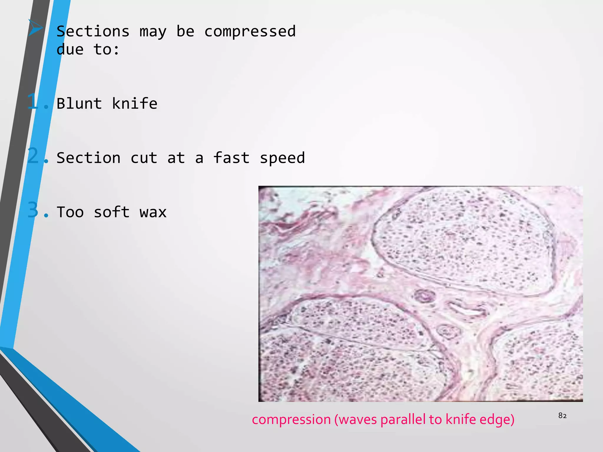
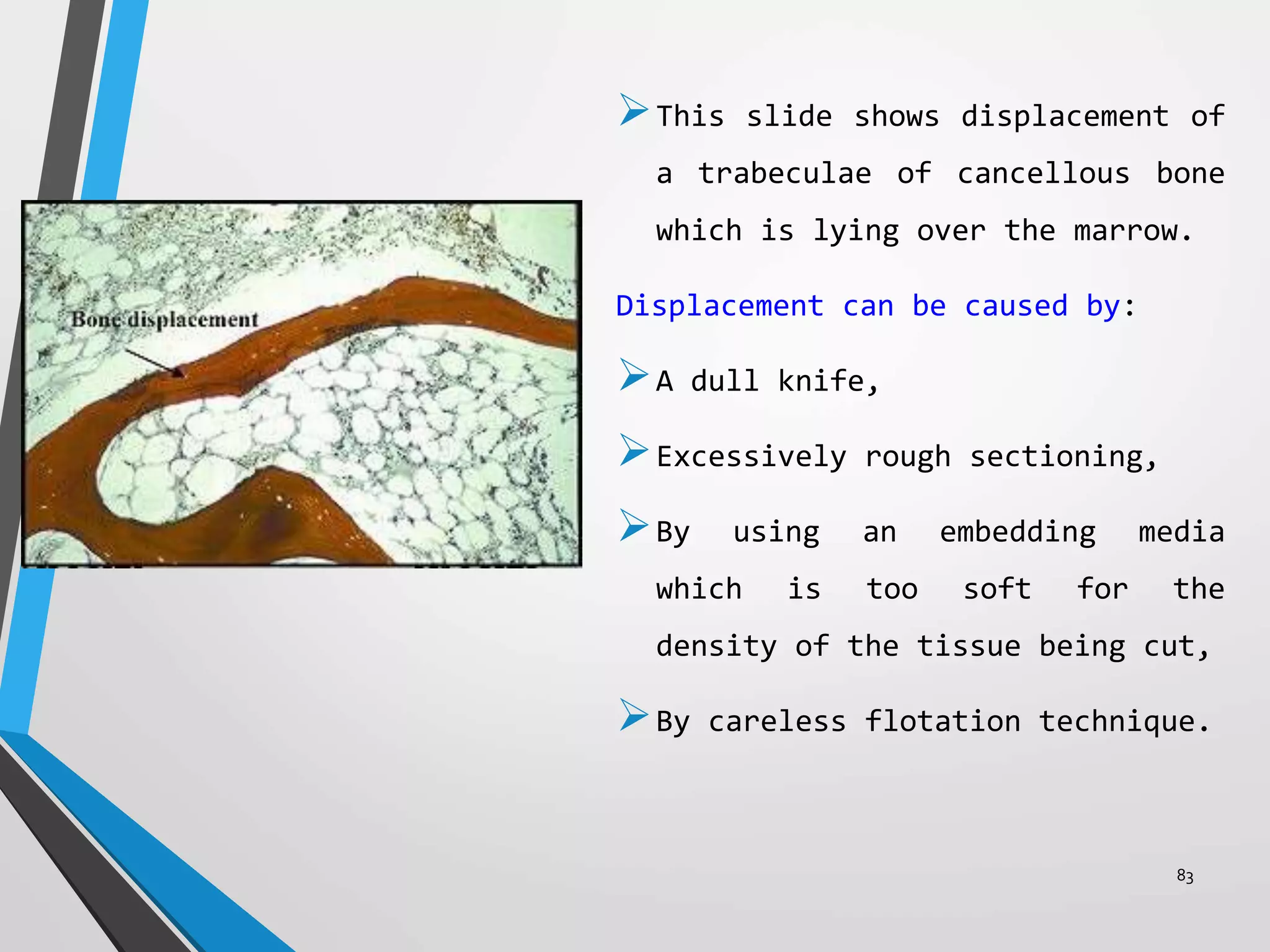
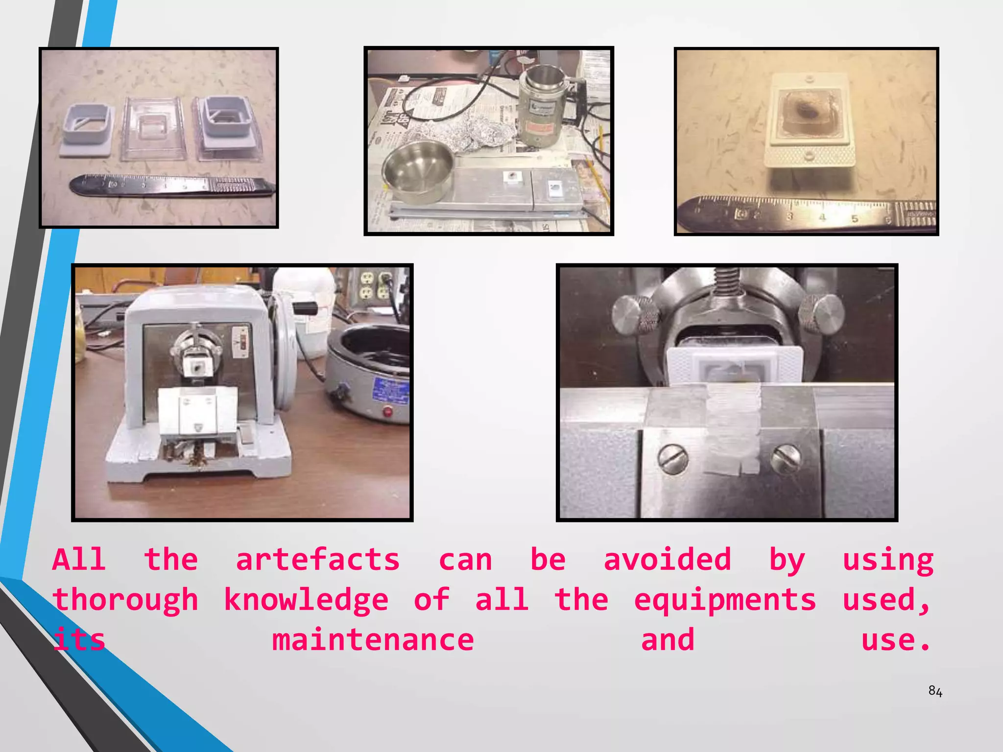
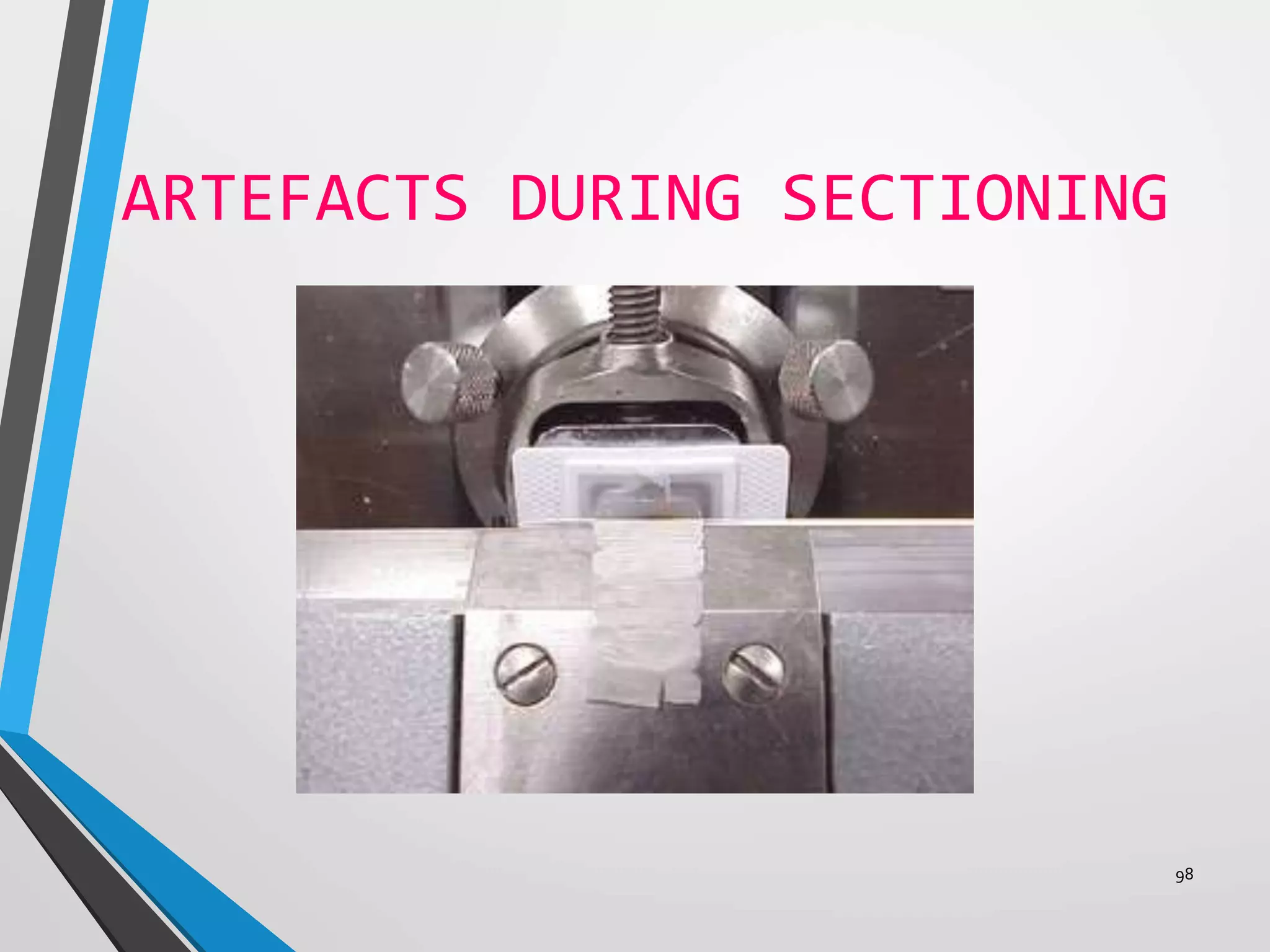
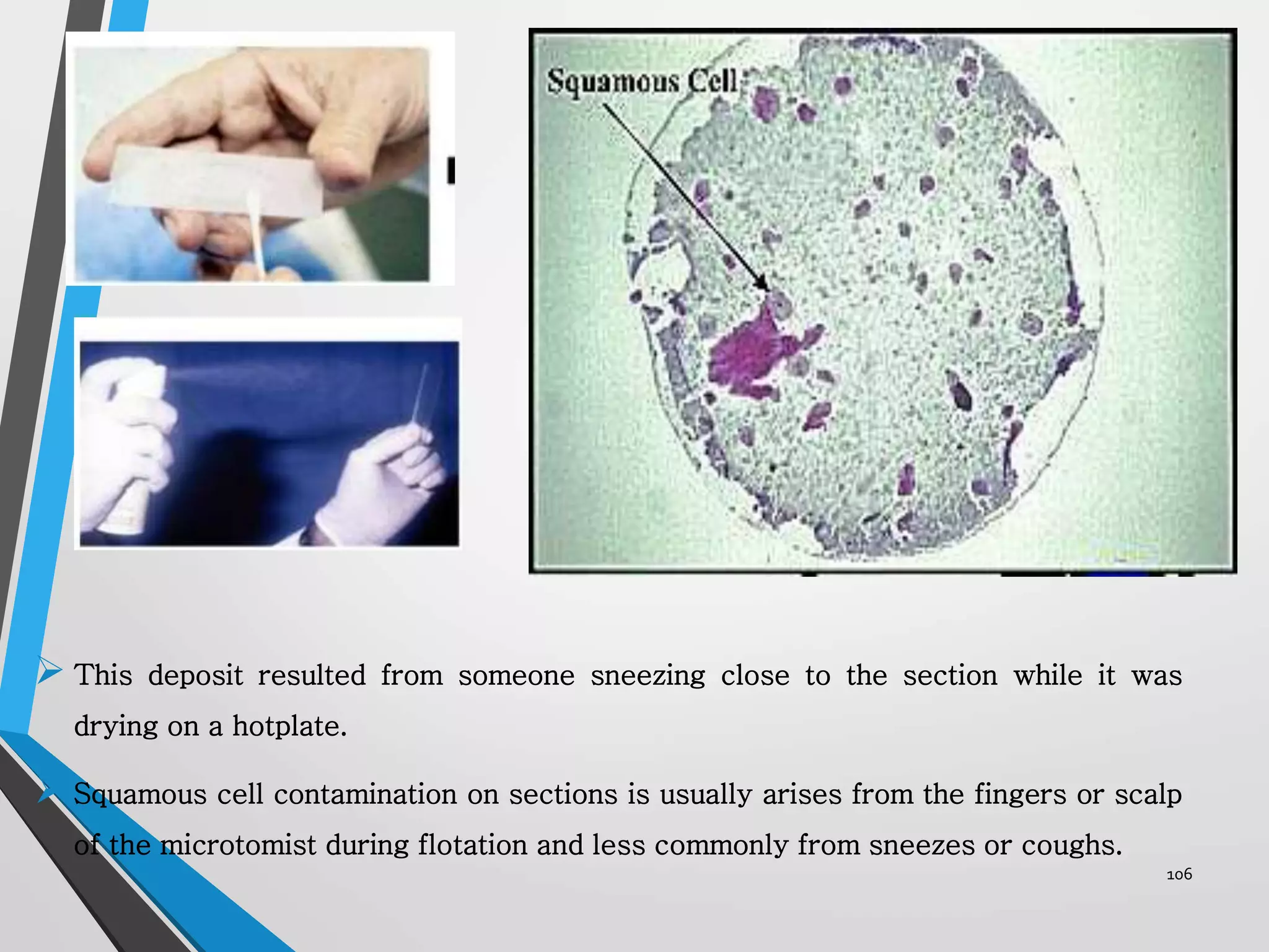
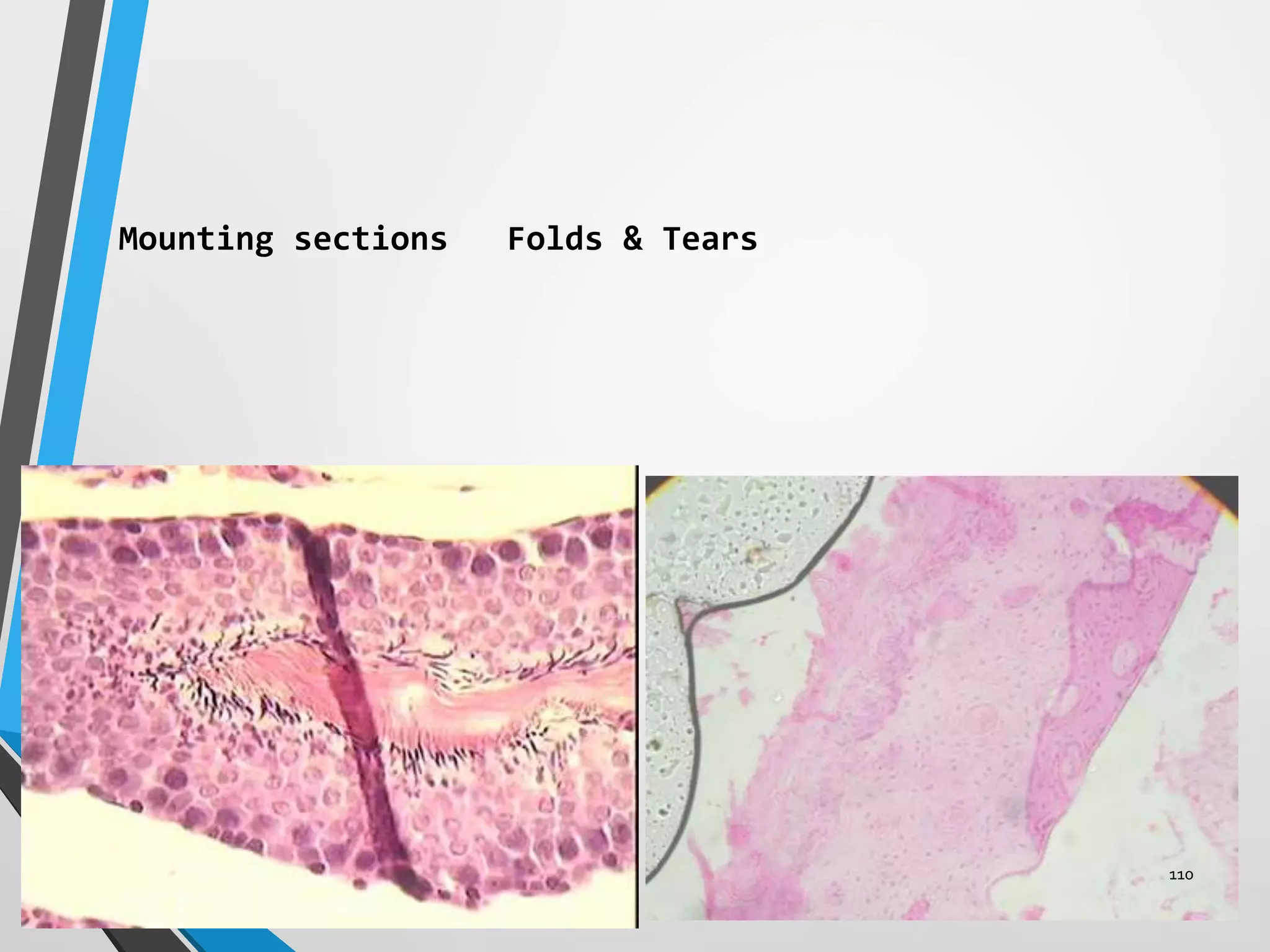
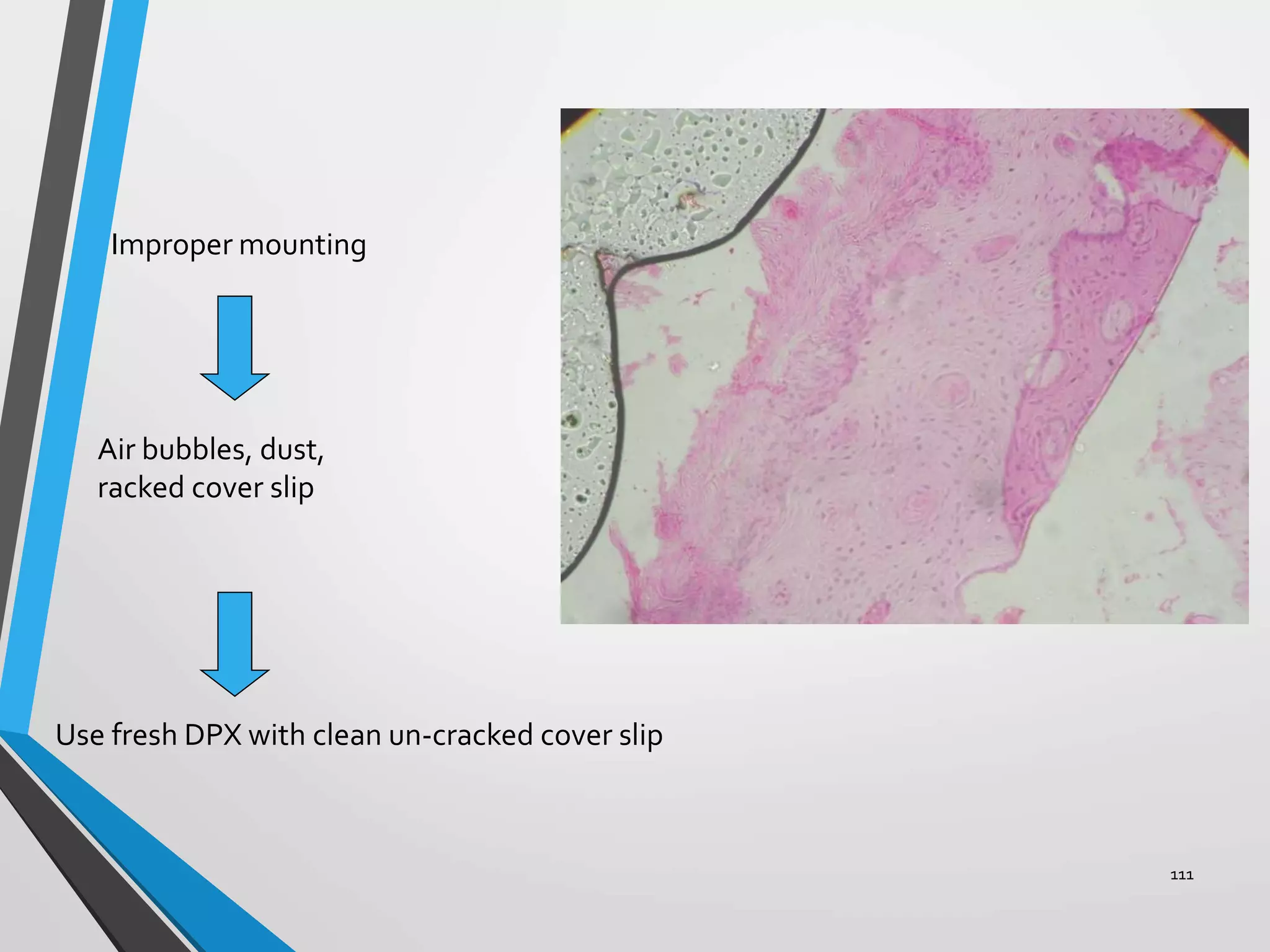
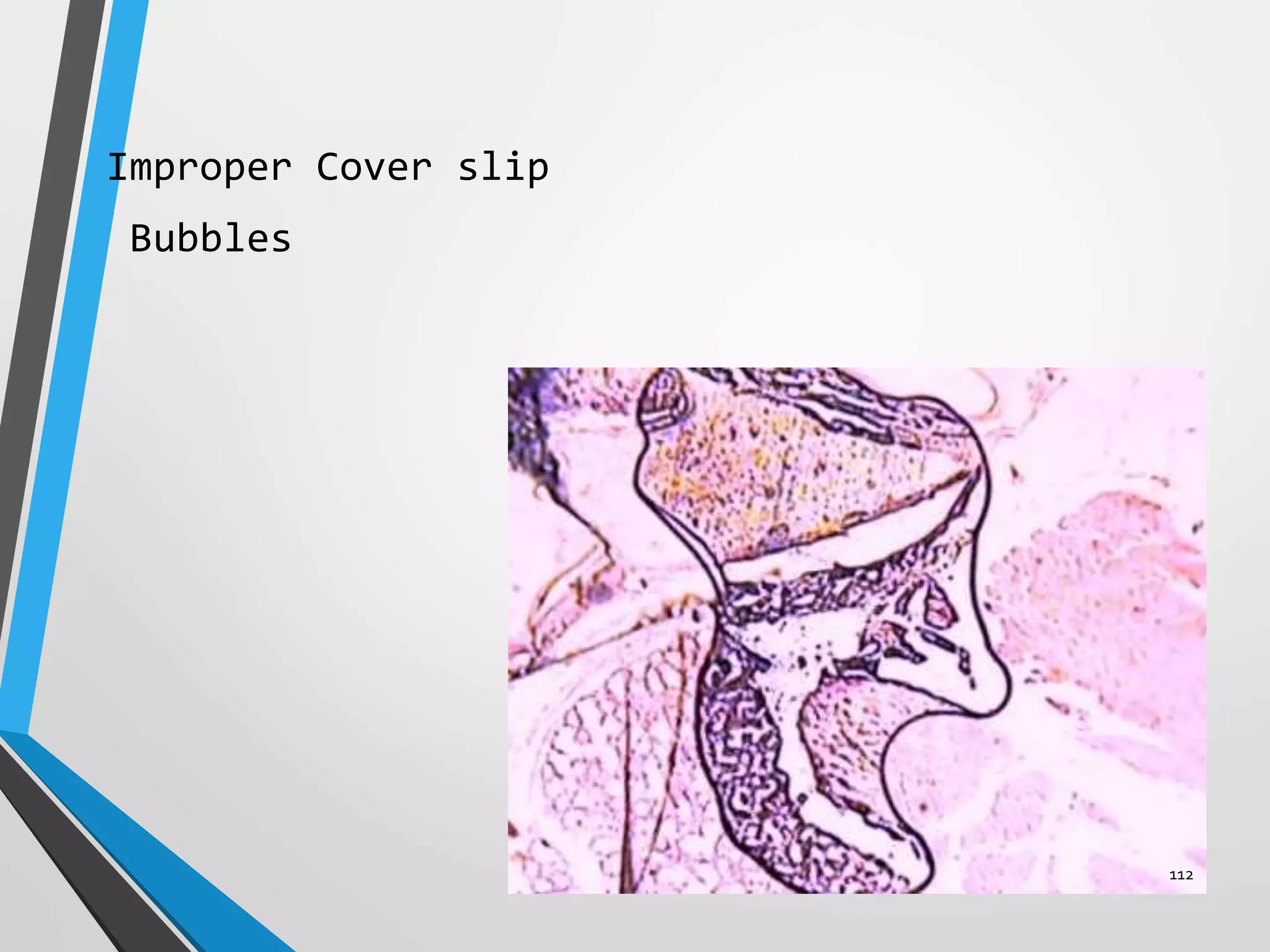
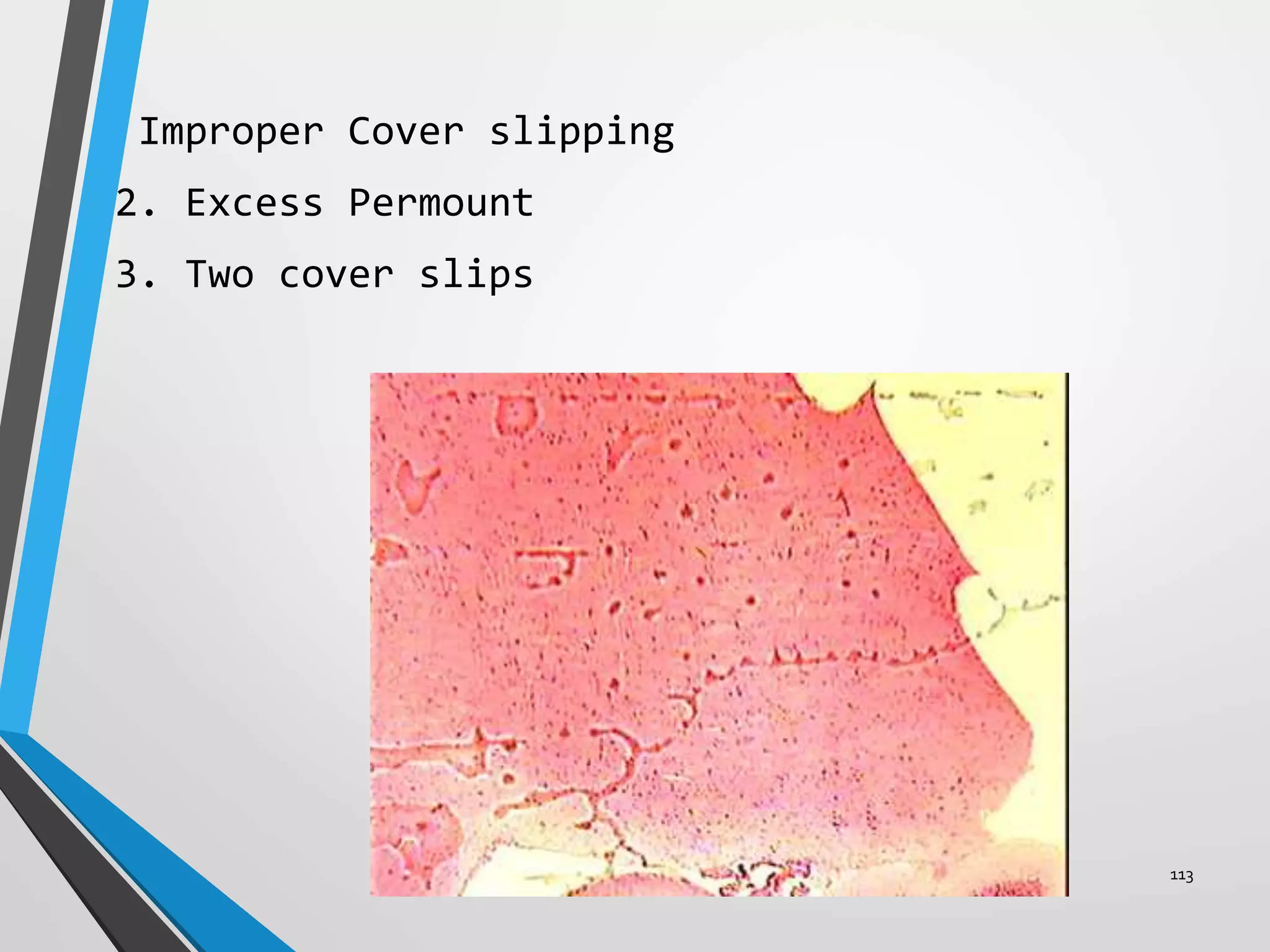
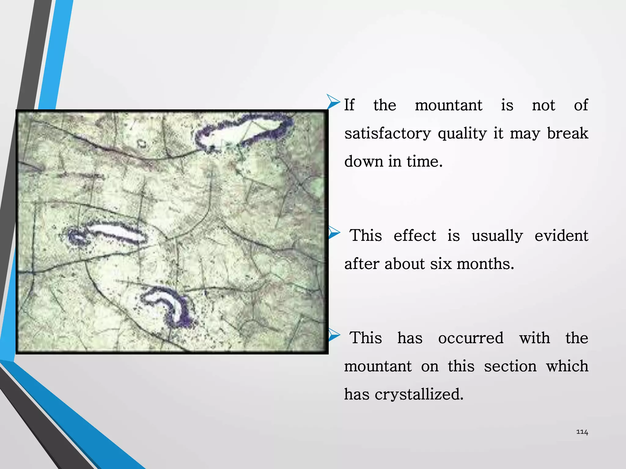
Akhil s
Post graduate student
SMIDS
1
INTRODUCTION
Biopsy of the tissue helps in formulating diagnosis
by permitting study of both the cells and their
relationship to the surrounding tissues.
Biopsy provides the information regarding the benign
and malignant nature of the tumor.
2
The specimen removed from the oral cavity
are often small, the chances of producing
artifacts are greater.
Sometimes the state of the tissue is so
severe that the tissue is useless for the
histopathological diagnosis.
3
These includes errors by the surgeon or
assistant in handling the tissue at the time of
biopsy
problems in the transport of the tissue to
laboratory & faulty tissue processing.
4
• Some artefacts are easily distinguished from
normal or pathological tissue components, and
some are difficult to distinguish from such
entities.
5
CONTENTS
DEFINITION
ARTIFACTS DURING TISSUE MANIPULATION
INJECTION
FORCEPS
FULGuRATION
IMPROPER SURGICAL REMOVAL
ARTIFACTS DUE TO IMPROPER SPECIMEN TRANSPORT
ARTIFACT DUE TO IMPROPER FIXATION
FREEZE ARTIFACTS
CURLING ARTIFACTS 6
 ARTIFACTS DUE TO IMPROPER
ORIENTATION
ARTIFACTS DUE TO
IMPROPER EMBEDDING
ARTEFACTS DUE TO
UNDECALCIFIED BODIES
ARTEFACTS DUE TO SECTIONING
FAULT
ARTEFACTS DURING
HISTOCHEMICAL PROCEDURE
ARTIFICIAL DEPOSITS
FOREIGN BODY ARTEFACTS
7
Latin. Ars- art or skill, factum- made
(any artificial product)
DEFINITION
is a defect or distortion that occurs as a result
of the way the tissue was handled right from the
time of the biopsy taking to the fixation process
including problems in the transportation of the
tissue to the lab and the histotechnical procedure
used for embedding and staining.
8
Artefacts occur at each of the following stages in
the
• During handling of the tissue
• during the fixation of tissues,
• during processing, paraffin embedding and
microtomy,
• during the mounting of tissue sections onto glass
slides,
• staining procedures and coverslipping.
9
Use of gloves: Starch powder is used as a lubricant
of surgical gloves
• Oral cytological smear & tissue sections
• Refractile, glassy, polygonal bodies (5-20microns in
diameter)
• Exhibit central dot/ Y shaped structure
• H&E – light blue
• Lugol solution – blue-black
• PAS +ve – Lilac red & diastase resistant
Artifacts by surgeons
10
Starch granules resembles atypical epithelial cells.
 Spore like structure with dark central area can resemble a
pyknotic nuclei
Polarised microscope –”Maltese cross*” configuration
EM – spherical, faceted balls (2.5 – 30microns)
*- cross where arms look like arrowheads pointing inwards/
Garden flower having brilliant red 5 parted flower. 11
• Starch also produces clinically significant
diseases (granulomatous reaction)
• It produces acute postsurgical reaction possibly
mediated by HyperSensitivity mechanisms.
12
INJECTION ARTEFACTS:
Local anesthesia when injected
into the areas to be biopsied can
produce 2 major tissue changes.
Needle insertion may produce
hemorrhage with extravasation of
the blood which will mask the
normal cellular architecture.
ARTEFACTS DURING MANIPULATION OFTHETISSUES
13
LA injected rapidly may results in separation of the
epithelium & CT or there may be separation of CT
bands with vacuolization.
Hence LA should be injected slowly away from the
area to be biopsied.
14
15
FORCEP / SQUEEZE / CRUSH ARTEFACTS
Most frequently encountered.
Occurs when the tissue have
been removed by using
toothed forceps or hemostat.
During surgical procedure the
tissue may be grasped with
excessive force during
removal.
16
When the teeth of the instruments penetrate the
tissue, results in voids/tears of the tissue along
with compression of the surrounding tissue.
The surface epithelium, may be forced through the
CT producing small pseudocyst/microcyst, which
distort the normal relationship of the tissue.
Compression of the tissue results in loss of
cytological detail with rupture of the nuclear and
cell membrane.
17
Excessive force with forceps – voids 18
Squeeze artifacts - pseudo cyst 19
Incorrect use of forceps -
pseudocysts 20
Tissue grasped with hemostat21
COMPRESION BY IMPROPER GRASP
22
Folded artefact
Cracked tissue artefact
Knife mark + folded arterfact
23
Crush artifact
At one margin the section shows darkly staining,
distorted cell nuclei, some obviously stretched out and
flattened, but maintaining an intense basophilia.
 Some are almost fiber-like. 24
Generally these squeeze artifacts are easily
recognized and there is no problem in,
arriving at proper diagnosis but in the
biopsies of the epithelial neoplasm there may
be problem in accurate diagnosis.
The problems can be avoided by handling the
tissue with a suture during incision.
25
The tissue forceps should be used gently for
handling the specimen.
If the use of the tissue forceps can’t be
avoided, Care must be taken to provide
adequate retraction and visualization of the
area
26
FULGURATION / HEAT ARTEFACTS
Electrosurgery has an advantage of producing
hemostasis at the biopsy site by coagulating
the severed blood vessel, this leads to a
profound artifactual alteration.
The heat produced may alter both the
epithelium and the connective tissue.
Microscopically: A broad band of basophilic
coagulum along the surgical margins giving
amorphous appearance.
27
The epithelium may have an appearance of the
detached surface and the nuclei assume a spindle
palisading configuration.
Separation of the epithelium from the basement
membrane.
Hard tissue biopsy should be removed with chisel
and not with surgical bur to avoid generation of
the heat which will damage the biopsy specimen.
28
Elongated
hyperchromatic
epithelium
29
Small biopsy – distorted by heat
– non- diagnostic
30
31
Hence the use of the electrosurgical procedure should not be
encouraged.
 If at all done it should be limited to the larger specimen as the
artifacts can obscure the details in the smaller specimen.
Care should be, taken to use the cutting and not the coagulation
electrode, so that the low current will be produced that will
allow cutting & liberation of the specimen.
32
The margin of the incision should, be far
away from the interface of the lesion and
normal tissue to prevent thermal changes in
that area.
Accidental contact of cutting tip with the
metal instrument used to hold the specimen
should be avoided as this will also cause
tissue changes.
33
Electrosurgery, when used in combination with the
scalpel procedures better results were seen.
The scalpel is used for the initial incision
around the lesion to be biopsied and the
electrosurgery is used to complete the removal of
the specimen.
This method has an advantage of producing superior
hemostasis and also reduces the amount of the
heat, to which the specimen is exposed.
34
Ideally single electrosurgery incision should
be made with a thin electrode at a speed of
7mm/sec and the successive incision should be
separated by cooling intervals of 8- 10 sec
avoid generation of the heat sufficient to
initiate an adverse affect.
35
ARTEFACTS DURING IMPROPER
SURGICAL REMOVAL
Biopsy specimen if shallow and minute, correct evaluation of
the epithelium & its interrelationship with the underlying
connective tissue is impossible.
36
37
Prevention:
Deeper elliptical incisions should be used
involving adjacent normal mucosa.
The clinician should know the history and
appearance, the basic type of the lesion with
which he is dealing and how the dimensions of
the specimen should be modified for each
clinical situation.
38
Example:
1. Pemphigus : A longer, broader and
shallow surface specimen should be taken
rather than deep, narrow and short specimen
2. SCC: A deeper incision should be taken
39
• Artifacts due to fixation occurs mainly due to:
Improper fixation
Delay in fixation
Use of inadequate volume of fixative
Use of inappropriate solution
Inadequate fixation time
ARTEFACTS DURING FIXATION
40
Adequate Fixation
41
Delay in fixation & inadequate
fixation time produces similar
changes
The specimen will lack the detail and loss of
cellularity due to autolysis.
The staining quality of the cells is altered
and the cell cytoplasm and nuclear structure
appears completely indistinct.
42
Fixation delayed 30 min – false hyperchromasia
Poor detail of fibroblast nuclei
Excellent indication - loss of detail of RBC
43
The cell appears shrunken showing clumping of the cytoplasm with
vacuole formation and there may be lysis of the cell membrane.
The nucleoli may not be seen.
SG interstitium – opaque & acellular
nuclei of individual units is not visible
44
Vascular, nervous & glandular structure shows loss of the
details which may give an impression of the scar formation.
In severe cases, the entire tissue undergoes autolysis and
appears fibrillar, eosinophilic and necrotic
Generation of fibrous component
Amorphous collagen bundles (scar formation)
45
Alcohol use results in poor staining of the
epithelium and improper fixation of the
connective tissue
 Water/saline fixative will alter the tissue
morphology and may result in an artifacts
simulating acantholytic disease (pemphigus)
making evaluation difficult.
Alcohol fixation –Acantholysis, hyperchromatic, disintegration of fibrous tissue
46
This slide shows areas which crystals once occupied but which
dissolved during fixation and processing.
The characteristic shape suggests cholesterol which is often
deposited as tapering needle-crystals. 47
48
Autolysis Of Nuclei
(Inadequate Fixation)
49
Effect Of Osmolality Of
Fixative
Crenation of tissue due
to hypertonic fixative
Normally fixed tissue
50
• To avoid fixation artifacts, specimen should
be immediately fixed after removal in 10%
formalin and the volume should be
approximately 20 times the volume of the
specimen & should be kept overnight.
51
Tissue In Fixative
Fixative is only 5 times the
volume of the specimen.
Fixative is 20 times the
volume of the specimen. 52
ARTEFACTS DUE TO IMPROPER
SPECIMEN TRANSPORT
53
Leakage or evaporation of the
formalin can results if the container
is not sealed properly.
 If such tissue is again rehydrated
and placed in other container results
in an artifacts resembling
acantholysis.
54
FREEZE ARTIFACTS
Occurs if the tissue is frozen prior to the
fixation.
Specimen received through mail during the month
of winter results in freeze artifacts.
Freeze temp, of the aqueous formalin is 12.2o F &
hence the temp in the winter season will be
sufficiently cool to freeze the formalin.
55
Biopsy which are left overnight in the mail
box.
The water within the tissue may be freezed and
form ice crystals.
The processed freeze artifacts is characterized
by the formation of interstitial vacuoles and
also within the cytoplasm mainly in the areas
of the ice crystals. 56
This slide shows ice-crystal artifact which appears as
intercellular clefts in highly cellular tissues and as
intracellular clefts and vacuoles in skeletal muscle.
57
So during the transportation of the tissue the freezing
should be avoided
Use the container which provides insulation from the
extreme cold.
58
The tissue sometimes may twist by its own and get fixed in
this unnatural position, after being excised.
Particularly seen in the smaller or the thin specimen such
as in case of delicate strip of the oral mucosa or the
gingival specimen.
 Formalin fixation of the tissue causes shrinkage and
curling which results in difficultly in the orientation of
the tissue during the process of embedding.
CURLINGARTEFACTS
59
60
61
HOW TO PREVENT?
The thin and small specimen, place on the cardboard/
gauze to maintain its form before immersing it into the
formalin.
Non corrosive pins used to secure the tissue in the
position over the cardboard. This results in the proper
orientation of the tissue.
62
Curling is less problematic when the lesion have
relatively thick keratotic surface.
The keratin layer serve as a back bone enabling the
specimen to retain the shape during the fixation
63
Avoid:
Use of dry gauze to place unfixed tissue, because
this may cause dehydration of the tissue due to water
absorption from tissue.
64
Flat lesion with ulcerated, bloody or thin
epithelium.
 The specimen which has similar dimension without
any labelling can result in difficult orientation.
ARTEFACTS DUETO IMPROPERORIENTATION
65
Once the specimen
placed in the
formalin the entire
surface will be
darkened, it becomes
difficult to identify
the surface specially
when it is ulcerated.
66
This leads to improper
orientation.
Subsequently incorrect
sectioning i.e. instead
of cutting the section
perpendicularly to the
epithelial surface it may
be cut parallel to the
epithelial surface.
67
PREVENTION
Identify the sides with
label or suture tags.
Suture tags placed either on
the top, bottom or the side
of the specimen.
This should be accompanied
with diagrammatic
representation which shows
the pathologist the proper
surface. 68
Over Dehydration
Cracking of
tissue
69
Incomplete Dehydration
Bubbles in cells
70
ARTEFACTS DUE TO IMPROPER
EMBEDDING
71
Embedding
A. Orient tissue
1. cross section
2. longitudinal section
B. Dissection orientation
Plane of section effects
72
Procedure
1. Place tissue cassette
in melted paraffin
2. Fill mold with paraffin
3. Place tissue in mold
4. Allow to cool
73
Tissue distortation seen especially
in the hard tissue like bone/
teeth, due to improper orientation
of specimen to the microtome blade.
This will obviate the jumping that
may occur if the blade had to meet
the entire hard tissue.
In case of the tough epithelial
surface as in palms; the blade
should first cut the softer
connective tissue then epithelial
surface
74
Hard tissue within the specimen can alter the
quality of the section.
Undecalcified tissue may result in the tears
and rips while sectioning which may cause
problem in proper interpretation.
ARTEFACTS DUETO UNDECALCIFIED BODIES
75
Undecalcified bone left in the soft tissue
The rip in the tissue is unavoidable
76
Indication of the possibility of calcification in
the soft tissue should be made depending on the
case history form of the patient that accompanies
the specimen so that decalcification can be
carried out prior to the processing of the
specimen.
77
ARTEFACTS DUE TO FAULTY SECTIONING (Knife
marks)
Block
Knife
78
Sections may show tears or rips in the
vertical direction due to damaged microtome
knife.
Knife may show small nicks along the
cutting edge and it require sharpening
79
Poor sectioning
1. knife marks (scratches perpendicular to knife
edge)
80
 Sections may have thin/thick
zones i.e. the section with no
uniform thickness which may be
due to:
Presence of the hard tissue
which needs treatment with
softening agents (Blocks soaked
in 4% phenol)
Adjust the screws of the
microtome which may be not
tight.
81
 Sections may be compressed
due to:
1.Blunt knife
2.Section cut at a fast speed
3.Too soft wax
compression (waves parallel to knife edge) 82
This slide shows displacement of
a trabeculae of cancellous bone
which is lying over the marrow.
Displacement can be caused by:
A dull knife,
Excessively rough sectioning,
By using an embedding media
which is too soft for the
density of the tissue being cut,
By careless flotation technique.
83
All the artefacts can be avoided by using
thorough knowledge of all the equipments used,
its maintenance and use.
84
A. Artifacts deposits
1.Formalin pigment
2.Mercury pigment
3.Chromate deposits
4.Paraffin wax
5.Dust contamination
6.Rust contamination
B. Foreign body artifacts
ARTEFACTS OCCURING DURING HISTOCHEMICAL PROCEDURE
85
Artifacts deposits
The deposits due to some reagents used the
processing of the tissue or section with tissue
components.
86
Formalin pigment
Formation of the acid formaldehyde, haematin within the
tissue due to action of acid formaldehyde solution on the
hemoglobin
Seen as brown/brownish black deposit
Morphology may vary but commonly seen as microcrystalline
deposits i.e. anisotropic (birefringent)
Found in association:
Blood within the tissue
Most commonly in spleen, liver, lung, bone marrow
Areas of the hemorrhage
Along with blood vessel
87
Formalin Pigment
88
Avoided by:
• Treating the unstained tissue sections with
saturated alcoholic picric acid
• Alcoholic solution of both Na/K hydroxide (but
deleterious effect on staining)
• Treat with 10% ammonium hydroxide in 70%
alcohol for 5-15 minutes
• Change fixative on regular basis.
Prevention: By fixing the tissue in non acid
formaldehyde (buffered formalin),
89
Mercury pigment
When the tissue fixed in the solution
containing mercuric chloride.
Appears as dark brown or grey granules or
irregular masses which are distributed
throughout the tissue.
Removed by oxidation with iodine to mercuric
iodide with subsequently removed with sodium
thiosulphate.
90
Chromate deposits
Tissue fixed in chromate containing solution yield
an yellowish-brown to black precipitate with in
the tissue.
 To prevent this tissue should be thoroughly
washed in water after fixation to prevent reaction
between chrome salts with dehydrating alcohol.
Removed by treating sections with 1% HCL in 70%
alcohol for 30 min 91
Paraffin wax
 Paraffin wax sometimes are retained within the
tissue, particularly in the nuclei
This can be recognized by
Its position
Its refractile appearance
Its birefringence
Removed by treating in xylene 92
A section of skin where the superficial
epidermis and keratin have failed to
stain with both H&E because of
residual wax.
The final clearing of the section prior
to cover slipping has removed all
traces of the wax leaving no evidence
of the cause of the patchy staining.
93
Dust contamination: Seen in H &E section
Rust contamination: Can occur from the metal caps
94
Foreign body artifacts
Presence of foreign bodies makes interpretation difficult
Examples:
1. Cotton in the section resemble eosinophilic amyloid like substance
95
Silk sutures around which has formed a granulomatous reaction,
the so called 'stitch granuloma'. The sutures of course are
birefringent.
96
The section shows cellulose fibers which are
birefringent.
• Same section under polarised
light
97
ARTEFACTS DURING SECTIONING
98
RIBBON & CONSECUTIVE SECTIONS ARE CURVED
CAUSES Solutions
Leading & trailing edges of block
not parallel.
Trim until they are parallel
Knife blunt in one area Sharpen/ use different part of knife.
Surplus area of wax at one side Trim excess wax.
Tissue of varying consistency Reorient the block by turning block
through 90o & Cool block with ice
regularly.
99
ALTERNATE THIN AND THICK
SECTION
Problem Solution
Wax too soft for that tissue Cool wax with ice/ re-embed in
high- melting point of wax.
Block/knife loose Tighten
Insufficient clearance angle Increase angle
100
SPLIITING OF SECTIONS AT RIGHT
ANGLE TO KNIFE
Problem Solution
Nick in the knife edge Sharpen
Hard particles in tissue Calcium deposits
remove with
sharp scalpel
Hard particles in wax Re-embed in fresh
filtered wax
101
AREAS OF TISSUE NOT PRESENT IN
SECTION
Incomplete impregnation of
tissue
Return tissue to vacuum
impregnation bath for few
hours
102
SECTION ROLL INTO A TIGHT COIL
INSTEAD OF REMAINING FLAT ON
KNIFE
Knife blunt
Sharpen
103
SECTION EXPAND AND DISINTEGRATE
ON WATER SUFACE
1. Poor impregnation of tissue
2. Water temperature too high
Return tissue to vacuum impregnation
bath for few hours
Cool
104
Micrograph shows a circular deposit adjacent to a section and
which contained recognizable squamous cells, cell debris,
some bacteria and an amorphous background which stains
weakly with hematoxylin 105
 This deposit resulted from someone sneezing close to the section while it was
drying on a hotplate.
 Squamous cell contamination on sections is usually arises from the fingers or scalp
of the microtomist during flotation and less commonly from sneezes or coughs.
106
Excess albumin (stain) 107
Improper staining
Over staining,
Use proper concentration of staining .
solutions with proper timing
Under staining,
108
Staining
1. Inadequate rehydration (uneven staining)
2. Too dark or too light
3. Inadequate agitation
109
Mounting sections Folds & Tears
110
Improper mounting
Air bubbles, dust,
racked cover slip
Use fresh DPX with clean un-cracked cover slip
111
Improper Cover slip
Bubbles
112
Improper Cover slipping
2. Excess Permount
3. Two cover slips
113
If the mountant is not of
satisfactory quality it may break
down in time.
 This effect is usually evident
after about six months.
 This has occurred with the
mountant on this section which
has crystallized.
114
Guidelines to avoid
artifacts
1. Good clinical judgment in selecting best area for biopsy.
2. Submit sufficient tissue or whole specimen.
3. Handle tissue with care, without pulling/crushing the
specimen. Minute specimen should be placed on the
cardboard prior to fixation.
4. Not to contaminate tissue with foreign material
115
5. Place specimen in the fixative immediately
after excision.
6. Label specimen properly with sufficient
clinical data to permit pathologist to return
with most definitive diagnosis
116
More than proper surgical technique is required to
facilitate the proper diagnosis of an oral biopsy specimen.
The proper preparation of the tissue for microscopic
analysis depend on steps taken by the surgeon, assistant
and histotechnician to reduce the inclusion of artifacts.
There are many ways that exact interpretation of tissue
specimen can be compromised.
117
REFERENCES
Cellular pathology technique by cullings. 4th ed
Theory and practice of histological techniques: Sheehan 2nd ed
Joseph E. “Artifacts in oral biopsy specimen”. J oral maxillofac surg: 1985. 43:
163-172:
Giuseppe Ficarra. “Artifacts created during oral biopsy procedure”. J cranio-max
surg. 1987. 15 : 35-38:
G.L. Lovas. “Starch artifacts in oral cytologic specimen” Oral Surg: August , 1985.
Elizabeth McInnes “Artefacts in histopathology” Comp Clin Path (2005) 13: 100–
108
118
• Chatterjee S. Artefacts in histopathology. J OralMaxillofac
Pathol 2014;18:111-6.
• Bindhu PR, Krishnapillai R, Thomas P, Jayanthi P. Facts
in artifacts. J Oral Maxillofac Pathol 2013;17:397-401.
• Kumar K,Shetty DC,Dua M.Biopsy and tissue processing
artifacts in oral mucosal tissues.Int J Head Neck Surg
2012:3(2):92-8.
119
120

Artifacts in Tissue processing

  • 1.
    Akhil s Post graduatestudent SMIDS 1
  • 2.
    INTRODUCTION Biopsy of thetissue helps in formulating diagnosis by permitting study of both the cells and their relationship to the surrounding tissues. Biopsy provides the information regarding the benign and malignant nature of the tumor. 2
  • 3.
    The specimen removedfrom the oral cavity are often small, the chances of producing artifacts are greater. Sometimes the state of the tissue is so severe that the tissue is useless for the histopathological diagnosis. 3
  • 4.
    These includes errorsby the surgeon or assistant in handling the tissue at the time of biopsy problems in the transport of the tissue to laboratory & faulty tissue processing. 4
  • 5.
    • Some artefactsare easily distinguished from normal or pathological tissue components, and some are difficult to distinguish from such entities. 5
  • 6.
    CONTENTS DEFINITION ARTIFACTS DURING TISSUEMANIPULATION INJECTION FORCEPS FULGuRATION IMPROPER SURGICAL REMOVAL ARTIFACTS DUE TO IMPROPER SPECIMEN TRANSPORT ARTIFACT DUE TO IMPROPER FIXATION FREEZE ARTIFACTS CURLING ARTIFACTS 6
  • 7.
     ARTIFACTS DUETO IMPROPER ORIENTATION ARTIFACTS DUE TO IMPROPER EMBEDDING ARTEFACTS DUE TO UNDECALCIFIED BODIES ARTEFACTS DUE TO SECTIONING FAULT ARTEFACTS DURING HISTOCHEMICAL PROCEDURE ARTIFICIAL DEPOSITS FOREIGN BODY ARTEFACTS 7
  • 8.
    Latin. Ars- artor skill, factum- made (any artificial product) DEFINITION is a defect or distortion that occurs as a result of the way the tissue was handled right from the time of the biopsy taking to the fixation process including problems in the transportation of the tissue to the lab and the histotechnical procedure used for embedding and staining. 8
  • 9.
    Artefacts occur ateach of the following stages in the • During handling of the tissue • during the fixation of tissues, • during processing, paraffin embedding and microtomy, • during the mounting of tissue sections onto glass slides, • staining procedures and coverslipping. 9
  • 10.
    Use of gloves:Starch powder is used as a lubricant of surgical gloves • Oral cytological smear & tissue sections • Refractile, glassy, polygonal bodies (5-20microns in diameter) • Exhibit central dot/ Y shaped structure • H&E – light blue • Lugol solution – blue-black • PAS +ve – Lilac red & diastase resistant Artifacts by surgeons 10
  • 11.
    Starch granules resemblesatypical epithelial cells.  Spore like structure with dark central area can resemble a pyknotic nuclei Polarised microscope –”Maltese cross*” configuration EM – spherical, faceted balls (2.5 – 30microns) *- cross where arms look like arrowheads pointing inwards/ Garden flower having brilliant red 5 parted flower. 11
  • 12.
    • Starch alsoproduces clinically significant diseases (granulomatous reaction) • It produces acute postsurgical reaction possibly mediated by HyperSensitivity mechanisms. 12
  • 13.
    INJECTION ARTEFACTS: Local anesthesiawhen injected into the areas to be biopsied can produce 2 major tissue changes. Needle insertion may produce hemorrhage with extravasation of the blood which will mask the normal cellular architecture. ARTEFACTS DURING MANIPULATION OFTHETISSUES 13
  • 14.
    LA injected rapidlymay results in separation of the epithelium & CT or there may be separation of CT bands with vacuolization. Hence LA should be injected slowly away from the area to be biopsied. 14
  • 15.
  • 16.
    FORCEP / SQUEEZE/ CRUSH ARTEFACTS Most frequently encountered. Occurs when the tissue have been removed by using toothed forceps or hemostat. During surgical procedure the tissue may be grasped with excessive force during removal. 16
  • 17.
    When the teethof the instruments penetrate the tissue, results in voids/tears of the tissue along with compression of the surrounding tissue. The surface epithelium, may be forced through the CT producing small pseudocyst/microcyst, which distort the normal relationship of the tissue. Compression of the tissue results in loss of cytological detail with rupture of the nuclear and cell membrane. 17
  • 18.
    Excessive force withforceps – voids 18
  • 19.
    Squeeze artifacts -pseudo cyst 19
  • 20.
    Incorrect use offorceps - pseudocysts 20
  • 21.
  • 22.
  • 23.
    Folded artefact Cracked tissueartefact Knife mark + folded arterfact 23
  • 24.
    Crush artifact At onemargin the section shows darkly staining, distorted cell nuclei, some obviously stretched out and flattened, but maintaining an intense basophilia.  Some are almost fiber-like. 24
  • 25.
    Generally these squeezeartifacts are easily recognized and there is no problem in, arriving at proper diagnosis but in the biopsies of the epithelial neoplasm there may be problem in accurate diagnosis. The problems can be avoided by handling the tissue with a suture during incision. 25
  • 26.
    The tissue forcepsshould be used gently for handling the specimen. If the use of the tissue forceps can’t be avoided, Care must be taken to provide adequate retraction and visualization of the area 26
  • 27.
    FULGURATION / HEATARTEFACTS Electrosurgery has an advantage of producing hemostasis at the biopsy site by coagulating the severed blood vessel, this leads to a profound artifactual alteration. The heat produced may alter both the epithelium and the connective tissue. Microscopically: A broad band of basophilic coagulum along the surgical margins giving amorphous appearance. 27
  • 28.
    The epithelium mayhave an appearance of the detached surface and the nuclei assume a spindle palisading configuration. Separation of the epithelium from the basement membrane. Hard tissue biopsy should be removed with chisel and not with surgical bur to avoid generation of the heat which will damage the biopsy specimen. 28
  • 29.
  • 30.
    Small biopsy –distorted by heat – non- diagnostic 30
  • 31.
  • 32.
    Hence the useof the electrosurgical procedure should not be encouraged.  If at all done it should be limited to the larger specimen as the artifacts can obscure the details in the smaller specimen. Care should be, taken to use the cutting and not the coagulation electrode, so that the low current will be produced that will allow cutting & liberation of the specimen. 32
  • 33.
    The margin ofthe incision should, be far away from the interface of the lesion and normal tissue to prevent thermal changes in that area. Accidental contact of cutting tip with the metal instrument used to hold the specimen should be avoided as this will also cause tissue changes. 33
  • 34.
    Electrosurgery, when usedin combination with the scalpel procedures better results were seen. The scalpel is used for the initial incision around the lesion to be biopsied and the electrosurgery is used to complete the removal of the specimen. This method has an advantage of producing superior hemostasis and also reduces the amount of the heat, to which the specimen is exposed. 34
  • 35.
    Ideally single electrosurgeryincision should be made with a thin electrode at a speed of 7mm/sec and the successive incision should be separated by cooling intervals of 8- 10 sec avoid generation of the heat sufficient to initiate an adverse affect. 35
  • 36.
    ARTEFACTS DURING IMPROPER SURGICALREMOVAL Biopsy specimen if shallow and minute, correct evaluation of the epithelium & its interrelationship with the underlying connective tissue is impossible. 36
  • 37.
  • 38.
    Prevention: Deeper elliptical incisionsshould be used involving adjacent normal mucosa. The clinician should know the history and appearance, the basic type of the lesion with which he is dealing and how the dimensions of the specimen should be modified for each clinical situation. 38
  • 39.
    Example: 1. Pemphigus :A longer, broader and shallow surface specimen should be taken rather than deep, narrow and short specimen 2. SCC: A deeper incision should be taken 39
  • 40.
    • Artifacts dueto fixation occurs mainly due to: Improper fixation Delay in fixation Use of inadequate volume of fixative Use of inappropriate solution Inadequate fixation time ARTEFACTS DURING FIXATION 40
  • 41.
  • 42.
    Delay in fixation& inadequate fixation time produces similar changes The specimen will lack the detail and loss of cellularity due to autolysis. The staining quality of the cells is altered and the cell cytoplasm and nuclear structure appears completely indistinct. 42
  • 43.
    Fixation delayed 30min – false hyperchromasia Poor detail of fibroblast nuclei Excellent indication - loss of detail of RBC 43
  • 44.
    The cell appearsshrunken showing clumping of the cytoplasm with vacuole formation and there may be lysis of the cell membrane. The nucleoli may not be seen. SG interstitium – opaque & acellular nuclei of individual units is not visible 44
  • 45.
    Vascular, nervous &glandular structure shows loss of the details which may give an impression of the scar formation. In severe cases, the entire tissue undergoes autolysis and appears fibrillar, eosinophilic and necrotic Generation of fibrous component Amorphous collagen bundles (scar formation) 45
  • 46.
    Alcohol use resultsin poor staining of the epithelium and improper fixation of the connective tissue  Water/saline fixative will alter the tissue morphology and may result in an artifacts simulating acantholytic disease (pemphigus) making evaluation difficult. Alcohol fixation –Acantholysis, hyperchromatic, disintegration of fibrous tissue 46
  • 47.
    This slide showsareas which crystals once occupied but which dissolved during fixation and processing. The characteristic shape suggests cholesterol which is often deposited as tapering needle-crystals. 47
  • 48.
  • 49.
  • 50.
    Effect Of OsmolalityOf Fixative Crenation of tissue due to hypertonic fixative Normally fixed tissue 50
  • 51.
    • To avoidfixation artifacts, specimen should be immediately fixed after removal in 10% formalin and the volume should be approximately 20 times the volume of the specimen & should be kept overnight. 51
  • 52.
    Tissue In Fixative Fixativeis only 5 times the volume of the specimen. Fixative is 20 times the volume of the specimen. 52
  • 53.
    ARTEFACTS DUE TOIMPROPER SPECIMEN TRANSPORT 53
  • 54.
    Leakage or evaporationof the formalin can results if the container is not sealed properly.  If such tissue is again rehydrated and placed in other container results in an artifacts resembling acantholysis. 54
  • 55.
    FREEZE ARTIFACTS Occurs ifthe tissue is frozen prior to the fixation. Specimen received through mail during the month of winter results in freeze artifacts. Freeze temp, of the aqueous formalin is 12.2o F & hence the temp in the winter season will be sufficiently cool to freeze the formalin. 55
  • 56.
    Biopsy which areleft overnight in the mail box. The water within the tissue may be freezed and form ice crystals. The processed freeze artifacts is characterized by the formation of interstitial vacuoles and also within the cytoplasm mainly in the areas of the ice crystals. 56
  • 57.
    This slide showsice-crystal artifact which appears as intercellular clefts in highly cellular tissues and as intracellular clefts and vacuoles in skeletal muscle. 57
  • 58.
    So during thetransportation of the tissue the freezing should be avoided Use the container which provides insulation from the extreme cold. 58
  • 59.
    The tissue sometimesmay twist by its own and get fixed in this unnatural position, after being excised. Particularly seen in the smaller or the thin specimen such as in case of delicate strip of the oral mucosa or the gingival specimen.  Formalin fixation of the tissue causes shrinkage and curling which results in difficultly in the orientation of the tissue during the process of embedding. CURLINGARTEFACTS 59
  • 60.
  • 61.
  • 62.
    HOW TO PREVENT? Thethin and small specimen, place on the cardboard/ gauze to maintain its form before immersing it into the formalin. Non corrosive pins used to secure the tissue in the position over the cardboard. This results in the proper orientation of the tissue. 62
  • 63.
    Curling is lessproblematic when the lesion have relatively thick keratotic surface. The keratin layer serve as a back bone enabling the specimen to retain the shape during the fixation 63
  • 64.
    Avoid: Use of drygauze to place unfixed tissue, because this may cause dehydration of the tissue due to water absorption from tissue. 64
  • 65.
    Flat lesion withulcerated, bloody or thin epithelium.  The specimen which has similar dimension without any labelling can result in difficult orientation. ARTEFACTS DUETO IMPROPERORIENTATION 65
  • 66.
    Once the specimen placedin the formalin the entire surface will be darkened, it becomes difficult to identify the surface specially when it is ulcerated. 66
  • 67.
    This leads toimproper orientation. Subsequently incorrect sectioning i.e. instead of cutting the section perpendicularly to the epithelial surface it may be cut parallel to the epithelial surface. 67
  • 68.
    PREVENTION Identify the sideswith label or suture tags. Suture tags placed either on the top, bottom or the side of the specimen. This should be accompanied with diagrammatic representation which shows the pathologist the proper surface. 68
  • 69.
  • 70.
  • 71.
    ARTEFACTS DUE TOIMPROPER EMBEDDING 71
  • 72.
    Embedding A. Orient tissue 1.cross section 2. longitudinal section B. Dissection orientation Plane of section effects 72
  • 73.
    Procedure 1. Place tissuecassette in melted paraffin 2. Fill mold with paraffin 3. Place tissue in mold 4. Allow to cool 73
  • 74.
    Tissue distortation seenespecially in the hard tissue like bone/ teeth, due to improper orientation of specimen to the microtome blade. This will obviate the jumping that may occur if the blade had to meet the entire hard tissue. In case of the tough epithelial surface as in palms; the blade should first cut the softer connective tissue then epithelial surface 74
  • 75.
    Hard tissue withinthe specimen can alter the quality of the section. Undecalcified tissue may result in the tears and rips while sectioning which may cause problem in proper interpretation. ARTEFACTS DUETO UNDECALCIFIED BODIES 75
  • 76.
    Undecalcified bone leftin the soft tissue The rip in the tissue is unavoidable 76
  • 77.
    Indication of thepossibility of calcification in the soft tissue should be made depending on the case history form of the patient that accompanies the specimen so that decalcification can be carried out prior to the processing of the specimen. 77
  • 78.
    ARTEFACTS DUE TOFAULTY SECTIONING (Knife marks) Block Knife 78
  • 79.
    Sections may showtears or rips in the vertical direction due to damaged microtome knife. Knife may show small nicks along the cutting edge and it require sharpening 79
  • 80.
    Poor sectioning 1. knifemarks (scratches perpendicular to knife edge) 80
  • 81.
     Sections mayhave thin/thick zones i.e. the section with no uniform thickness which may be due to: Presence of the hard tissue which needs treatment with softening agents (Blocks soaked in 4% phenol) Adjust the screws of the microtome which may be not tight. 81
  • 82.
     Sections maybe compressed due to: 1.Blunt knife 2.Section cut at a fast speed 3.Too soft wax compression (waves parallel to knife edge) 82
  • 83.
    This slide showsdisplacement of a trabeculae of cancellous bone which is lying over the marrow. Displacement can be caused by: A dull knife, Excessively rough sectioning, By using an embedding media which is too soft for the density of the tissue being cut, By careless flotation technique. 83
  • 84.
    All the artefactscan be avoided by using thorough knowledge of all the equipments used, its maintenance and use. 84
  • 85.
    A. Artifacts deposits 1.Formalinpigment 2.Mercury pigment 3.Chromate deposits 4.Paraffin wax 5.Dust contamination 6.Rust contamination B. Foreign body artifacts ARTEFACTS OCCURING DURING HISTOCHEMICAL PROCEDURE 85
  • 86.
    Artifacts deposits The depositsdue to some reagents used the processing of the tissue or section with tissue components. 86
  • 87.
    Formalin pigment Formation ofthe acid formaldehyde, haematin within the tissue due to action of acid formaldehyde solution on the hemoglobin Seen as brown/brownish black deposit Morphology may vary but commonly seen as microcrystalline deposits i.e. anisotropic (birefringent) Found in association: Blood within the tissue Most commonly in spleen, liver, lung, bone marrow Areas of the hemorrhage Along with blood vessel 87
  • 88.
  • 89.
    Avoided by: • Treatingthe unstained tissue sections with saturated alcoholic picric acid • Alcoholic solution of both Na/K hydroxide (but deleterious effect on staining) • Treat with 10% ammonium hydroxide in 70% alcohol for 5-15 minutes • Change fixative on regular basis. Prevention: By fixing the tissue in non acid formaldehyde (buffered formalin), 89
  • 90.
    Mercury pigment When thetissue fixed in the solution containing mercuric chloride. Appears as dark brown or grey granules or irregular masses which are distributed throughout the tissue. Removed by oxidation with iodine to mercuric iodide with subsequently removed with sodium thiosulphate. 90
  • 91.
    Chromate deposits Tissue fixedin chromate containing solution yield an yellowish-brown to black precipitate with in the tissue.  To prevent this tissue should be thoroughly washed in water after fixation to prevent reaction between chrome salts with dehydrating alcohol. Removed by treating sections with 1% HCL in 70% alcohol for 30 min 91
  • 92.
    Paraffin wax  Paraffinwax sometimes are retained within the tissue, particularly in the nuclei This can be recognized by Its position Its refractile appearance Its birefringence Removed by treating in xylene 92
  • 93.
    A section ofskin where the superficial epidermis and keratin have failed to stain with both H&E because of residual wax. The final clearing of the section prior to cover slipping has removed all traces of the wax leaving no evidence of the cause of the patchy staining. 93
  • 94.
    Dust contamination: Seenin H &E section Rust contamination: Can occur from the metal caps 94
  • 95.
    Foreign body artifacts Presenceof foreign bodies makes interpretation difficult Examples: 1. Cotton in the section resemble eosinophilic amyloid like substance 95
  • 96.
    Silk sutures aroundwhich has formed a granulomatous reaction, the so called 'stitch granuloma'. The sutures of course are birefringent. 96
  • 97.
    The section showscellulose fibers which are birefringent. • Same section under polarised light 97
  • 98.
  • 99.
    RIBBON & CONSECUTIVESECTIONS ARE CURVED CAUSES Solutions Leading & trailing edges of block not parallel. Trim until they are parallel Knife blunt in one area Sharpen/ use different part of knife. Surplus area of wax at one side Trim excess wax. Tissue of varying consistency Reorient the block by turning block through 90o & Cool block with ice regularly. 99
  • 100.
    ALTERNATE THIN ANDTHICK SECTION Problem Solution Wax too soft for that tissue Cool wax with ice/ re-embed in high- melting point of wax. Block/knife loose Tighten Insufficient clearance angle Increase angle 100
  • 101.
    SPLIITING OF SECTIONSAT RIGHT ANGLE TO KNIFE Problem Solution Nick in the knife edge Sharpen Hard particles in tissue Calcium deposits remove with sharp scalpel Hard particles in wax Re-embed in fresh filtered wax 101
  • 102.
    AREAS OF TISSUENOT PRESENT IN SECTION Incomplete impregnation of tissue Return tissue to vacuum impregnation bath for few hours 102
  • 103.
    SECTION ROLL INTOA TIGHT COIL INSTEAD OF REMAINING FLAT ON KNIFE Knife blunt Sharpen 103
  • 104.
    SECTION EXPAND ANDDISINTEGRATE ON WATER SUFACE 1. Poor impregnation of tissue 2. Water temperature too high Return tissue to vacuum impregnation bath for few hours Cool 104
  • 105.
    Micrograph shows acircular deposit adjacent to a section and which contained recognizable squamous cells, cell debris, some bacteria and an amorphous background which stains weakly with hematoxylin 105
  • 106.
     This depositresulted from someone sneezing close to the section while it was drying on a hotplate.  Squamous cell contamination on sections is usually arises from the fingers or scalp of the microtomist during flotation and less commonly from sneezes or coughs. 106
  • 107.
  • 108.
    Improper staining Over staining, Useproper concentration of staining . solutions with proper timing Under staining, 108
  • 109.
    Staining 1. Inadequate rehydration(uneven staining) 2. Too dark or too light 3. Inadequate agitation 109
  • 110.
  • 111.
    Improper mounting Air bubbles,dust, racked cover slip Use fresh DPX with clean un-cracked cover slip 111
  • 112.
  • 113.
    Improper Cover slipping 2.Excess Permount 3. Two cover slips 113
  • 114.
    If the mountantis not of satisfactory quality it may break down in time.  This effect is usually evident after about six months.  This has occurred with the mountant on this section which has crystallized. 114
  • 115.
    Guidelines to avoid artifacts 1.Good clinical judgment in selecting best area for biopsy. 2. Submit sufficient tissue or whole specimen. 3. Handle tissue with care, without pulling/crushing the specimen. Minute specimen should be placed on the cardboard prior to fixation. 4. Not to contaminate tissue with foreign material 115
  • 116.
    5. Place specimenin the fixative immediately after excision. 6. Label specimen properly with sufficient clinical data to permit pathologist to return with most definitive diagnosis 116
  • 117.
    More than propersurgical technique is required to facilitate the proper diagnosis of an oral biopsy specimen. The proper preparation of the tissue for microscopic analysis depend on steps taken by the surgeon, assistant and histotechnician to reduce the inclusion of artifacts. There are many ways that exact interpretation of tissue specimen can be compromised. 117
  • 118.
    REFERENCES Cellular pathology techniqueby cullings. 4th ed Theory and practice of histological techniques: Sheehan 2nd ed Joseph E. “Artifacts in oral biopsy specimen”. J oral maxillofac surg: 1985. 43: 163-172: Giuseppe Ficarra. “Artifacts created during oral biopsy procedure”. J cranio-max surg. 1987. 15 : 35-38: G.L. Lovas. “Starch artifacts in oral cytologic specimen” Oral Surg: August , 1985. Elizabeth McInnes “Artefacts in histopathology” Comp Clin Path (2005) 13: 100– 108 118
  • 119.
    • Chatterjee S.Artefacts in histopathology. J OralMaxillofac Pathol 2014;18:111-6. • Bindhu PR, Krishnapillai R, Thomas P, Jayanthi P. Facts in artifacts. J Oral Maxillofac Pathol 2013;17:397-401. • Kumar K,Shetty DC,Dua M.Biopsy and tissue processing artifacts in oral mucosal tissues.Int J Head Neck Surg 2012:3(2):92-8. 119
  • 120.

Editor's Notes

  • #3 Hence the extra care should be taken while handling a biopsy specimen in order to minimize the artifacts.
  • #27 According to Meghna and Ahemad Mujib,PUNCH BIOPSY produced fewer artifacts than scalpel biopsy.