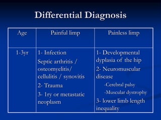
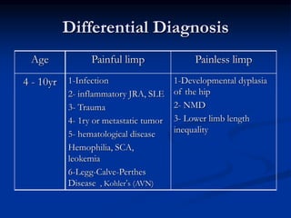
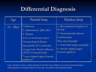
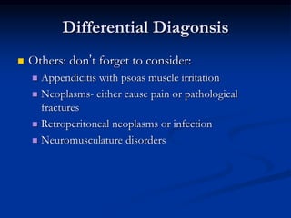
Approach to a limping
child
BY
Magdy Shafik Ramadan
Senior Pediatric and Neonatology consultant
M.S, Diploma, Ph.D of P ediatrics
Pathophysiology
 Three major factors cause a child to limp: pain,
weakness, and structural or mechanical
abnormalities of the spine, pelvis, and lower
extremities (Clark, 1997; deBoeck & Vorlat, 2003; Lawrence, 1998).
 A normal gait is composed of symmetrical, alternating,
rhythmical motions involving two phases: stance and
swing. The stance phase normally encompasses 60%
of the gait cycle. The type of gait may be helpful in
identifying the etiology of the limp .
Some Abnormal Gaits
 An antalgic gait results from pain in one extremity
that causes the patient to shorten the stance phase
on that side with a resultant increase in the swing
phase. The most common causes of an antalgic
gait are trauma or infection .
 A Trendelenburg gait is a downward pelvic tilt
away from the affected hip during the swing phase
as a result of weakness of the contralateral gluteus
medius muscle .
 The gait disturbance is commonly observed in
children with developmental dysplasia of the hip,
Legg-Calves-Perthes disease, or slipped capital
femoral epiphysis. If the involvement is bilateral, a
waddling gait results
 A steppage (equinus) gait is a result of the
inability to actively dorsiflex the foot, with
exaggerated hip and knee flexion during the
swing phase. A steppage gait is seen in
children with neuromuscular diseases (eg,
cerebral palsy) that cause impairment of
dorsiflexion of the ankle.
 A vaulting gait (‫مشية‬‫القفز‬)
 occurs when the knee is hyperextended and
locked at the end of the stance phase and the
child vaults over the extremity .A vaulting gait
is seen in children with limb length
discrepancy or abnormal knee mobility.
 A stooped gait is characterized by walking
with bilaterally increased hip flexion A
stooped gait is common in children with pelvic
or lower abdominal pain.
Differential Diagnosis
Painless limpPainful limpAge
1- Developmental
dyplasia of the hip
2- Neuromuscular
disease
-Cerebral palsy
-Muscular dystrophy
3- lower limb length
inequality
1- Infection
Septic arthritis /
osteomyelitis/
cellulitis / synovitis
2- Trauma
3- 1ry or metastatic
neoplasm
1-3yr
Differential Diagnosis
Painless limpPainful limpAge
1-Developmental dyplasia
of the hip
2- NMD
3- Lower limb length
inequality
1-Infection
2- inflammatory JRA, SLE
3- Trauma
4- 1ry or metastatic tumor
5- hematological disease
Hemophilia, SCA,
leokemia
6-Legg-Calve-Perthes
Disease , Kohler’s (AVN)
4 - 10yr
Differential Diagnosis
Painless limpPainful limpAge
1- Developmental dyplasia of
the hip
2- Neuromuscular disease
Cerebral palsy
Muscular dystrophy
3- lower limb length inequality
4- chronic slipped upper
femoral epiphysis
1-Infection
2- inflammatory :JRA, SLE
3- Trauma
4-1ry or metastatic tumor
5-hematological disease
Hemophilia, SCA, leukemia
6-Legg-Calve-Perthes Disease
(AVN of femoral head)
7-acute slipped upper femoral
epiphysis*
11- 18yr
*very tall and/or obese. Limp and pain in the hip. Leg is held in an extemal rotation position.
Often painful on internal rotation of the hip. Association with hypothyroidism
Differential Diagonsis
 Others: don’t forget to consider:
 Appendicitis with psoas muscle irritation
 Neoplasms- either cause pain or pathological
fractures
 Retroperitoneal neoplasms or infection
 Neuromusculature disorders
Approach
 History
 Examination
 Investigation
 Management
History
 Age
 Sex
 Onset
 Painful or painless? ( analysis…)
 Acute or chronic
 History of trauma
 Association : Night pain, arthralgia, swelling,
morning stiffness, backache
History
 Systemic review
 Recent illness : URTI
 Weight loss, anorexia
 Fever, chills
 Unexplained rash or bruising
 Voiding problem
History
 Past history
 Medical : chronic illness
 Drugs : steroids, antibiotic
 Allergies
 Developmental
 Nutritional
 Vaccination ( site, MMR vaccine)
 Family history
 Hemoglobinopathy, CTD, IBD, NMD
 Social history
Examination
 General inspection + Gait
 Vital signs & anthropometric measurements
 Musculoskeletal examination +Back exam
 Neurological examination
 Evaluate leg lengths- anterior iliac spine to
medial mallelous
Investigations
 CBC
 ESR, CRP
 Blood culture
 Sickle test
 Coagulation test
 Peripheral smear
 Immunological : RF, ANA, etc
Investigations
 Imaging studies
 Plain x ray
 U/S
 CT
 MRI
 Radionuclide studies
 Bone scan
Investigations
 Synovial
fluid
aspiration
Septic
Arthriti
s
JRAtraumat
ic
normal
PurulentCloudy
yellow
Bloody to
straw
colored
Clear to
yellow
appearance
50,000-
200,000
5,000-
80,000
<5,000<200WBC
75-100%50-75%<25%<25%Polymorph
s
Bacterial
culture
positive
Low
glucose
High
protien
High RBC
count
other

Approach to limping child 2

  • 1.
    Approach to alimping child BY Magdy Shafik Ramadan Senior Pediatric and Neonatology consultant M.S, Diploma, Ph.D of P ediatrics
  • 2.
    Pathophysiology  Three majorfactors cause a child to limp: pain, weakness, and structural or mechanical abnormalities of the spine, pelvis, and lower extremities (Clark, 1997; deBoeck & Vorlat, 2003; Lawrence, 1998).  A normal gait is composed of symmetrical, alternating, rhythmical motions involving two phases: stance and swing. The stance phase normally encompasses 60% of the gait cycle. The type of gait may be helpful in identifying the etiology of the limp .
  • 3.
    Some Abnormal Gaits An antalgic gait results from pain in one extremity that causes the patient to shorten the stance phase on that side with a resultant increase in the swing phase. The most common causes of an antalgic gait are trauma or infection .  A Trendelenburg gait is a downward pelvic tilt away from the affected hip during the swing phase as a result of weakness of the contralateral gluteus medius muscle .  The gait disturbance is commonly observed in children with developmental dysplasia of the hip, Legg-Calves-Perthes disease, or slipped capital femoral epiphysis. If the involvement is bilateral, a waddling gait results
  • 4.
     A steppage(equinus) gait is a result of the inability to actively dorsiflex the foot, with exaggerated hip and knee flexion during the swing phase. A steppage gait is seen in children with neuromuscular diseases (eg, cerebral palsy) that cause impairment of dorsiflexion of the ankle.  A vaulting gait (‫مشية‬‫القفز‬)  occurs when the knee is hyperextended and locked at the end of the stance phase and the child vaults over the extremity .A vaulting gait is seen in children with limb length discrepancy or abnormal knee mobility.
  • 5.
     A stoopedgait is characterized by walking with bilaterally increased hip flexion A stooped gait is common in children with pelvic or lower abdominal pain.
  • 6.
    Differential Diagnosis Painless limpPainfullimpAge 1- Developmental dyplasia of the hip 2- Neuromuscular disease -Cerebral palsy -Muscular dystrophy 3- lower limb length inequality 1- Infection Septic arthritis / osteomyelitis/ cellulitis / synovitis 2- Trauma 3- 1ry or metastatic neoplasm 1-3yr
  • 7.
    Differential Diagnosis Painless limpPainfullimpAge 1-Developmental dyplasia of the hip 2- NMD 3- Lower limb length inequality 1-Infection 2- inflammatory JRA, SLE 3- Trauma 4- 1ry or metastatic tumor 5- hematological disease Hemophilia, SCA, leokemia 6-Legg-Calve-Perthes Disease , Kohler’s (AVN) 4 - 10yr
  • 8.
    Differential Diagnosis Painless limpPainfullimpAge 1- Developmental dyplasia of the hip 2- Neuromuscular disease Cerebral palsy Muscular dystrophy 3- lower limb length inequality 4- chronic slipped upper femoral epiphysis 1-Infection 2- inflammatory :JRA, SLE 3- Trauma 4-1ry or metastatic tumor 5-hematological disease Hemophilia, SCA, leukemia 6-Legg-Calve-Perthes Disease (AVN of femoral head) 7-acute slipped upper femoral epiphysis* 11- 18yr *very tall and/or obese. Limp and pain in the hip. Leg is held in an extemal rotation position. Often painful on internal rotation of the hip. Association with hypothyroidism
  • 9.
    Differential Diagonsis  Others:don’t forget to consider:  Appendicitis with psoas muscle irritation  Neoplasms- either cause pain or pathological fractures  Retroperitoneal neoplasms or infection  Neuromusculature disorders
  • 10.
    Approach  History  Examination Investigation  Management
  • 11.
    History  Age  Sex Onset  Painful or painless? ( analysis…)  Acute or chronic  History of trauma  Association : Night pain, arthralgia, swelling, morning stiffness, backache
  • 12.
    History  Systemic review Recent illness : URTI  Weight loss, anorexia  Fever, chills  Unexplained rash or bruising  Voiding problem
  • 13.
    History  Past history Medical : chronic illness  Drugs : steroids, antibiotic  Allergies  Developmental  Nutritional  Vaccination ( site, MMR vaccine)  Family history  Hemoglobinopathy, CTD, IBD, NMD  Social history
  • 14.
    Examination  General inspection+ Gait  Vital signs & anthropometric measurements  Musculoskeletal examination +Back exam  Neurological examination  Evaluate leg lengths- anterior iliac spine to medial mallelous
  • 15.
    Investigations  CBC  ESR,CRP  Blood culture  Sickle test  Coagulation test  Peripheral smear  Immunological : RF, ANA, etc
  • 16.
    Investigations  Imaging studies Plain x ray  U/S  CT  MRI  Radionuclide studies  Bone scan
  • 17.
    Investigations  Synovial fluid aspiration Septic Arthriti s JRAtraumat ic normal PurulentCloudy yellow Bloody to straw colored Clearto yellow appearance 50,000- 200,000 5,000- 80,000 <5,000<200WBC 75-100%50-75%<25%<25%Polymorph s Bacterial culture positive Low glucose High protien High RBC count other