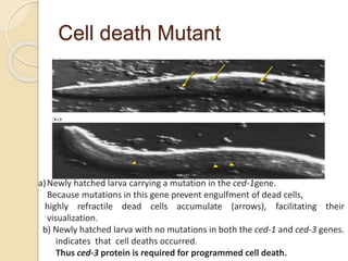
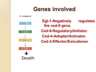
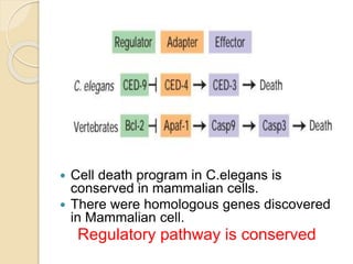
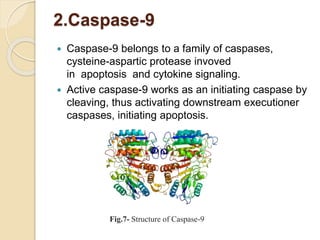
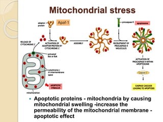
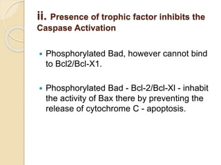
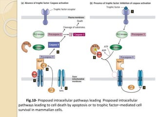
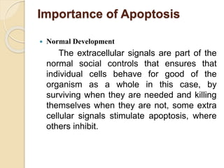
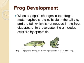
Anil.Kulkarni
DOS in Zoology
University of Mysore, Mysuru
‘To be or not to be’- Caspase as a Switch
Contents
 INTRODUCTION
 DISCOVERY
 GENES INVOLVED
 CASPASES AS A CENTRAL SWITCH-CASPASE CASCADE
 IMPOTANT CASPASES INVOLVED IN APOPTOSIS
 INTRINSIC PATHWAY OF REGULATION
i. MITOCHONDRIAL STRESS
 EXTRINSIC PATHWAYS OF REGULATION OF
APOPTOSIS
CELL DEATH SIGNAL –FAS PATHWAY
SURVIVAL SIGNAL- TROPHIC FACTORS
 IMPORTANCE APOPTOSIS IN NORMAL DEVELOPMENT
 APOPTOSIS IN DISEASES
 SUMMARY
Introduction
 In growth and development of the plant and
animal the cell death plays crucial role.
 In healthy adult human, billions of cells die in
the bone marrow and intestine etc, every hour.
 These cell deaths are suicides, which are
caused by activation of inter and intra cellular
death program.
 Programmed cell death-Apoptosis
Discovery
Fig .1 – The Scientists won the Nobel Prize in Medicine in 2002,
for their pioneering research identifying genes that control apoptosis ,
the genes were identified by studies in the nematode Caenorhabdities elegans.
Sydney Brenner Horvitz John .E.Sulston
 Programmed cell death was discovered
during cell lineage studies in C.elegans.
 947non gonadal cells131cells undergo
apoptosis as a part of developmental
program.
Genetic basis ?
As a part of developmental
program.
Genetic screening in
C.elegans.
Cell death Mutant
a)Newly hatched larva carrying a mutation in the ced-1gene.
Because mutations in this gene prevent engulfment of dead cells,
highly refractile dead cells accumulate (arrows), facilitating their
visualization.
b) Newly hatched larva with no mutations in both the ced-1 and ced-3 genes.
indicates that cell deaths occurred.
Thus ced-3 protein is required for programmed cell death.
Genes involved
Egl-1-Negatively regulates
the ced-9 gene.
Ced-9-Regulatory/Initiator.
Ced-4-Adopter/Activator.
Ced-3-Effector/Exicutioner
Several such genes were
discovered in different
organisms.
Different organisms have
various regulatory proteins .
 Cell death program in C.elegans is
conserved in mammalian cells.
 There were homologous genes discovered
in Mammalian cell.
Regulatory pathway is conserved
Caspases as a Central switch
•Proteolytic enzyme.
•C for cytein and asp for
aspartic acid.
•Procaspase
B. Activation of initiator caspase
Loaded Gun just Require to pull the trigger .
A.Procaspase cleavage
Some important Caspases
involved in the Apoptosis
 1.Caspase -3
The CASP3 protein is a member of the cysteine-
aspartic acid protease (caspase) family. Sequential
activation of caspases plays a central role in the
execution-phase of cell apoptosis
Fig.6 Structure of Caspase-3
2.Caspase-9
 Caspase-9 belongs to a family of caspases,
cysteine-aspartic protease invoved
in apoptosis and cytokine signaling.
 Active caspase-9 works as an initiating caspase by
cleaving, thus activating downstream executioner
caspases, initiating apoptosis.
Fig.7- Structure of Caspase-9
3.Caspase-10
 This gene encodes a protein that is a
member of the cysteine-aspartic acid
protease (caspase) family.
 Sequential activation of caspases plays a
central role in the execution-phase of cell
apoptosis.
 This protein cleaves and activates
caspases 3 and 7, and the protein itself is
processed by caspase 8.They are involved
in the extrinsic death receptor pathway in
humans.
Loaded Gun
 Who operates/pull the trigger???
 The two best understood
signaling pathways that can
activate a caspases.
i. Intrinsic Pathway
ii. Extrinsic Pathway
The intrinsic pathway of
regulation
a.Mitochonrial stress
 Excessive oxidation of
nutrients.
 Metabolic diaseases
Mitochondrial stress
 Apoptotic proteins - mitochondria by causing
mitochondrial swelling -increase the
permeability of the mitochondrial membrane -
apoptotic effect
EXTRINSIC PATHWAYS OF
REGULATION OF APOPTOSIS
1.Death signal –Fas mediated
2.Survival signal- Trophic factor
 Binding of Fas ligand-adaptor
proteins/activator proteins which in turn
recruit initiator procaspases that is
procaspase -8-procaspase-10 forming
death –including signaling complex (DISC) .
 Downstream executioner procaspases to
induce apoptosis.
Fig.9- The extrinsic path way of apoptosis activated through Fas
death receptor
Survival signal –Trophic factor
i. Caspase activation in the absence of Trophic
Factor
 Absence of trophic factor Bad(Bcl-2-
associated death promote), binds to the
anti-apoptotic proteins Bcl-2 and Bcl-Xl.
 Bad prevents the anti-apoptotic from
interacting with Bax a membrane bound
pro-apoptotic protein.
 As a consequence Bax forms channels in
the membrane that medicate ion flux.
 The release of cytochrome C into the
cytosol, binds the adaptor protein Apaf-
1(Apoptotic protease activating factor-1)-
cell death.
ii. Presence of trophic factor inhibits the
Caspase Activation
 Phosphorylated Bad, however cannot bind
to Bcl2/Bcl-X1.
 Phosphorylated Bad - Bcl-2/Bcl-Xl - inhabit
the activity of Bax there by preventing the
release of cytochrome C - apoptosis.
Fig.10- Proposed intracellular pathways leading Proposed intracellular
pathways leading to cell death by apoptosis or to trophic factor–mediated cell
survival in mammalian cells.
Importance of Apoptosis
 Normal Development
The extracellular signals are part of the
normal social controls that ensures that
individual cells behave for good of the
organism as a whole in this case, by
surviving when they are needed and killing
themselves when they are not, some extra
cellular signals stimulate apoptosis, where
others inhibit.
Frog Development
 When a tadpole changes in to a frog at
metamorphosis, the cells die in the tail die,
and the tail, which is not needed in the frog,
disappears. In these case, the unneeded
cells die by apoptosis.
Fig.11- Apoptosis during the metamorphosis of a tadpole into a frog.
Digit Formation
 While locally produced bone morphogenic proteins
stimulate cells between developing fingers and
toes to kill themselves.
Fig.12-Sculpting the digits in the developing paw mouse by
apoptosis
Neuronal Development
 Nerve cells that receive enough of the appropriate
type survival signal live, while others die. In this
way, the number of surviving neurons is
automatically adjusted so that it is appropriate for
the number of target cells they connect with.
Fig.13- The role of survival factor and cell death in adjusting the number of
developing nerve cells to the amount of target tissue.
Apoptosis In Diseases
1.Why Cancerous cells do not Undergo
Apoptosis.
P53- Protein induces cell death when DNA is
damaged.
Cancer-P53 is mutated.
Bcl-2-Acts as a regulator.
Cancer-Bcl2 is mutated.
Neurodegenerative Disease
 Parkinson’s disease.
 Alzheimer’s disease.
 Huntington’s disease.
Summary
 Apoptosis depends on the proteolytic
enzymes called Caspases, which cleave
specific intracellular proteins to kill the cell.
 Caspases are present in all nucleated animal
cells as inactive precursors called
procaspases.
 Initiator procaspases are activated when
brought into proximity in activation
complexes, once activated, they cleave and
activate downstream executioner
procaspases, which activate other
executioner procaspases and other various
target proteins in the cell, producing an
 Cells use at least two distinct pathways to
activate initiator procaspases and trigger a
caspase cascade leading to apoptosis.
 The extrinsic pathway is activated by
extracellular ligand binding to cell surface
death receptor.
 The intrinsic pathway is activated by
intracellular signals generated when cells are
stressed.
 Every cell do contain the components of
apoptotic pathway and will be subject to
regulation of the balance between activation
and repression signal cell death.
References
Alberts,B., Bray,D., Hopkin,K., Johnson,A., Lewis,J., Raff,M.,
Roberts,K., and.Walter,P.,(2010). Essential Cell Biology, 3rd
edition . Garland Science, Taylor & Francis Group, LLC. pp-
638-650
Alberts,B., Johnson,A., Lewis,J., Raff,M., Roberts,K.,
Walter,P.,(2014). Molecular Biology Of Cell,5thediton . Garland
Science, Taylor & Francis Group, LLC. pp-1115-1129
Lodish,H., Berk,A., Kaiser,C.A., Ploegh,H., Amom,A., and
Scott,M.P. (2000). Molecular Cell Biology,7thedition.New York:
W.H Freeman and Company. pp-924-934
Websites.
https://www.nature.com/articles/4400476
https://www.ncbi.nlm.nih.gov/pubmed/9604928
https://www.ncbi.nlm.nih.gov/pmc/articles/PMC4762029/
Thank you

Apoptosis Caspases As A Switch

  • 1.
    Anil.Kulkarni DOS in Zoology Universityof Mysore, Mysuru ‘To be or not to be’- Caspase as a Switch
  • 2.
    Contents  INTRODUCTION  DISCOVERY GENES INVOLVED  CASPASES AS A CENTRAL SWITCH-CASPASE CASCADE  IMPOTANT CASPASES INVOLVED IN APOPTOSIS  INTRINSIC PATHWAY OF REGULATION i. MITOCHONDRIAL STRESS  EXTRINSIC PATHWAYS OF REGULATION OF APOPTOSIS CELL DEATH SIGNAL –FAS PATHWAY SURVIVAL SIGNAL- TROPHIC FACTORS  IMPORTANCE APOPTOSIS IN NORMAL DEVELOPMENT  APOPTOSIS IN DISEASES  SUMMARY
  • 3.
    Introduction  In growthand development of the plant and animal the cell death plays crucial role.  In healthy adult human, billions of cells die in the bone marrow and intestine etc, every hour.  These cell deaths are suicides, which are caused by activation of inter and intra cellular death program.  Programmed cell death-Apoptosis
  • 4.
    Discovery Fig .1 –The Scientists won the Nobel Prize in Medicine in 2002, for their pioneering research identifying genes that control apoptosis , the genes were identified by studies in the nematode Caenorhabdities elegans. Sydney Brenner Horvitz John .E.Sulston
  • 5.
     Programmed celldeath was discovered during cell lineage studies in C.elegans.  947non gonadal cells131cells undergo apoptosis as a part of developmental program.
  • 6.
    Genetic basis ? Asa part of developmental program. Genetic screening in C.elegans.
  • 7.
    Cell death Mutant a)Newlyhatched larva carrying a mutation in the ced-1gene. Because mutations in this gene prevent engulfment of dead cells, highly refractile dead cells accumulate (arrows), facilitating their visualization. b) Newly hatched larva with no mutations in both the ced-1 and ced-3 genes. indicates that cell deaths occurred. Thus ced-3 protein is required for programmed cell death.
  • 8.
    Genes involved Egl-1-Negatively regulates theced-9 gene. Ced-9-Regulatory/Initiator. Ced-4-Adopter/Activator. Ced-3-Effector/Exicutioner
  • 9.
    Several such geneswere discovered in different organisms. Different organisms have various regulatory proteins .
  • 10.
     Cell deathprogram in C.elegans is conserved in mammalian cells.  There were homologous genes discovered in Mammalian cell. Regulatory pathway is conserved
  • 11.
    Caspases as aCentral switch •Proteolytic enzyme. •C for cytein and asp for aspartic acid. •Procaspase
  • 12.
    B. Activation ofinitiator caspase Loaded Gun just Require to pull the trigger . A.Procaspase cleavage
  • 13.
    Some important Caspases involvedin the Apoptosis  1.Caspase -3 The CASP3 protein is a member of the cysteine- aspartic acid protease (caspase) family. Sequential activation of caspases plays a central role in the execution-phase of cell apoptosis Fig.6 Structure of Caspase-3
  • 14.
    2.Caspase-9  Caspase-9 belongsto a family of caspases, cysteine-aspartic protease invoved in apoptosis and cytokine signaling.  Active caspase-9 works as an initiating caspase by cleaving, thus activating downstream executioner caspases, initiating apoptosis. Fig.7- Structure of Caspase-9
  • 15.
    3.Caspase-10  This geneencodes a protein that is a member of the cysteine-aspartic acid protease (caspase) family.  Sequential activation of caspases plays a central role in the execution-phase of cell apoptosis.  This protein cleaves and activates caspases 3 and 7, and the protein itself is processed by caspase 8.They are involved in the extrinsic death receptor pathway in humans.
  • 16.
    Loaded Gun  Whooperates/pull the trigger???  The two best understood signaling pathways that can activate a caspases. i. Intrinsic Pathway ii. Extrinsic Pathway
  • 17.
    The intrinsic pathwayof regulation a.Mitochonrial stress  Excessive oxidation of nutrients.  Metabolic diaseases
  • 18.
    Mitochondrial stress  Apoptoticproteins - mitochondria by causing mitochondrial swelling -increase the permeability of the mitochondrial membrane - apoptotic effect
  • 19.
    EXTRINSIC PATHWAYS OF REGULATIONOF APOPTOSIS 1.Death signal –Fas mediated 2.Survival signal- Trophic factor
  • 20.
     Binding ofFas ligand-adaptor proteins/activator proteins which in turn recruit initiator procaspases that is procaspase -8-procaspase-10 forming death –including signaling complex (DISC) .  Downstream executioner procaspases to induce apoptosis.
  • 21.
    Fig.9- The extrinsicpath way of apoptosis activated through Fas death receptor
  • 22.
    Survival signal –Trophicfactor i. Caspase activation in the absence of Trophic Factor  Absence of trophic factor Bad(Bcl-2- associated death promote), binds to the anti-apoptotic proteins Bcl-2 and Bcl-Xl.  Bad prevents the anti-apoptotic from interacting with Bax a membrane bound pro-apoptotic protein.  As a consequence Bax forms channels in the membrane that medicate ion flux.  The release of cytochrome C into the cytosol, binds the adaptor protein Apaf- 1(Apoptotic protease activating factor-1)- cell death.
  • 23.
    ii. Presence oftrophic factor inhibits the Caspase Activation  Phosphorylated Bad, however cannot bind to Bcl2/Bcl-X1.  Phosphorylated Bad - Bcl-2/Bcl-Xl - inhabit the activity of Bax there by preventing the release of cytochrome C - apoptosis.
  • 24.
    Fig.10- Proposed intracellularpathways leading Proposed intracellular pathways leading to cell death by apoptosis or to trophic factor–mediated cell survival in mammalian cells.
  • 25.
    Importance of Apoptosis Normal Development The extracellular signals are part of the normal social controls that ensures that individual cells behave for good of the organism as a whole in this case, by surviving when they are needed and killing themselves when they are not, some extra cellular signals stimulate apoptosis, where others inhibit.
  • 26.
    Frog Development  Whena tadpole changes in to a frog at metamorphosis, the cells die in the tail die, and the tail, which is not needed in the frog, disappears. In these case, the unneeded cells die by apoptosis. Fig.11- Apoptosis during the metamorphosis of a tadpole into a frog.
  • 27.
    Digit Formation  Whilelocally produced bone morphogenic proteins stimulate cells between developing fingers and toes to kill themselves. Fig.12-Sculpting the digits in the developing paw mouse by apoptosis
  • 28.
    Neuronal Development  Nervecells that receive enough of the appropriate type survival signal live, while others die. In this way, the number of surviving neurons is automatically adjusted so that it is appropriate for the number of target cells they connect with. Fig.13- The role of survival factor and cell death in adjusting the number of developing nerve cells to the amount of target tissue.
  • 29.
    Apoptosis In Diseases 1.WhyCancerous cells do not Undergo Apoptosis. P53- Protein induces cell death when DNA is damaged. Cancer-P53 is mutated. Bcl-2-Acts as a regulator. Cancer-Bcl2 is mutated.
  • 30.
    Neurodegenerative Disease  Parkinson’sdisease.  Alzheimer’s disease.  Huntington’s disease.
  • 31.
    Summary  Apoptosis dependson the proteolytic enzymes called Caspases, which cleave specific intracellular proteins to kill the cell.  Caspases are present in all nucleated animal cells as inactive precursors called procaspases.  Initiator procaspases are activated when brought into proximity in activation complexes, once activated, they cleave and activate downstream executioner procaspases, which activate other executioner procaspases and other various target proteins in the cell, producing an
  • 32.
     Cells useat least two distinct pathways to activate initiator procaspases and trigger a caspase cascade leading to apoptosis.  The extrinsic pathway is activated by extracellular ligand binding to cell surface death receptor.  The intrinsic pathway is activated by intracellular signals generated when cells are stressed.  Every cell do contain the components of apoptotic pathway and will be subject to regulation of the balance between activation and repression signal cell death.
  • 33.
    References Alberts,B., Bray,D., Hopkin,K.,Johnson,A., Lewis,J., Raff,M., Roberts,K., and.Walter,P.,(2010). Essential Cell Biology, 3rd edition . Garland Science, Taylor & Francis Group, LLC. pp- 638-650 Alberts,B., Johnson,A., Lewis,J., Raff,M., Roberts,K., Walter,P.,(2014). Molecular Biology Of Cell,5thediton . Garland Science, Taylor & Francis Group, LLC. pp-1115-1129 Lodish,H., Berk,A., Kaiser,C.A., Ploegh,H., Amom,A., and Scott,M.P. (2000). Molecular Cell Biology,7thedition.New York: W.H Freeman and Company. pp-924-934 Websites. https://www.nature.com/articles/4400476 https://www.ncbi.nlm.nih.gov/pubmed/9604928 https://www.ncbi.nlm.nih.gov/pmc/articles/PMC4762029/
  • 34.