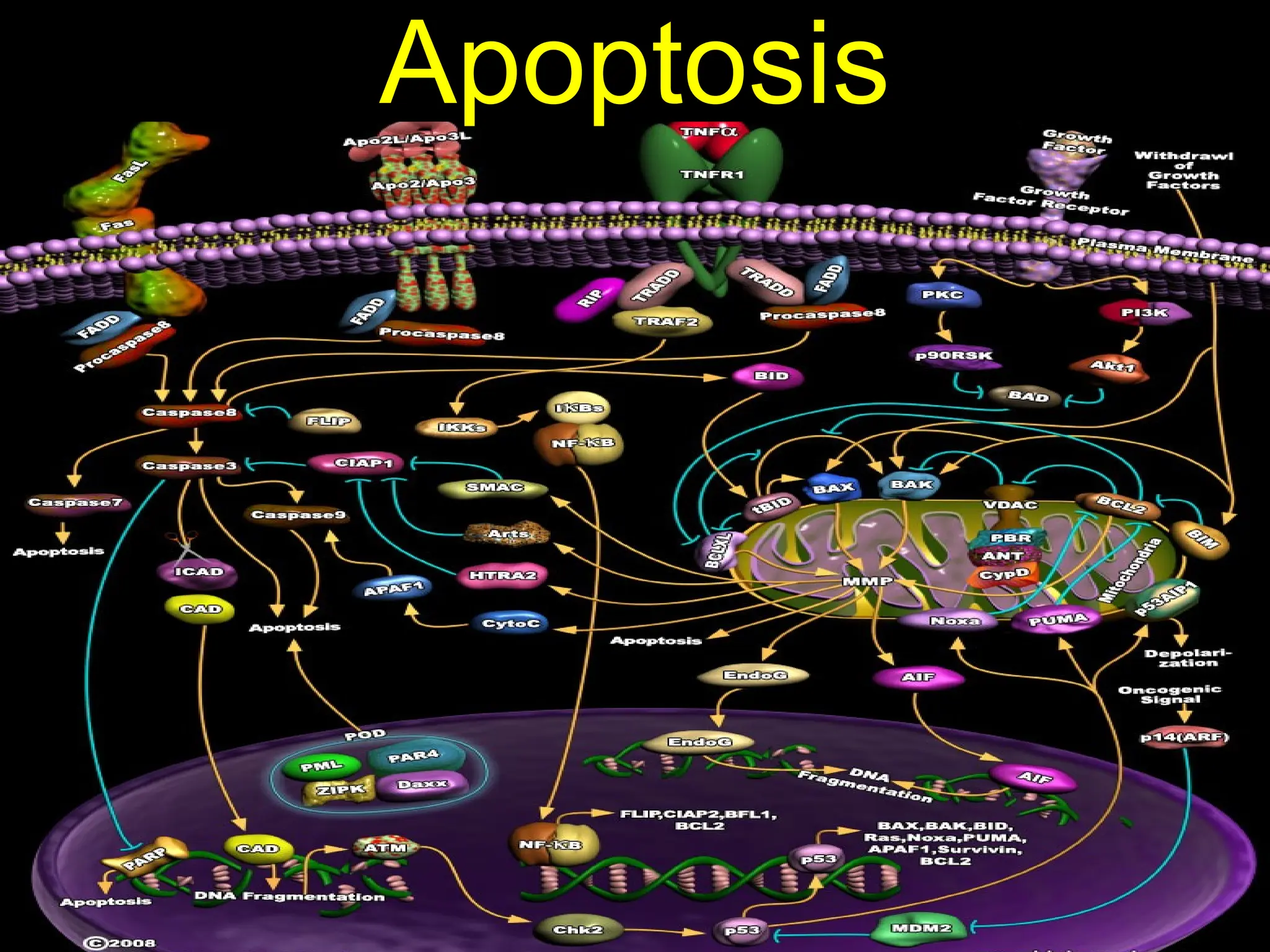
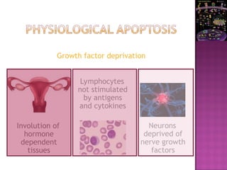
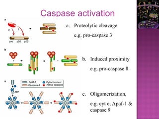
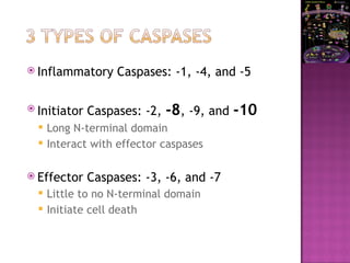
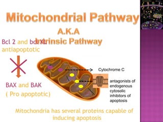
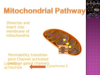
Apoptosis
Literal sense
 Programmed cell death
 Internally controlled “suicide” program
 Cells are removed with minimal disruption
of the surrounding tissue
Pathological
Physiological
Physiological
 Potentially harmful cells
 Outlived their usefulness
 Maintain a steady number of cells
Pathological
 Damaged beyond repair
EMBRYOGENESIS
Growth factor deprivation
Involution of
hormone
dependent
tissues
Lymphocytes
not stimulated
by antigens
and cytokines
Neurons
deprived of
nerve growth
factors
Cell loss in proliferating populations
• Eliminate self reactive lymphocytes
• Cell death by induced cytotoxic T lymphocytes
(defense against virus / tumorogenisis)
 Genetically altered beyond repair
 DNA damage (Direct or Free radical)
 Radiation
 Cytotoxic drugs
 Extreme tempetature
 Hypoxia
 Accumulation of misfolded proteins
 Arise because of mutations in the genes coding
these proteins or extrensic factors like free
radicals
 Cell injury in infections
 Atropy in parynchyma after duct obstruction
G1 S G2 M
p53
Repair Attempted
NO
YES
 Chaperones control normal folding
 Misfolded polypeptides are ubiquitinated and
targeted for proteolysis
 If accumulated ER stress occurs
 That triggers unfolded protein response i.e
increase production of chaperones and retard
protein translation
 If unable to cope caspase activation and
apoptosis occurs
CASPASE
Mitochondrial pathway Death receptor pathway
Both pathways are not mutually exclusive with p53 causing cross over to
death receptor pathway.
Cystene protease that cleave proteins after aspartic residues
Cell Death by Apoptosis
Activation of caspase is the fundamental event of apoptosis
Caspase-1 (ICE)
Caspase-2 (ICH-1, Nedd-2)
Caspase-3 (CPP32, Apopain, Yama)
Caspase-4 (ICH-2, TX, ICEreıı)
Caspase-5 (ICErelııı, TY)
Caspase-6 (Mch2)
Caspase-7 (ICE-LAP3, Mch3, CMH-1)
Caspase-8 (FLICE, Mch5, MACH)
Caspace-9 (Mch6, ICE-LAP6)
Caspase-10 (Mch4)
Caspase activation
a. Proteolytic cleavage
e.g. pro-caspase 3
b. Induced proximity
e.g. pro-caspase 8
c. Oligomerization,
e.g. cyt c, Apaf-1 &
caspase 9
 Inflammatory Caspases: -1, -4, and -5
 Initiator Caspases: -2, -8, -9, and -10
 Long N-terminal domain
 Interact with effector caspases
 Effector Caspases: -3, -6, and -7
 Little to no N-terminal domain
 Initiate cell death
Cytochrome C
antagonists of
endogenous
cytosolic
inhibitors of
apoptosis
Mitochondria has several proteins capable of
inducing apoptosis
Bcl 2 and bcl XL
antiapoptotic
BAX and BAK
( Pro apoptotic)
Dimerize and
insert into
membrane of
mitochondria
Permiability transition
pore Channel activated
(voltage gated channel)
Cytochrome C
CASPASE
ACTIVATION
Surface molecules trigger apoptosis
Members of TNF family
Ex: Type 1 TNF and Fas (CD95)
Normally present in cytoplasmic region
On Activation move to surface
FasLigand
membrane
protein on
activated T
lymphocytes
activate caspase-8 activating caspase cascade by activating
member of Bcl-2 family called Bid
Bind to Fas
 Active enzymatic process
 Nucleoproteins
are broken down
 Cell organelles are
fragmented
 Nuclei show various stages of condensation
and aggregation ultimately karyorrhexis.
 Plasma membrane remains intact
 But become avid targets to phagocytosis by flip
out of phosphotidyl serine on the inner leaflet of
plasma membrane
 These are recognised by macrophages
 Also secrete soluble factors that recruit
phagocytes
 Express adhesive glycoproteins recognised by
phagocytes and macrophages
 Dead cells cleared rapidly before contents
leak out
 Hence does not elicit inflammatory
reaction
 Hence differs from necrosis
CASPASE
.
Cleavage of nuclear LAMINS is involved in chromatin condensation
and nuclear shrinkage.
Cleavage of the inhibitor of the DNase CAD (caspase-activated
deoxyribonuclease), ICAD causes the release of the endonuclease,
which fragment DNA.
Cleavage of cytoskeletal proteins such as actin, plectin, Rho
kinase 1 (ROCK1) and gelsolin leads to cell fragmentation, blebbing
and the formation of apoptotic bodies.
 Constant number of cells in an organism.
 Cell death = Cell proliferation
Cell
Death
Growth
Survival
Proliferation
What happens when things become imbalanced?
Cell
Death
Growth
Survival
Proliferation
Cell
Death
Growth
Survival
Proliferation
Alzheimer’s
Parkinson’s
Cancer
 Resistance of tumour cells to apoptosis is an
essential feature of cancer development
 Loss of apoptosis can promote tumor
 Initiation
 Progression
 Treatment resistence
 Chemotherapy, irradiation and other stimuli
can initiate apoptosis through the
mitochondrial (intrinsic) pathway.
 Pro-apoptotic BCL2 family proteins are
activated by treatment ---- caspase cascade
---- death of cell
 Disrupted apoptotic cycle can effect sensitivity of
cancer drugs
 As multiple drugs effect same mechanism it
causes multidrug resistance
 Sufficient doses of almost all anticancer drugs
induce apoptosis by alternate pathway
indipendent of p53 pathway
 Contribution of p53 is dependent on
 Agent
 Dose
 mutational background
 The best-defined mechanism of therapy-induced
cellular stress induced cance cell death.
 Chemotherapeutic drugs (for example, the
nucleotide analogue 5-FU) induce CD95 by a
transcriptionally regulated, p53-dependent
mechanism.
 They also engage the SAPK/JNK pathway
 Leads to upregulation of CD95L
 Allows the cells to either commit suicide or kill
neighbouring cells.
 p53 induce the expression of proteins involved in
 the mitochondrial pathway — BAX, NOXA, PUMA and
P53aip1
 death receptor pathway — CD95, TRAIL-R1 and TRAIL-
R2.
 When p53 is damaged it cannot induce apoptosis
so cell survive
 Subsequent mutations develop
 Progresses to malignancy
 overexpression of anti-apoptotic genes
 follicular B-cell lymphoma t(14;18), couples BCL2
gene to immunoglobulin heavy chain locus, leading
to enhanced BCL2 expression.
 EBV and HHV8 encode proteins that are
homologues of BCL2.
 BHRF1 from EBV and KSbcl-2 (vBcl-2) from HHV8 —
have an anti-apoptotic function and enhance
survival of the infected cells.
 soluble receptors that act as decoys for death
ligands.
 soluble CD95 (sCD95) and decoy receptor 3 (DcR3)
shown to competitively inhibit CD95 signalling.
 sCD95 is expressed in various malignancies, and
elevated levels can be found in the sera of cancer
patients.
 .
 associated with poor prognosis in melanoma
patients
 It is genetically amplified in several lung and
colon carcinomas
 overexpressed in several adenocarcinomas,
glioma cell lines and glioblastomas.
 the pro-apoptotic BCL2 family member BAX is
mutated.
 Frameshift mutations
 loss of expression and function.
 Reduced BAX expression is associated with a
poor response rate to chemotherapy and
shorter survival
 Bcl2 shows discrepencies in effect on tumor
management
 Studies have shown a correlation between
high levels of BCL2 expression and the
severity of malignancy of human tumours.
 a high level of BCL2 expression is associated
with a poor response to chemotherapy and
seems to be predictive of shorter, disease-
free survival.
 While in some cases Bcl-2 has been shown to
cause chemoresistence in other settings it is
shown to improve prognosis.
 Clinical studies examine single alterations
 Cannot exclude extragenic mutations in the same
pathway
 Thus almost impossible to determine negative
results
 Ex: murine lymphomas harbouring INK4a/ARF
have defective p53 but harbour wild type p53
hence classified as p53 normal
 Extracellular survival factors affect cell
death
 Cell density
 microenvironment
 Deregulated proliferation alone is not sufficient
for tumour formation
 Overexpression of growth-promoting oncogenes
— such as c-MYC, E1A or E2F1 — sensitizes cells
to apoptosis.
 Besides the expression of proteins that promote
cell proliferation, tumour progression requires
the expression of anti-apoptotic proteins or the
inactivation of essential pro-apoptotic proteins.
 Apoptosis limited role in solid tumors
 Side effects of cancer drugs are because
along with cancer cells they also target
normal cells like intestine and bone marrow
 Caspase 10 mutation lymphoproliferative
syndrome type 2
 Frameshift in caspase5 HNPCC, GI and
endometrial tumors
 Death receptors Fas/CD95, TNFR1, DR3, DR4, DR5
 TNFalpha trimerizes its ligand TNFR1
 Recruts signal transducing TRADD
 TRADD Recruits RIP and TRAF-2
 Activates NF-kB which supress TNFa induced apoptosis
 TRADD activate FADD thatactivate caspase 8 --- apoptosis
 FADD disruption can prevent apoptosis (does not occur in
oncogenic mutations but occurs in tumor development )
 Cytokines like IL6 can inhibit apoptosis
 Adenovirus induce cells to bypass apoptosis
and replicate by E1B oncoprotein inducing
cells to enter s phase
 Most mutations occur upstream
 Machinery is retained
 Tumor specific alterations in apoptotic
programmes
 Ex: Adenoviral gene transfer in ovarian cancer
 Ad-DF3-Bax eradicated >99% of these tumors in nude mice
 Ex2: Taxanes known to phosphorylate and inactivate bcl2
 inactivating NF-kb enhances chemo induced cell death
 Restoring of pro-apoptotic p53
 Activating death ligands hence p53 indipendent
death as normal cells are resistent to TRAIL
induced apoptosis in view of decoy receptors that
compete with DR4 and DR5
 Oncogene induced apoptosis
 Ras and its appln

Apoptosis content.ppt

  • 1.
  • 2.
  • 3.
     Programmed celldeath  Internally controlled “suicide” program  Cells are removed with minimal disruption of the surrounding tissue
  • 4.
  • 5.
    Physiological  Potentially harmfulcells  Outlived their usefulness  Maintain a steady number of cells Pathological  Damaged beyond repair
  • 6.
  • 7.
    Growth factor deprivation Involutionof hormone dependent tissues Lymphocytes not stimulated by antigens and cytokines Neurons deprived of nerve growth factors
  • 8.
    Cell loss inproliferating populations • Eliminate self reactive lymphocytes • Cell death by induced cytotoxic T lymphocytes (defense against virus / tumorogenisis)
  • 9.
     Genetically alteredbeyond repair  DNA damage (Direct or Free radical)  Radiation  Cytotoxic drugs  Extreme tempetature  Hypoxia
  • 10.
     Accumulation ofmisfolded proteins  Arise because of mutations in the genes coding these proteins or extrensic factors like free radicals  Cell injury in infections  Atropy in parynchyma after duct obstruction
  • 12.
    G1 S G2M p53 Repair Attempted NO YES
  • 13.
     Chaperones controlnormal folding  Misfolded polypeptides are ubiquitinated and targeted for proteolysis  If accumulated ER stress occurs  That triggers unfolded protein response i.e increase production of chaperones and retard protein translation  If unable to cope caspase activation and apoptosis occurs
  • 14.
    CASPASE Mitochondrial pathway Deathreceptor pathway Both pathways are not mutually exclusive with p53 causing cross over to death receptor pathway. Cystene protease that cleave proteins after aspartic residues Cell Death by Apoptosis Activation of caspase is the fundamental event of apoptosis
  • 15.
    Caspase-1 (ICE) Caspase-2 (ICH-1,Nedd-2) Caspase-3 (CPP32, Apopain, Yama) Caspase-4 (ICH-2, TX, ICEreıı) Caspase-5 (ICErelııı, TY) Caspase-6 (Mch2) Caspase-7 (ICE-LAP3, Mch3, CMH-1) Caspase-8 (FLICE, Mch5, MACH) Caspace-9 (Mch6, ICE-LAP6) Caspase-10 (Mch4)
  • 16.
    Caspase activation a. Proteolyticcleavage e.g. pro-caspase 3 b. Induced proximity e.g. pro-caspase 8 c. Oligomerization, e.g. cyt c, Apaf-1 & caspase 9
  • 17.
     Inflammatory Caspases:-1, -4, and -5  Initiator Caspases: -2, -8, -9, and -10  Long N-terminal domain  Interact with effector caspases  Effector Caspases: -3, -6, and -7  Little to no N-terminal domain  Initiate cell death
  • 18.
    Cytochrome C antagonists of endogenous cytosolic inhibitorsof apoptosis Mitochondria has several proteins capable of inducing apoptosis Bcl 2 and bcl XL antiapoptotic BAX and BAK ( Pro apoptotic)
  • 19.
    Dimerize and insert into membraneof mitochondria Permiability transition pore Channel activated (voltage gated channel) Cytochrome C CASPASE ACTIVATION
  • 20.
    Surface molecules triggerapoptosis Members of TNF family Ex: Type 1 TNF and Fas (CD95) Normally present in cytoplasmic region On Activation move to surface
  • 21.
    FasLigand membrane protein on activated T lymphocytes activatecaspase-8 activating caspase cascade by activating member of Bcl-2 family called Bid Bind to Fas
  • 22.
     Active enzymaticprocess  Nucleoproteins are broken down  Cell organelles are fragmented  Nuclei show various stages of condensation and aggregation ultimately karyorrhexis.
  • 24.
     Plasma membraneremains intact  But become avid targets to phagocytosis by flip out of phosphotidyl serine on the inner leaflet of plasma membrane  These are recognised by macrophages  Also secrete soluble factors that recruit phagocytes  Express adhesive glycoproteins recognised by phagocytes and macrophages
  • 25.
     Dead cellscleared rapidly before contents leak out  Hence does not elicit inflammatory reaction  Hence differs from necrosis
  • 26.
    CASPASE . Cleavage of nuclearLAMINS is involved in chromatin condensation and nuclear shrinkage. Cleavage of the inhibitor of the DNase CAD (caspase-activated deoxyribonuclease), ICAD causes the release of the endonuclease, which fragment DNA. Cleavage of cytoskeletal proteins such as actin, plectin, Rho kinase 1 (ROCK1) and gelsolin leads to cell fragmentation, blebbing and the formation of apoptotic bodies.
  • 28.
     Constant numberof cells in an organism.  Cell death = Cell proliferation Cell Death Growth Survival Proliferation
  • 29.
    What happens whenthings become imbalanced? Cell Death Growth Survival Proliferation Cell Death Growth Survival Proliferation Alzheimer’s Parkinson’s Cancer
  • 30.
     Resistance oftumour cells to apoptosis is an essential feature of cancer development  Loss of apoptosis can promote tumor  Initiation  Progression  Treatment resistence
  • 31.
     Chemotherapy, irradiationand other stimuli can initiate apoptosis through the mitochondrial (intrinsic) pathway.  Pro-apoptotic BCL2 family proteins are activated by treatment ---- caspase cascade ---- death of cell
  • 32.
     Disrupted apoptoticcycle can effect sensitivity of cancer drugs  As multiple drugs effect same mechanism it causes multidrug resistance  Sufficient doses of almost all anticancer drugs induce apoptosis by alternate pathway indipendent of p53 pathway  Contribution of p53 is dependent on  Agent  Dose  mutational background
  • 33.
     The best-definedmechanism of therapy-induced cellular stress induced cance cell death.  Chemotherapeutic drugs (for example, the nucleotide analogue 5-FU) induce CD95 by a transcriptionally regulated, p53-dependent mechanism.  They also engage the SAPK/JNK pathway  Leads to upregulation of CD95L  Allows the cells to either commit suicide or kill neighbouring cells.
  • 34.
     p53 inducethe expression of proteins involved in  the mitochondrial pathway — BAX, NOXA, PUMA and P53aip1  death receptor pathway — CD95, TRAIL-R1 and TRAIL- R2.  When p53 is damaged it cannot induce apoptosis so cell survive  Subsequent mutations develop  Progresses to malignancy
  • 35.
     overexpression ofanti-apoptotic genes  follicular B-cell lymphoma t(14;18), couples BCL2 gene to immunoglobulin heavy chain locus, leading to enhanced BCL2 expression.  EBV and HHV8 encode proteins that are homologues of BCL2.  BHRF1 from EBV and KSbcl-2 (vBcl-2) from HHV8 — have an anti-apoptotic function and enhance survival of the infected cells.
  • 36.
     soluble receptorsthat act as decoys for death ligands.  soluble CD95 (sCD95) and decoy receptor 3 (DcR3) shown to competitively inhibit CD95 signalling.  sCD95 is expressed in various malignancies, and elevated levels can be found in the sera of cancer patients.  .
  • 37.
     associated withpoor prognosis in melanoma patients  It is genetically amplified in several lung and colon carcinomas  overexpressed in several adenocarcinomas, glioma cell lines and glioblastomas.
  • 38.
     the pro-apoptoticBCL2 family member BAX is mutated.  Frameshift mutations  loss of expression and function.  Reduced BAX expression is associated with a poor response rate to chemotherapy and shorter survival
  • 39.
     Bcl2 showsdiscrepencies in effect on tumor management  Studies have shown a correlation between high levels of BCL2 expression and the severity of malignancy of human tumours.  a high level of BCL2 expression is associated with a poor response to chemotherapy and seems to be predictive of shorter, disease- free survival.  While in some cases Bcl-2 has been shown to cause chemoresistence in other settings it is shown to improve prognosis.
  • 40.
     Clinical studiesexamine single alterations  Cannot exclude extragenic mutations in the same pathway  Thus almost impossible to determine negative results  Ex: murine lymphomas harbouring INK4a/ARF have defective p53 but harbour wild type p53 hence classified as p53 normal  Extracellular survival factors affect cell death  Cell density  microenvironment
  • 41.
     Deregulated proliferationalone is not sufficient for tumour formation  Overexpression of growth-promoting oncogenes — such as c-MYC, E1A or E2F1 — sensitizes cells to apoptosis.  Besides the expression of proteins that promote cell proliferation, tumour progression requires the expression of anti-apoptotic proteins or the inactivation of essential pro-apoptotic proteins.
  • 42.
     Apoptosis limitedrole in solid tumors  Side effects of cancer drugs are because along with cancer cells they also target normal cells like intestine and bone marrow
  • 43.
     Caspase 10mutation lymphoproliferative syndrome type 2  Frameshift in caspase5 HNPCC, GI and endometrial tumors
  • 44.
     Death receptorsFas/CD95, TNFR1, DR3, DR4, DR5  TNFalpha trimerizes its ligand TNFR1  Recruts signal transducing TRADD  TRADD Recruits RIP and TRAF-2  Activates NF-kB which supress TNFa induced apoptosis  TRADD activate FADD thatactivate caspase 8 --- apoptosis  FADD disruption can prevent apoptosis (does not occur in oncogenic mutations but occurs in tumor development )
  • 45.
     Cytokines likeIL6 can inhibit apoptosis  Adenovirus induce cells to bypass apoptosis and replicate by E1B oncoprotein inducing cells to enter s phase
  • 46.
     Most mutationsoccur upstream  Machinery is retained  Tumor specific alterations in apoptotic programmes  Ex: Adenoviral gene transfer in ovarian cancer  Ad-DF3-Bax eradicated >99% of these tumors in nude mice  Ex2: Taxanes known to phosphorylate and inactivate bcl2  inactivating NF-kb enhances chemo induced cell death  Restoring of pro-apoptotic p53  Activating death ligands hence p53 indipendent death as normal cells are resistent to TRAIL induced apoptosis in view of decoy receptors that compete with DR4 and DR5
  • 47.
     Oncogene inducedapoptosis  Ras and its appln

Editor's Notes

  • #3 Entire mechanism triggered inside the cell. There is no inflammation, no residual or any sign that the cell existed. Infact its close to impossible to pick up under a microscope.
  • #14 Fundamental event is activation of caspases
  • #18 Mito membrane permeability important More than 20 proteins Prototype is Bcl-2
  • #21 Binding of death ligands (CD95L is used here as an example) to their receptor leads to the formation of the death-inducing signalling complex (DISC). In the DISC, the initiator procaspase-8 is recruited by FADD (FAS-associated death domain protein) and is activated by autocatalytic cleavage. Death-receptor-mediated apoptosis can be inhibited at several levels by anti-apoptotic proteins: CD95L can be prevented from binding to CD95 by soluble 'decoy' receptors, such as soluble CD95 (sCD95) or DcR3 (decoy receptor 3). FLICE-inhibitory proteins (FLIPs) bind to the DISC and prevent the activation of caspase-8; and inhibitors of apoptosis proteins (IAPs) bind to and inhibit caspases. FLIPL and FLIPS refer to long and short forms of FLIP, respectively. Viruses produce homologus of FLIP hence keeping cells alive This process mainly involved in eliminating self reactive lymphocytes and killing targets by cytoxic T cells.