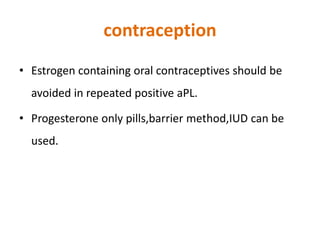
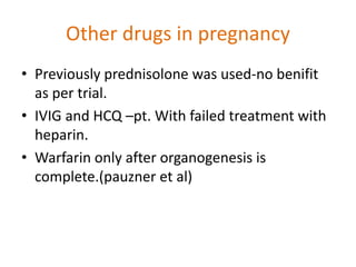
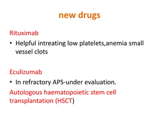
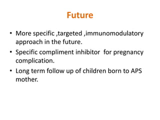
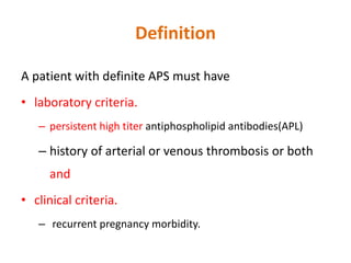
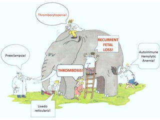
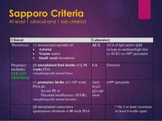
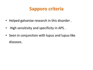
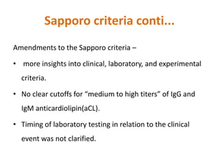
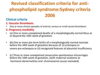
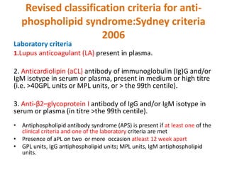
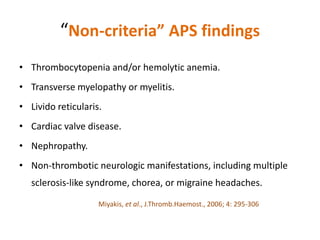
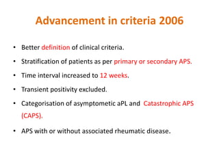
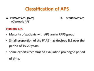
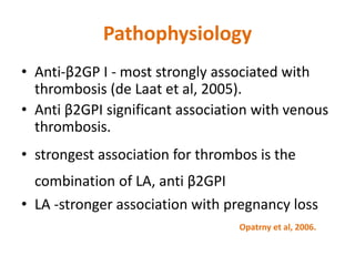
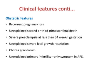
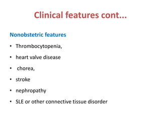
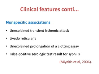
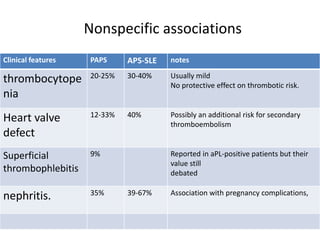
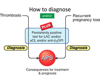
.
Antiphospholipid syndrome
Dr. Suman Kumari
MS OBGY
Graham R.V. Hughes
First discribed by Graham R.V. Hughes in 1983
Introduction
• known as Hughes syndrome.
• Autoimmune disorder associated with acquired
thrombophilia.
• Most common association is SLE.
• Asherson’s syndrome-Catastrophic APS.
• Obstetric complications are “hallmark” of APS.
• Severe FGR,preeclampsia,prematurity imp. Complications.
Introduction conti..
• It is the most common thrombotic defect leading to fetal
wastage.
• Responsible for frequent miscarriages in young female.
• Obstetric care based on medical- obs high risk combined
management.
• Prevalence of antiphospholipid antibodies is 2% in overall
pregnancy.
• 15% of women with recurrent miscarriage.(RCOG)
Introduction conti..
Definition
A patient with definite APS must have
• laboratory criteria.
– persistent high titer antiphospholipid antibodies(APL)
– history of arterial or venous thrombosis or both
and
• clinical criteria.
– recurrent pregnancy morbidity.
Talk Plan
Sapporo criteria
• Diagnostic criteria
Sydney criteria
primary APS
• Classification of APS
• Epidemiology Secondary APS
• Pathophysiology
• Clinical features
• Investigation Preconception
• Management Antenatal
• Follow up Postnatal
• Recommendation
International consensus conference held in
Sapporo(98)
Sapporo criteria
• Helped galvanize research in this disorder .
• High sensitivity and specificity in APS .
• Seen in conjunction with lupus and lupus-like
diseases.
Sapporo criteria conti...
Amendments to the Sapporo criteria –
• more insights into clinical, laboratory, and experimental
criteria.
• No clear cutoffs for “medium to high titers” of IgG and
IgM anticardiolipin(aCL).
• Timing of laboratory testing in relation to the clinical
event was not clarified.
Revised classification criteria for anti-
phospholipid syndrome:Sydney criteria
2006
Clinical criteria
1. Vascular thrombosis
• One or more clinical episodes of arterial, venous or small vessel thrombosis
2. Pregnancy morbidity
• (a) One or more unexplained deaths of a morphologically normal fetus at
or beyond the 10th week of gestation
• (b) One or more pre-term births of a morphologically normal neonate
before the 34th week of gestation because of: (i) eclampsia or
severe pre-eclampsia or (ii) recognized features of placental insufficiency
• (c) Three or more unexplained consecutive spontaneous miscarriages
before the 10th week of gestation, (with maternal anatomic or
hormonal abnormalities and chromosomal causes excluded)
Revised classification criteria for anti-
phospholipid syndrome:Sydney criteria
2006
Laboratory criteria
1.Lupus anticoagulant (LA) present in plasma.
2. Anticardiolipin (aCL) antibody of immunoglobulin (Ig)G and/or
IgM isotype in serum or plasma, present in medium or high titre
(i.e. >40GPL units or MPL units, or > the 99th centile).
3. Anti-β2–glycoprotein I antibody of IgG and/or IgM isotype in
serum or plasma (in titre >the 99th centile).
• Antiphospholipid antibody syndrome (APS) is present if at least one of the
clinical criteria and one of the laboratory criteria are met
• Presence of aPL on two or more occasion atleast 12 week apart
• GPL units, IgG antiphospholipid units; MPL units, IgM antiphospholipid
units.
“Non-criteria” APS findings
• Thrombocytopenia and/or hemolytic anemia.
• Transverse myelopathy or myelitis.
• Livido reticularis.
• Cardiac valve disease.
• Nephropathy.
• Non-thrombotic neurologic manifestations, including multiple
sclerosis-like syndrome, chorea, or migraine headaches.
Miyakis, et al., J.Thromb.Haemost., 2006; 4: 295-306
Advancement in criteria 2006
• Better definition of clinical criteria.
• Stratification of patients as per primary or secondary APS.
• Time interval increased to 12 weeks.
• Transient positivity excluded.
• Categorisation of asymptometic aPL and Catastrophic APS
(CAPS).
• APS with or without associated rheumatic disease.
Classification of APS
A. PRIMARY APS (PAPS) B. SECONDARY APS
(Obstetric APS)
PRIMARY APS
• Majority of patients with APS are in PAPS group.
• Small proportion of the PAPS may devlops SLE over the
period of 15-20 years.
• some experts recommend evaluation prolonged period
of time.
Classification of APS....
B. SECONDARY APS
1. autoimmune disorder 3. Malignancy
SLE 25-50% Ca cervix and ovary
Sjogren syndrome-42% Lymphoma
Rheumatoid arthritis-33% leukemia
DM
Autoimmune thyroid disease.
2 .Drug induced condition 4. infectious disease
ocp syphlis, HIV
Epidemiology
• Women : men is 5:1
• Mean age of onset is 31 yrs.
• Risk of thrombosis is 0.5-30 %
• Artheritis,livedo ,migrane more common in female.
• MI,epilepsy arterial thrombosis more common in
male.
Mechanisms by which APS causes pregnancy
morbidity
early pregnancy late pregnancy
inhibition to
trophoblastic function
and differentiation
thrombosis of the
Uteroplacental
vasulature
Activation of compliment
system at the maternal
fetal interface --local
inflammatory response.
Pathophysiology
• aPLs –acquired antibodies(IgG,IgM,and/orIgA) -binds to
negatively charged phospholipids.
• Beta2GP-I is major target molecule,present on the surface of
trophoblastic cell membrane.
• Beta 2 GP-1 helps in syncytial trophoblast formation from
cytotrophoblast.
• Inhibit prothrombin to thrombin conversion and activation of
intrensic coagulation pathway and thus inhibits thrombosis.
Pathophysiology
• Anti-β2GP I - most strongly associated with
thrombosis (de Laat et al, 2005).
• Anti β2GPI significant association with venous
thrombosis.
• strongest association for thrombos is the
combination of LA, anti β2GPI
• LA -stronger association with pregnancy loss
Opatrny et al, 2006.
Mechanisms for thrombosis in APS
• Increased expression of tissue factor on monocytes and
endothelial cells (Branch & Rodgers, 1993)
• Interference in the protein C anticoagulant pathway
(Malia et al, 1990)
• Inhibition of fibrinolysis (Atsumi et al, 1998b)
• Pregnancy failure may be due to thrombosis in the
placental bed.
Clinical features
• Can affect any organ of the body including the lung,
skin, brain, liver,kidneys, adrenal glands, heart, and
eyes.
• ‘‘classic’’ clinical presentations include peripheral
deep venous thrombosis (DVT), pulmonary embolism
(PE), and/or arterial thrombotic events
Clinical features cont...
• Venous thrombosis in APS is commonly- lower limb deep
vein thrombosis (DVT) and/or pulmonary embolism(PE)
• Any part of the venous system may be involved.
• The most frequent site of arterial thrombosis in APS is in
the cerebral vasculature resulting in transient cerebral
ischaemia/stroke.
Clinical features conti...
• APS -only a minor contribution to the overall burden
of disease fromVTE and stroke.
• Microvascular thrombosis in APS is least common
seen in‘catastrophic antiphospholipid syndrome’
(CAPS)potentially lethal .
• In CAPS -multiorgan failure ,but lungs, brain and
kidneys are spared.
Clinical features conti...
Obstetric features
• Recurrent pregnancy loss
• Unexplained second or third trimester fetal death
• Severe preeclampsia at less than 34 weeks’ gestation
• Unexplained severe fetal growth restriction.
• Chorea gravidarum
• Unexplained primary infertility –only symptom in APS.
Clinical features cont...
Nonobstetric features
• Thrombocytopenia,
• heart valve disease
• chorea,
• stroke
• nephropathy
• SLE or other connective tissue disorder
Clinical features conti...
Nonspecific associations
• Unexplained transient ischemic attack
• Livedo reticularis
• Unexplained prolongation of a clotting assay
• False-positive serologic test result for syphilis
(Miyakis et al, 2006).
Nonspecific associations
Clinical features PAPS APS-SLE notes
thrombocytope
nia
20-25% 30-40% Usually mild
No protective effect on thrombotic risk.
Heart valve
defect
12-33% 40% Possibly an additional risk for secondary
thromboembolism
Superficial
thrombophlebitis
9% Reported in aPL-positive patients but their
value still
debated
nephritis. 35% 39-67% Association with pregnancy complications,
How to diagnose
Who should be tested for aPL???
• recurrent first-trimester miscarriage
• one or more unexplained second-trimester or third-
trimester loss
• Early onset,severe preeclampsia.
• Severe IUGR
Who should be tested for aPL???
• Autoimmune disease or connective tissue
disease.
• False positive serological test for syphilis.
• Positive autoantibody tests.
RCOG GUIDELINES
Investigation
• Antiphospholipid (aPL) antibodies
solid-phase enzyme-linked immunoassays.
• Results -reported in semiquantitative terms such as negative, low positive,
medium positive, or high positive.
• Systemic lupus erythematosus (SLE) -lower serum complement levels,
total hemolytic complement
activity (CH50) complement C3 and C4,
Ds DNA, ANA ,LE cell test are the basic test for SLE.
Investigation plan
If APS suspected-as per the laboratory
diagnostic criteria following aPL are tested.
• Lupus anti coagulant(LA/LAC)
• Anticardiolipin antibody(aCL).
• Anti beta2 GP-1(β2GP- 1)
Investigation conti...
Lupus anticoagulant(LAC)
• Misnomer-associated with hypercoagulable state and
frequently found in patient without SLE.
• An immunoglobulin (IgG/IgM/Both) –interfere with one or
more phospholipid-dependent test of in vitro coagulation.
• Most common test that identify LAC includes
aPTT-activated partial thromboplastin time
KCT-the kaolin clot time test
dRVVT-dilute Russell viper venom time
PCT-plasma clot time.
Screening test for lupus anticoagulants(LAC)
test In pregnancy advantage disadvantage sensitivity
aPTT increased
factor VIII may
mask the
result.
Readily
available,easily
automated.
Vary in
sensitivity
widely
least
KCT Not affected Very sensitive
to LAC if on
oral
anticoagulant
Not readily
automated
most
dRVVT Not affected Easy to
perform
,readily
available
Affected by ocp
and heparin
good
PCT Increased
factor VII –
blunt LAC
effect.
Requires no
reagents/equip
ment.
Must be
performed on
freshly drawn
blood.
good
Confirmation of LAC
Prolonged phospholipid-dependent coagulation
mixing with normal plasma
Failure to correct the prolong coagulation time .
addition of excess phospholipid
Shortening/correction of prolonged coagulation
time
Exclusion of other coagulopathy.
Anti cardiolipin antibody(aCL).
• Cardiolipin(phospholipid) –on membrane.
• ELISA –gold standard.
• IgG,IgM,and IgA-Standard sera available.
• Ability to bind 1 ug of cardiolipin and result in titres.
• Value decline in pregnancy as increase in plasma
volume causes dilution of phohpholipid.
Management
• Pre conception
• Antepartum
• Postpartum
• Follow up
Preconception management
• Pregnant women with APS are considered high-risk
obstetric patients.
• Patients should be counselled regarding symptoms of
thrombosis and thromboembolism.
• Therapeutic abortions -not indicated in pregnant
women with autoimmune disease.
Preconception management cont..
• With no treatment-only 20-30% of patient with aPL antibody
has successful delivery.
• Combination of unfractionated heparin with low dose aspirin
–provide the highest success rate in terms of live birth.
• Low dose heparin with low dose aspirin reduces the
pregnancy loss by 54%
Preconception management cont..
• Low dose aspirin 75 mg should be initiated before
conception and discontinued 4 weeks before EDD.
• M.O.A of aspirin-selective inhibition of thromboxane
production-restoration of balance with
prostaglandin.
Antepartum surveillance
• If h/o first trimester losses-do USG weekly till progress beyond
the point of their prior losses.
• If h/o second /third trimester fetal loss-serial antepartum
testing required.
• close fetal survillance needed in form of 30-32 wk onword
nonstress test-twice weekly.
Biophysical profile weekly.
Daily fetal kick count,
Management
Feature Pregnant Nonpregnant
APS with prior fetal death or
recurrent pregnancy loss
Heparin in prophylactic doses
with low-dose aspirin.
Calcium and vitamin D
supplementation
Optimal management ,no
treatment or treatment with
low-dose aspirin.
APS with prior thrombosis or
stroke
Heparin in therapeutic doses.
Calcium and vitamin D
supplementation
Warfarin administered daily in
doses to maintain
international normalized ratio
of =3
APS without prior pregnancy
loss or thrombosis
No treatment /low-dose
aspirin / prophylactic doses of
heparin plus low-dose aspirin.
No treatment or daily
treatment with low-dose
aspirin.
Anticoagulation therapy
• Heparin should be continued until the onset of spontaneous
labor.
• Or night before scheduled induction/operative delivery.
• If the patient is fully anticoagulated and delivery is emergent-
1%protamine sulphate ,iv over 10 min.
Anticoagulation therapy (heparin)
• After confirmation of pregnancy.
5000 units every 12 hr.-
• Platelets and PTT –
base line
every week for 2 weeks
Once ineach trimester throughout pregnancy .
Anticoagulation therapy ( heparin)
• With prior h/o thromboembolism –
therapeutic dose required10000-12000 IU sc/day.
• Does not cross the placenta
• Aim to keep aPTT at the upper end of normal range.
Anticoagulation therapy
• Side effect of heparin-
 Heparin induced thrombocytopenia (HIT)-
o incidence 1% at dose >15000Iu/day(combined with low platelets in
pregnancy.)
o bleeding risks-If platelets counts decreases significantly ,heparin
dosage should be reduced.
o Splenectomy -- early second trimester or at the time of CS may be
considered in patients with thrombocytopenia refractory to
glucocorticoid therapy.
Anticoagulation therapy
 Heparin induced osteopenia –
o dose >15000 IU/day(combined with normal osteoporosis
associated with pregnancy .
o Heparin-induced osteoporosis occurs in 1-2% of cases
risks of osteopenia-
o Bone density studies should be considered
Anticoagulation therapy
• Warfarin may be substituted for heparin during the
postpartum period to limit further risk of heparin-
induced osteoporosis and bone fracture
UFH vs LMWH
• LMWH-have therapeutic effect with less side
effect.
• LMWH-single daily dose.
Prophyllactic LMWH- enoxaparin -40mg,s/c once/day
dalteparin-5000u s/c once/day
Therapeutic LMWH-enaxaparin-1mg/kg/12 hr
dalteparin-200u/kg once daily.
Prophyllactic UFH-5000 u sc every 12 hr.
Therapeutic UFH-10,000 u s/c every 12 hr.
Calcium and vitamin-D
The heparinised pregnant patient
• increase her calcium to 600 mg BD
• along with vitamin D to optimise the absorption of calcium
and
• reduce the risk of osteopenia.
Delivery plan
• Elective termination of pregnancy at term.
• Caesarean delivery associated with higher rate
of peripartum thrombosis.
• Vaginal delivery is preferred.
Obs gynecol clin N Am 2007
• Epidural anaesthesia is not recommended in
marked drop in the maternal platelet count
Postpartum surveillance
• Post vaginal delivery-ambulation as soon as possible.
• Post cesarean delivery-pneumatic compression
stocking untill ambulation.
• Aspirin low dose –reinitiated.
• Heparin should be restarted post partum at lowest
predelivery dosage –continue for 4-6 week.
• In pt. With thromboembolic events-full anticoagulant -
6 wk.
postpartum surveillance
• Supplemental calcium to be continued as long as pt
is on heparin.
• No evidence indicates adverse effects related to
breastfeeding with low dose aspirin and heparin.
• Breastfeeding is not recommended if high doses of
cytotoxic or immunosuppressive agents are
required(secondary APS)
Consultations and Follow-up
• Once the diagnosis-diagnosis for life long.
(inspite of resolution).
• Informed about potential maternal and obstetric problems.
• Consultation with specialists in Maternal-Fetal Medicine and
Rheumatology .
• APS and 1 or more prior thrombotic events, lifelong
anticoagulation with warfarin - to avoid recurrent thrombosis.
Consultations and Follow-up
cont..
• Life style modification
maintain a normal weight.
Increased cholesterol –correction.
Avoid tobbaco related product.
Lifelong aspirin (low dose)/day-should be
continued.
contraception
• Estrogen containing oral contraceptives should be
avoided in repeated positive aPL.
• Progesterone only pills,barrier method,IUD can be
used.
Other drugs in pregnancy
• Previously prednisolone was used-no benifit
as per trial.
• IVIG and HCQ –pt. With failed treatment with
heparin.
• Warfarin only after organogenesis is
complete.(pauzner et al)
new drugs
Rituximab
• Helpful intreating low platelets,anemia small
vessel clots
Eculizumab
• In refractory APS-under evaluation.
Autologous haematopoietic stem cell
transplantation (HSCT)
Future
• More specific ,targeted ,immunomodulatory
approach in the future.
• Specific compliment inhibitor for pregnancy
complication.
• Long term follow up of children born to APS
mother.
Recommendations
• Women with recurrent pregnancy loss before 10 weeks
gestation should be screened for aPL.
• For women with APS with recurrent pregnancy loss, antenatal
administration of heparin combined with low dose aspirin is
recommended throughout pregnancy .
• Treatment should begin as soon as pregnancy is confirmed.
Recommendations
• For women with APS and a history of pre-
eclampsia or FGR, low dose aspirin is
recommended.
• Women with aPL should be considered for
post-partum thromboprophylaxis.
Take home message
Be APS minded
• It is a correctable cause of RPL,so one has to be APS
minded in patients presenting with pregnancy
morbidity.
Identify the at risk women
• To identify the at risk women and screen out patient
who warrant thromboprophylaxis and thus we can
prevent potentially life threatening complications of
thrombosis.
• thanks
Thank you

Aps 7 th aug 2015

  • 1.
  • 2.
    Graham R.V. Hughes Firstdiscribed by Graham R.V. Hughes in 1983
  • 3.
    Introduction • known asHughes syndrome. • Autoimmune disorder associated with acquired thrombophilia. • Most common association is SLE. • Asherson’s syndrome-Catastrophic APS. • Obstetric complications are “hallmark” of APS. • Severe FGR,preeclampsia,prematurity imp. Complications.
  • 4.
    Introduction conti.. • Itis the most common thrombotic defect leading to fetal wastage. • Responsible for frequent miscarriages in young female. • Obstetric care based on medical- obs high risk combined management. • Prevalence of antiphospholipid antibodies is 2% in overall pregnancy. • 15% of women with recurrent miscarriage.(RCOG)
  • 5.
  • 6.
    Definition A patient withdefinite APS must have • laboratory criteria. – persistent high titer antiphospholipid antibodies(APL) – history of arterial or venous thrombosis or both and • clinical criteria. – recurrent pregnancy morbidity.
  • 8.
    Talk Plan Sapporo criteria •Diagnostic criteria Sydney criteria primary APS • Classification of APS • Epidemiology Secondary APS • Pathophysiology • Clinical features • Investigation Preconception • Management Antenatal • Follow up Postnatal • Recommendation
  • 9.
  • 11.
    Sapporo criteria • Helpedgalvanize research in this disorder . • High sensitivity and specificity in APS . • Seen in conjunction with lupus and lupus-like diseases.
  • 12.
    Sapporo criteria conti... Amendmentsto the Sapporo criteria – • more insights into clinical, laboratory, and experimental criteria. • No clear cutoffs for “medium to high titers” of IgG and IgM anticardiolipin(aCL). • Timing of laboratory testing in relation to the clinical event was not clarified.
  • 13.
    Revised classification criteriafor anti- phospholipid syndrome:Sydney criteria 2006 Clinical criteria 1. Vascular thrombosis • One or more clinical episodes of arterial, venous or small vessel thrombosis 2. Pregnancy morbidity • (a) One or more unexplained deaths of a morphologically normal fetus at or beyond the 10th week of gestation • (b) One or more pre-term births of a morphologically normal neonate before the 34th week of gestation because of: (i) eclampsia or severe pre-eclampsia or (ii) recognized features of placental insufficiency • (c) Three or more unexplained consecutive spontaneous miscarriages before the 10th week of gestation, (with maternal anatomic or hormonal abnormalities and chromosomal causes excluded)
  • 14.
    Revised classification criteriafor anti- phospholipid syndrome:Sydney criteria 2006 Laboratory criteria 1.Lupus anticoagulant (LA) present in plasma. 2. Anticardiolipin (aCL) antibody of immunoglobulin (Ig)G and/or IgM isotype in serum or plasma, present in medium or high titre (i.e. >40GPL units or MPL units, or > the 99th centile). 3. Anti-β2–glycoprotein I antibody of IgG and/or IgM isotype in serum or plasma (in titre >the 99th centile). • Antiphospholipid antibody syndrome (APS) is present if at least one of the clinical criteria and one of the laboratory criteria are met • Presence of aPL on two or more occasion atleast 12 week apart • GPL units, IgG antiphospholipid units; MPL units, IgM antiphospholipid units.
  • 15.
    “Non-criteria” APS findings •Thrombocytopenia and/or hemolytic anemia. • Transverse myelopathy or myelitis. • Livido reticularis. • Cardiac valve disease. • Nephropathy. • Non-thrombotic neurologic manifestations, including multiple sclerosis-like syndrome, chorea, or migraine headaches. Miyakis, et al., J.Thromb.Haemost., 2006; 4: 295-306
  • 17.
    Advancement in criteria2006 • Better definition of clinical criteria. • Stratification of patients as per primary or secondary APS. • Time interval increased to 12 weeks. • Transient positivity excluded. • Categorisation of asymptometic aPL and Catastrophic APS (CAPS). • APS with or without associated rheumatic disease.
  • 18.
    Classification of APS A.PRIMARY APS (PAPS) B. SECONDARY APS (Obstetric APS) PRIMARY APS • Majority of patients with APS are in PAPS group. • Small proportion of the PAPS may devlops SLE over the period of 15-20 years. • some experts recommend evaluation prolonged period of time.
  • 19.
    Classification of APS.... B.SECONDARY APS 1. autoimmune disorder 3. Malignancy SLE 25-50% Ca cervix and ovary Sjogren syndrome-42% Lymphoma Rheumatoid arthritis-33% leukemia DM Autoimmune thyroid disease. 2 .Drug induced condition 4. infectious disease ocp syphlis, HIV
  • 20.
    Epidemiology • Women :men is 5:1 • Mean age of onset is 31 yrs. • Risk of thrombosis is 0.5-30 % • Artheritis,livedo ,migrane more common in female. • MI,epilepsy arterial thrombosis more common in male.
  • 21.
    Mechanisms by whichAPS causes pregnancy morbidity early pregnancy late pregnancy inhibition to trophoblastic function and differentiation thrombosis of the Uteroplacental vasulature Activation of compliment system at the maternal fetal interface --local inflammatory response.
  • 22.
    Pathophysiology • aPLs –acquiredantibodies(IgG,IgM,and/orIgA) -binds to negatively charged phospholipids. • Beta2GP-I is major target molecule,present on the surface of trophoblastic cell membrane. • Beta 2 GP-1 helps in syncytial trophoblast formation from cytotrophoblast. • Inhibit prothrombin to thrombin conversion and activation of intrensic coagulation pathway and thus inhibits thrombosis.
  • 24.
    Pathophysiology • Anti-β2GP I- most strongly associated with thrombosis (de Laat et al, 2005). • Anti β2GPI significant association with venous thrombosis. • strongest association for thrombos is the combination of LA, anti β2GPI • LA -stronger association with pregnancy loss Opatrny et al, 2006.
  • 26.
    Mechanisms for thrombosisin APS • Increased expression of tissue factor on monocytes and endothelial cells (Branch & Rodgers, 1993) • Interference in the protein C anticoagulant pathway (Malia et al, 1990) • Inhibition of fibrinolysis (Atsumi et al, 1998b) • Pregnancy failure may be due to thrombosis in the placental bed.
  • 29.
    Clinical features • Canaffect any organ of the body including the lung, skin, brain, liver,kidneys, adrenal glands, heart, and eyes. • ‘‘classic’’ clinical presentations include peripheral deep venous thrombosis (DVT), pulmonary embolism (PE), and/or arterial thrombotic events
  • 30.
    Clinical features cont... •Venous thrombosis in APS is commonly- lower limb deep vein thrombosis (DVT) and/or pulmonary embolism(PE) • Any part of the venous system may be involved. • The most frequent site of arterial thrombosis in APS is in the cerebral vasculature resulting in transient cerebral ischaemia/stroke.
  • 31.
    Clinical features conti... •APS -only a minor contribution to the overall burden of disease fromVTE and stroke. • Microvascular thrombosis in APS is least common seen in‘catastrophic antiphospholipid syndrome’ (CAPS)potentially lethal . • In CAPS -multiorgan failure ,but lungs, brain and kidneys are spared.
  • 32.
    Clinical features conti... Obstetricfeatures • Recurrent pregnancy loss • Unexplained second or third trimester fetal death • Severe preeclampsia at less than 34 weeks’ gestation • Unexplained severe fetal growth restriction. • Chorea gravidarum • Unexplained primary infertility –only symptom in APS.
  • 33.
    Clinical features cont... Nonobstetricfeatures • Thrombocytopenia, • heart valve disease • chorea, • stroke • nephropathy • SLE or other connective tissue disorder
  • 34.
    Clinical features conti... Nonspecificassociations • Unexplained transient ischemic attack • Livedo reticularis • Unexplained prolongation of a clotting assay • False-positive serologic test result for syphilis (Miyakis et al, 2006).
  • 35.
    Nonspecific associations Clinical featuresPAPS APS-SLE notes thrombocytope nia 20-25% 30-40% Usually mild No protective effect on thrombotic risk. Heart valve defect 12-33% 40% Possibly an additional risk for secondary thromboembolism Superficial thrombophlebitis 9% Reported in aPL-positive patients but their value still debated nephritis. 35% 39-67% Association with pregnancy complications,
  • 36.
  • 37.
    Who should betested for aPL??? • recurrent first-trimester miscarriage • one or more unexplained second-trimester or third- trimester loss • Early onset,severe preeclampsia. • Severe IUGR
  • 38.
    Who should betested for aPL??? • Autoimmune disease or connective tissue disease. • False positive serological test for syphilis. • Positive autoantibody tests. RCOG GUIDELINES
  • 39.
    Investigation • Antiphospholipid (aPL)antibodies solid-phase enzyme-linked immunoassays. • Results -reported in semiquantitative terms such as negative, low positive, medium positive, or high positive. • Systemic lupus erythematosus (SLE) -lower serum complement levels, total hemolytic complement activity (CH50) complement C3 and C4, Ds DNA, ANA ,LE cell test are the basic test for SLE.
  • 40.
    Investigation plan If APSsuspected-as per the laboratory diagnostic criteria following aPL are tested. • Lupus anti coagulant(LA/LAC) • Anticardiolipin antibody(aCL). • Anti beta2 GP-1(β2GP- 1)
  • 41.
    Investigation conti... Lupus anticoagulant(LAC) •Misnomer-associated with hypercoagulable state and frequently found in patient without SLE. • An immunoglobulin (IgG/IgM/Both) –interfere with one or more phospholipid-dependent test of in vitro coagulation. • Most common test that identify LAC includes aPTT-activated partial thromboplastin time KCT-the kaolin clot time test dRVVT-dilute Russell viper venom time PCT-plasma clot time.
  • 42.
    Screening test forlupus anticoagulants(LAC) test In pregnancy advantage disadvantage sensitivity aPTT increased factor VIII may mask the result. Readily available,easily automated. Vary in sensitivity widely least KCT Not affected Very sensitive to LAC if on oral anticoagulant Not readily automated most dRVVT Not affected Easy to perform ,readily available Affected by ocp and heparin good PCT Increased factor VII – blunt LAC effect. Requires no reagents/equip ment. Must be performed on freshly drawn blood. good
  • 43.
    Confirmation of LAC Prolongedphospholipid-dependent coagulation mixing with normal plasma Failure to correct the prolong coagulation time . addition of excess phospholipid Shortening/correction of prolonged coagulation time Exclusion of other coagulopathy.
  • 44.
    Anti cardiolipin antibody(aCL). •Cardiolipin(phospholipid) –on membrane. • ELISA –gold standard. • IgG,IgM,and IgA-Standard sera available. • Ability to bind 1 ug of cardiolipin and result in titres. • Value decline in pregnancy as increase in plasma volume causes dilution of phohpholipid.
  • 46.
    Management • Pre conception •Antepartum • Postpartum • Follow up
  • 47.
    Preconception management • Pregnantwomen with APS are considered high-risk obstetric patients. • Patients should be counselled regarding symptoms of thrombosis and thromboembolism. • Therapeutic abortions -not indicated in pregnant women with autoimmune disease.
  • 48.
    Preconception management cont.. •With no treatment-only 20-30% of patient with aPL antibody has successful delivery. • Combination of unfractionated heparin with low dose aspirin –provide the highest success rate in terms of live birth. • Low dose heparin with low dose aspirin reduces the pregnancy loss by 54%
  • 49.
    Preconception management cont.. •Low dose aspirin 75 mg should be initiated before conception and discontinued 4 weeks before EDD. • M.O.A of aspirin-selective inhibition of thromboxane production-restoration of balance with prostaglandin.
  • 50.
    Antepartum surveillance • Ifh/o first trimester losses-do USG weekly till progress beyond the point of their prior losses. • If h/o second /third trimester fetal loss-serial antepartum testing required. • close fetal survillance needed in form of 30-32 wk onword nonstress test-twice weekly. Biophysical profile weekly. Daily fetal kick count,
  • 51.
    Management Feature Pregnant Nonpregnant APSwith prior fetal death or recurrent pregnancy loss Heparin in prophylactic doses with low-dose aspirin. Calcium and vitamin D supplementation Optimal management ,no treatment or treatment with low-dose aspirin. APS with prior thrombosis or stroke Heparin in therapeutic doses. Calcium and vitamin D supplementation Warfarin administered daily in doses to maintain international normalized ratio of =3 APS without prior pregnancy loss or thrombosis No treatment /low-dose aspirin / prophylactic doses of heparin plus low-dose aspirin. No treatment or daily treatment with low-dose aspirin.
  • 52.
    Anticoagulation therapy • Heparinshould be continued until the onset of spontaneous labor. • Or night before scheduled induction/operative delivery. • If the patient is fully anticoagulated and delivery is emergent- 1%protamine sulphate ,iv over 10 min.
  • 53.
    Anticoagulation therapy (heparin) •After confirmation of pregnancy. 5000 units every 12 hr.- • Platelets and PTT – base line every week for 2 weeks Once ineach trimester throughout pregnancy .
  • 54.
    Anticoagulation therapy (heparin) • With prior h/o thromboembolism – therapeutic dose required10000-12000 IU sc/day. • Does not cross the placenta • Aim to keep aPTT at the upper end of normal range.
  • 55.
    Anticoagulation therapy • Sideeffect of heparin-  Heparin induced thrombocytopenia (HIT)- o incidence 1% at dose >15000Iu/day(combined with low platelets in pregnancy.) o bleeding risks-If platelets counts decreases significantly ,heparin dosage should be reduced. o Splenectomy -- early second trimester or at the time of CS may be considered in patients with thrombocytopenia refractory to glucocorticoid therapy.
  • 56.
    Anticoagulation therapy  Heparininduced osteopenia – o dose >15000 IU/day(combined with normal osteoporosis associated with pregnancy . o Heparin-induced osteoporosis occurs in 1-2% of cases risks of osteopenia- o Bone density studies should be considered
  • 57.
    Anticoagulation therapy • Warfarinmay be substituted for heparin during the postpartum period to limit further risk of heparin- induced osteoporosis and bone fracture
  • 58.
    UFH vs LMWH •LMWH-have therapeutic effect with less side effect. • LMWH-single daily dose. Prophyllactic LMWH- enoxaparin -40mg,s/c once/day dalteparin-5000u s/c once/day Therapeutic LMWH-enaxaparin-1mg/kg/12 hr dalteparin-200u/kg once daily. Prophyllactic UFH-5000 u sc every 12 hr. Therapeutic UFH-10,000 u s/c every 12 hr.
  • 59.
    Calcium and vitamin-D Theheparinised pregnant patient • increase her calcium to 600 mg BD • along with vitamin D to optimise the absorption of calcium and • reduce the risk of osteopenia.
  • 60.
    Delivery plan • Electivetermination of pregnancy at term. • Caesarean delivery associated with higher rate of peripartum thrombosis. • Vaginal delivery is preferred. Obs gynecol clin N Am 2007 • Epidural anaesthesia is not recommended in marked drop in the maternal platelet count
  • 61.
    Postpartum surveillance • Postvaginal delivery-ambulation as soon as possible. • Post cesarean delivery-pneumatic compression stocking untill ambulation. • Aspirin low dose –reinitiated. • Heparin should be restarted post partum at lowest predelivery dosage –continue for 4-6 week. • In pt. With thromboembolic events-full anticoagulant - 6 wk.
  • 62.
    postpartum surveillance • Supplementalcalcium to be continued as long as pt is on heparin. • No evidence indicates adverse effects related to breastfeeding with low dose aspirin and heparin. • Breastfeeding is not recommended if high doses of cytotoxic or immunosuppressive agents are required(secondary APS)
  • 63.
    Consultations and Follow-up •Once the diagnosis-diagnosis for life long. (inspite of resolution). • Informed about potential maternal and obstetric problems. • Consultation with specialists in Maternal-Fetal Medicine and Rheumatology . • APS and 1 or more prior thrombotic events, lifelong anticoagulation with warfarin - to avoid recurrent thrombosis.
  • 64.
    Consultations and Follow-up cont.. •Life style modification maintain a normal weight. Increased cholesterol –correction. Avoid tobbaco related product. Lifelong aspirin (low dose)/day-should be continued.
  • 65.
    contraception • Estrogen containingoral contraceptives should be avoided in repeated positive aPL. • Progesterone only pills,barrier method,IUD can be used.
  • 66.
    Other drugs inpregnancy • Previously prednisolone was used-no benifit as per trial. • IVIG and HCQ –pt. With failed treatment with heparin. • Warfarin only after organogenesis is complete.(pauzner et al)
  • 67.
    new drugs Rituximab • Helpfulintreating low platelets,anemia small vessel clots Eculizumab • In refractory APS-under evaluation. Autologous haematopoietic stem cell transplantation (HSCT)
  • 68.
    Future • More specific,targeted ,immunomodulatory approach in the future. • Specific compliment inhibitor for pregnancy complication. • Long term follow up of children born to APS mother.
  • 69.
    Recommendations • Women withrecurrent pregnancy loss before 10 weeks gestation should be screened for aPL. • For women with APS with recurrent pregnancy loss, antenatal administration of heparin combined with low dose aspirin is recommended throughout pregnancy . • Treatment should begin as soon as pregnancy is confirmed.
  • 70.
    Recommendations • For womenwith APS and a history of pre- eclampsia or FGR, low dose aspirin is recommended. • Women with aPL should be considered for post-partum thromboprophylaxis.
  • 71.
    Take home message BeAPS minded • It is a correctable cause of RPL,so one has to be APS minded in patients presenting with pregnancy morbidity. Identify the at risk women • To identify the at risk women and screen out patient who warrant thromboprophylaxis and thus we can prevent potentially life threatening complications of thrombosis.
  • 72.