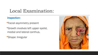
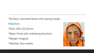
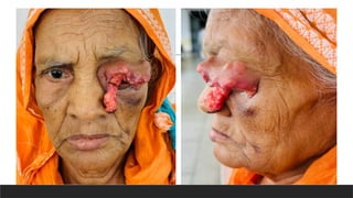
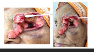
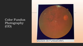
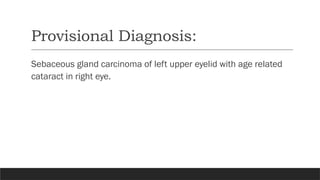
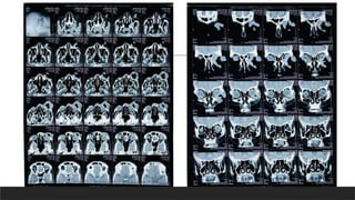
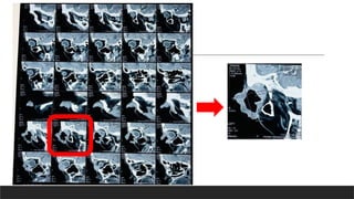
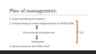
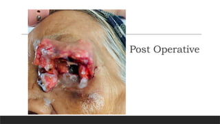
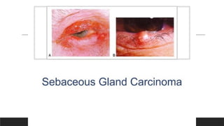
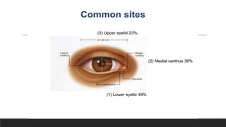
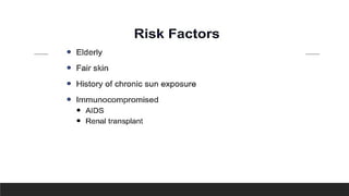
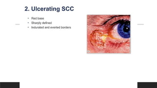
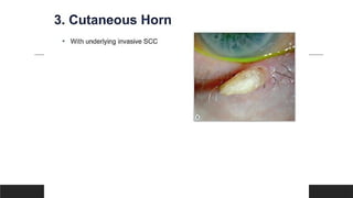
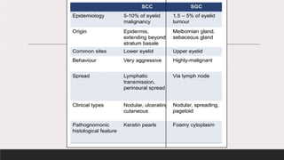
DR.MALIHA NAWAR
Resident, Phase-B
Community
Ophthalmology
Bangabandhu Sheikh
Mujib Medical
University
CASE
PRSENTATION
Particulars of the patient:
ļ‚§Name- Rahima Begum
ļ‚§Age- 80 years
ļ‚§Sex- female
ļ‚§Religion- Islam
ļ‚§ Occupation- Housewife
ļ‚§Address- Khilgoan,Dhaka.
ļ‚§Date of examination- 04/06/2024
Case Summary:
Mrs. Rahima Begum,80 years old female residing in
khilgoan,Dhaka came to BSMMU Community Ophthalmology OPD
with complaints of growth over left upper eyelid for 1.5 years. She
was reasonably well 1.5 years back then she developed a small
nodule on left upper eyelid which was painless and slowly growing
in nature. She gave history of taking homeopathy for the growth in
hope of cure, instead of visiting doctors.
But in last 5 months the growth increased rapidly. She also
complaint of loss of appetite for same duration. Now she has
come to BSMMU for better management. She is diabetic and
hypertensive. She has no history of ocular trauma or surgery. None
of her family members have such type of illness.
General Examination:
ļ‚§Appearance- ill – looking
ļ‚§Pulse- 68b/min
ļ‚§Blood pressure 90/60mmHg
ļ‚§Temp. 98F
ļ‚§Lymph node- Not palpable
ļ‚§Other vital parameters are within normal limit
Local Examination:
Inspection:
ļ‚§Facial asymmetry present
ļ‚§Growth involves left upper eyelid,
medial and lateral canthus.
ļ‚§Shape: Irregular
ļ‚§Surface: ulcerated lesion with oozing margin
Palpation:
ļ‚§Size: (30⨉12⨉2)mm
ļ‚§Base :Fixed with underlying structure
ļ‚§Margin: Irregular
ļ‚§Mobility: Non-mobile
Ocular Examination:
R/E L/E
Visual acuity: (unaided)
Distant
Near
Pinhole
HM
N12
6/60
NPL
BCVA 6/36,+1.50Ds*+1.00Dcyl@
90˚
N8, +2.50D sph
Could
Not
Pupillary Light Reflex Brisk Be
Ocular motility Full in all gazes Evaluated
Color vision Trichromatic
Slit Lamp Examination:
OD OS
Eyelid and eyelashes Normal Growth involves left upper eyelid,
medial and lateral
canthus(30 12 2)mm, Eyelash
⨉ ⨉
absent
Conjunctiva Not congested Could not be assed
Cornea Transparent Growth involves 6 to 12 o’clock
position
Anterior chamber Normal in depth both centrally and
peripherally.
Could
Iris Normal in color and pattern Not
Pupil Round, regular and reacting Be
Lens Nuclear sclerosis grade
IV+cortical+PSC
Evaluated
IOP(GAT on 04/16/2024@ 11.10am)
mmHg
13
Fundoscopic examination:
OD OS
Media Clear
Optic disc C:D= 0.3:1
Pink
Margin well defined.
Could
Blood vessel Number of blood vessels
normal.
Not
Be
NRR Healthy Evaluated
Background fundus Tessellated,RPE atrophy
present
Macula Healthy
Color Fundus
Photography
(OD)
Provisional Diagnosis:
Sebaceous gland carcinoma of left upper eyelid with age related
cataract in right eye.
Differential Diagnosis:
Squamous cell carcinoma.
Investigations:
ļ‚§CBC with ESR.
ļ‚§RBS
ļ‚§Blood grouping and Rh typing
ļ‚§Serum creatinine
ļ‚§SGPT
ļ‚§Urine R/E
ļ‚§ECG
ļ‚§ECHO
ļ‚§HbsAg
ļ‚§Anti-HCV
ļ‚§CT scan of orbit with contrast
Plan of management:
1. Proper counselling of the patient
2. Incisional biopsy to confirm diagnosis( Done on 06/06/2024)
Chemo therapy (to reduce size) L/E
Exenteration
3. Cataract extraction with PCIOL in R/E
Post Operative
Thank You

Sebaceous carcinoma case presentation...

  • 1.
    DR.MALIHA NAWAR Resident, Phase-B Community Ophthalmology BangabandhuSheikh Mujib Medical University CASE PRSENTATION
  • 2.
    Particulars of thepatient: ļ‚§Name- Rahima Begum ļ‚§Age- 80 years ļ‚§Sex- female ļ‚§Religion- Islam ļ‚§ Occupation- Housewife ļ‚§Address- Khilgoan,Dhaka. ļ‚§Date of examination- 04/06/2024
  • 3.
    Case Summary: Mrs. RahimaBegum,80 years old female residing in khilgoan,Dhaka came to BSMMU Community Ophthalmology OPD with complaints of growth over left upper eyelid for 1.5 years. She was reasonably well 1.5 years back then she developed a small nodule on left upper eyelid which was painless and slowly growing in nature. She gave history of taking homeopathy for the growth in hope of cure, instead of visiting doctors.
  • 4.
    But in last5 months the growth increased rapidly. She also complaint of loss of appetite for same duration. Now she has come to BSMMU for better management. She is diabetic and hypertensive. She has no history of ocular trauma or surgery. None of her family members have such type of illness.
  • 5.
    General Examination: ļ‚§Appearance- ill– looking ļ‚§Pulse- 68b/min ļ‚§Blood pressure 90/60mmHg ļ‚§Temp. 98F ļ‚§Lymph node- Not palpable ļ‚§Other vital parameters are within normal limit
  • 6.
    Local Examination: Inspection: ļ‚§Facial asymmetrypresent ļ‚§Growth involves left upper eyelid, medial and lateral canthus. ļ‚§Shape: Irregular
  • 7.
    ļ‚§Surface: ulcerated lesionwith oozing margin Palpation: ļ‚§Size: (30⨉12⨉2)mm ļ‚§Base :Fixed with underlying structure ļ‚§Margin: Irregular ļ‚§Mobility: Non-mobile
  • 10.
    Ocular Examination: R/E L/E Visualacuity: (unaided) Distant Near Pinhole HM N12 6/60 NPL BCVA 6/36,+1.50Ds*+1.00Dcyl@ 90˚ N8, +2.50D sph Could Not Pupillary Light Reflex Brisk Be Ocular motility Full in all gazes Evaluated Color vision Trichromatic
  • 11.
    Slit Lamp Examination: ODOS Eyelid and eyelashes Normal Growth involves left upper eyelid, medial and lateral canthus(30 12 2)mm, Eyelash ⨉ ⨉ absent Conjunctiva Not congested Could not be assed Cornea Transparent Growth involves 6 to 12 o’clock position Anterior chamber Normal in depth both centrally and peripherally. Could Iris Normal in color and pattern Not Pupil Round, regular and reacting Be Lens Nuclear sclerosis grade IV+cortical+PSC Evaluated IOP(GAT on 04/16/2024@ 11.10am) mmHg 13
  • 12.
    Fundoscopic examination: OD OS MediaClear Optic disc C:D= 0.3:1 Pink Margin well defined. Could Blood vessel Number of blood vessels normal. Not Be NRR Healthy Evaluated Background fundus Tessellated,RPE atrophy present Macula Healthy
  • 13.
  • 14.
    Provisional Diagnosis: Sebaceous glandcarcinoma of left upper eyelid with age related cataract in right eye.
  • 15.
  • 16.
    Investigations: ļ‚§CBC with ESR. ļ‚§RBS ļ‚§Bloodgrouping and Rh typing ļ‚§Serum creatinine ļ‚§SGPT ļ‚§Urine R/E ļ‚§ECG ļ‚§ECHO ļ‚§HbsAg ļ‚§Anti-HCV ļ‚§CT scan of orbit with contrast
  • 19.
    Plan of management: 1.Proper counselling of the patient 2. Incisional biopsy to confirm diagnosis( Done on 06/06/2024) Chemo therapy (to reduce size) L/E Exenteration 3. Cataract extraction with PCIOL in R/E
  • 21.
  • 43.