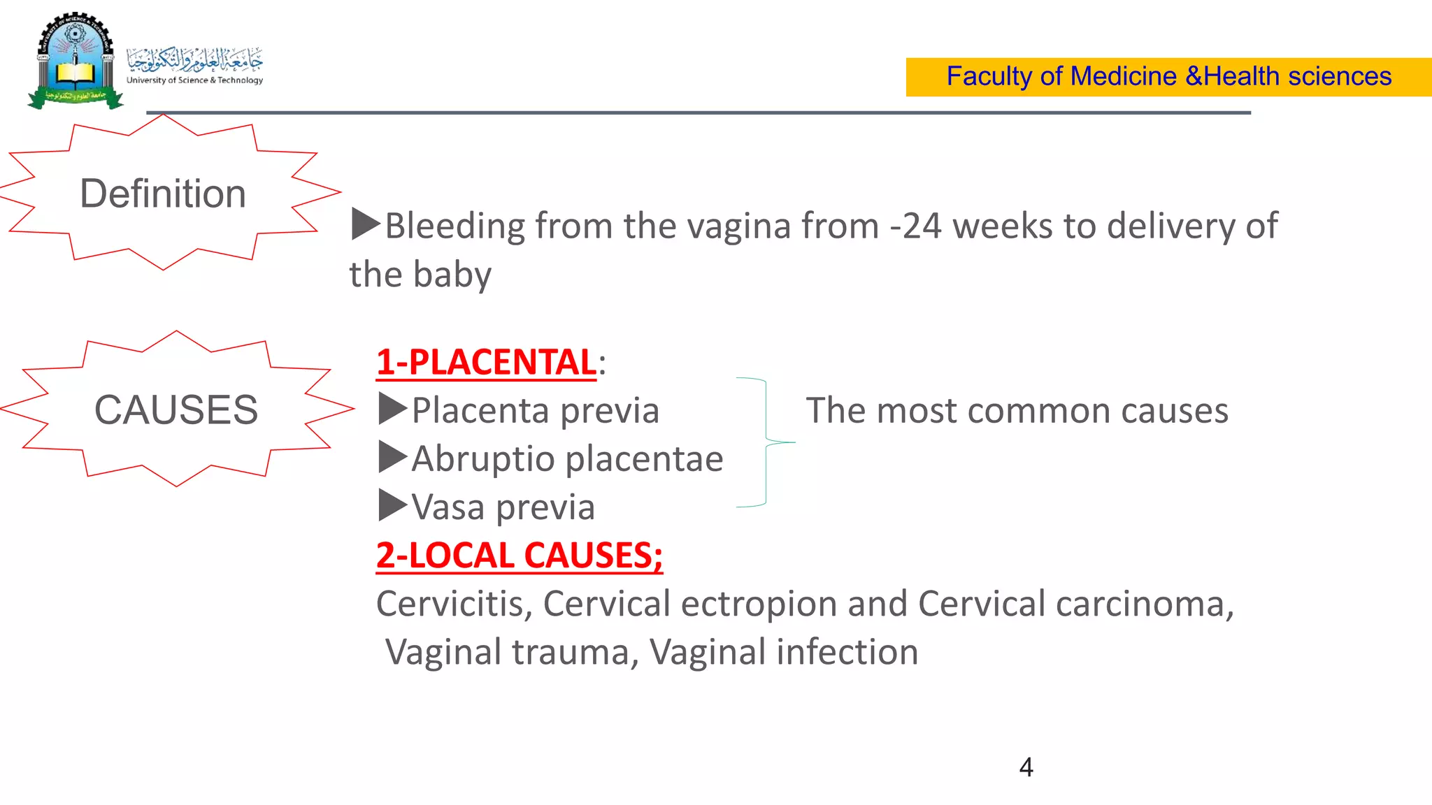
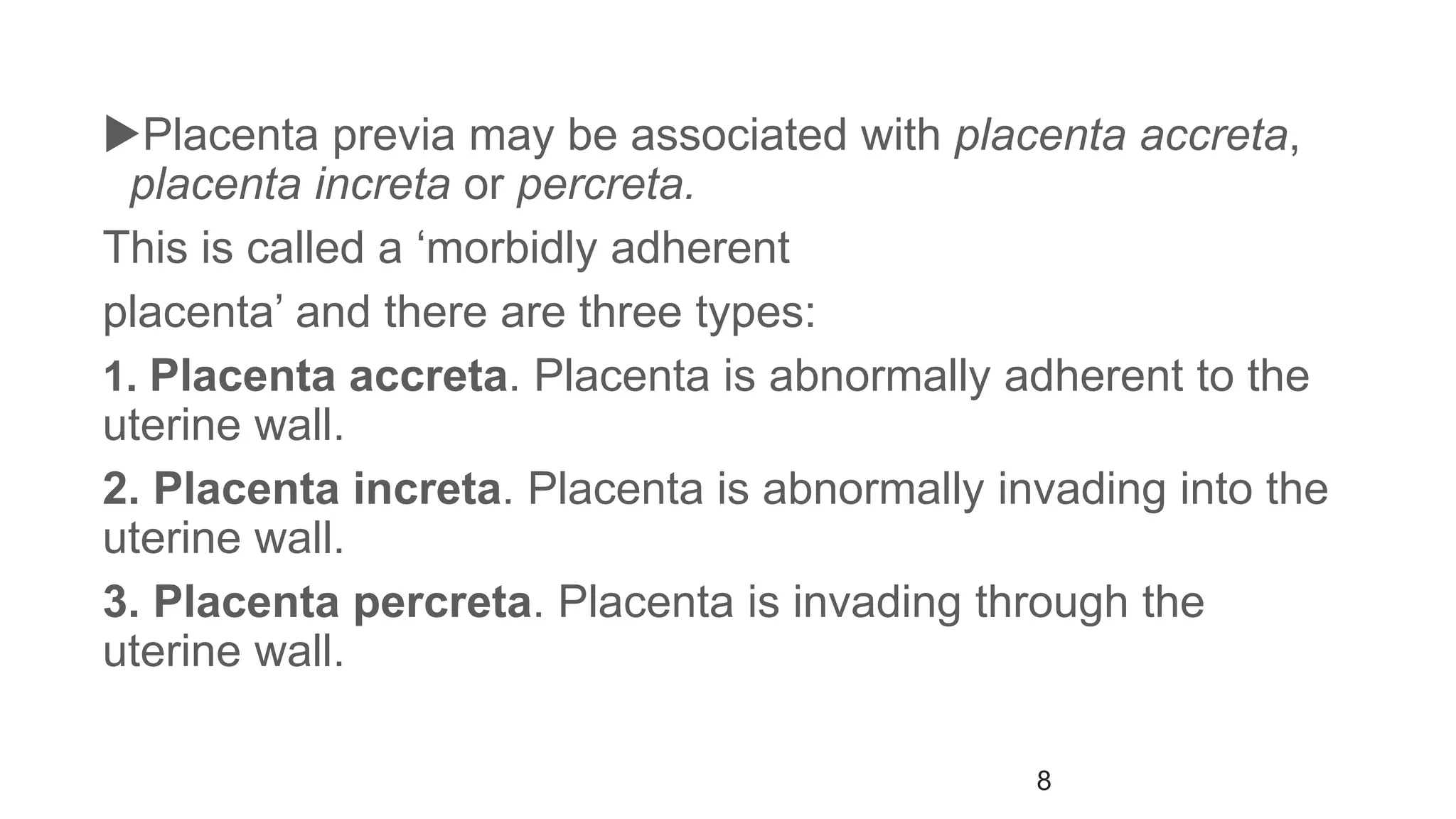
- OB&GY BLOCK(5th –level- O&G
DEPARTMENT)
Course Code -BMD 36
Faculty of medicine& Health sciences
Obstetrics & Gynecological Department
Bachelor of Medicine & Surgery
Dr. Maha.Abdulaziz
E-mail: m.abdulaziz2@ust.edu
1
Faculty of Medicine &Health sciences
Learning
outcomes
Be able to manage a case of miscarriage
Have a detailed knowledge about the clinical
presentation of miscarriage
IDENTIFY CAUSES OF APH.
Evaluate a case of APH.
DIFFERENTIAT BETWEEN DIFFERENT TYPES OF APH.
DIAGNOSE APH.
MANAGE A CASE OF APH
By the end of this topic you should
:
Objectives
Faculty of Medicine &Health sciences
2
ANTIPARTU
M
HEMORRHA
GE
Lecture contents
CONCLUSION
APH
DefinitionS
Causes &RISK FACTORS
Faculty of Medicine &Health sciences
CLINICAL PICTURES
MANAGEMENT
3
Definition
Faculty of Medicine &Health sciences
Bleeding from the vagina from -24 weeks to delivery of
the baby
CAUSES
1-PLACENTAL:
Placenta previa The most common causes
Abruptio placentae
Vasa previa
2-LOCAL CAUSES;
Cervicitis, Cervical ectropion and Cervical carcinoma,
Vaginal trauma, Vaginal infection
4
5
The incidence
 3 % of all pregnancies.1 % to placenta praevia, 1 % placental abruption
and the remaining 1 % is from other causes
An OBSTETRIC EMERGENCY!
One of the leading causes of:
antenatal hospitalization
maternal morbidity and mortality
operative intervention
Faculty of Medicine &Health sciences
5
6
Evaluation of APH?
History Examination Investigations
Faculty of Medicine &Health sciences
6
Placenta
previa
Faculty of Medicine &Health sciences
7
the implantation of the placenta in the lower
uterine segment with different grades of
encroachment on the cervix.
Placenta previa may be associated with placenta accreta,
placenta increta or percreta.
This is called a ‘morbidly adherent
placenta’ and there are three types:
1. Placenta accreta. Placenta is abnormally adherent to the
uterine wall.
2. Placenta increta. Placenta is abnormally invading into the
uterine wall.
3. Placenta percreta. Placenta is invading through the
uterine wall.
8
9
1. Low-lying placenta.
2. Marginal placenta previa.
3. Partial placenta previa.
4. Total placenta previa.
Grading (types)
Diagnosis of Placenta Previa)
Faculty of Medicine &Health sciences
• Clinical
presentation
•1-History- painless, vaginal bleeding
•2- Examination.
•-General examination
•-Abdominal obstetric palpation.
-Vaginal examination is contraindicated
U/S Localization of placenta
Examination in the
theatre
Double set up examination
10
11
Placenta Previa
Management)
Faculty of Medicine &Health sciences
11
Dependent on:
Gestational age of fetus
Amount of bleeding
Fetal condition
The bleeding is relatively minor and the fetus uncompromised
Admitted for observation for at least 24 hours has passed without
further bleeding.
Women with placenta praevia centralise who have had recurrent
bleeding should be admitted as inpatients from 34 weeks careful
risk assessment
12
Preterm with resolution
of bleeding
Faculty of Medicine &Health sciences
12
Bedrest, Rh-immune globulin, Tocolytics(Magnesium sulfate)
Corticosteroids
The indications for delivery
Reaching 37–38 weeks gestation.
Massive (1500 mL) bleed.
Continuing significant bleeding
CESAREAN DELIVERY
Complete previa at term
Persistent bleeding in pre-term patient
13
Faculty of Medicine &Health sciences
13
Premature separation of the normally implanted
placenta after 24th week of pregnancy (50%) and
before birth of the baby(15%).
Occurs in 0.4 and 2.0 % of pregnancies.
•Recurs in 10-15% of cases
•In 5% of these women DIC occurs
Abruption placenta
14
Faculty of Medicine &Health sciences
14
RISK FACTORS
Preeclampsia
External trauma
Increased parity
Over distension
of the uterus.
Sudden
decompression of
the uterus.
Cigarette
smoking
Cocaine use
Prior abruption
Chronic
hypertension
Anticoagulant
therapy
Bleeding may be
Bleeding may be
15
Faculty of Medicine &Health sciences
Clinical presentation History :The hallmark symptom of placental
abruption is pain
•Vaginal bleeding (dark).
•Symptoms of hypovolemia
•Symptoms of pre-eclampsia.
•The presence or absence of fetal movements.
•History of trauma.
Physical examination General examination:-
Abdominal examination:
A uterus that is very hard on palpation is known as
a Couvelaire uterus
Laboratory investigations: -ABO blood group and Rh type,Cross match at
2 units of blood ,Fibrinogen, PTT, PT,CT,Serum
creatinine &Urine analysis for protein and RBCs
15
DIAGNOSIS
16
Faculty of Medicine &Health sciences
16
TREATMENT
Treatment for placental abruption varies depending on gestational age and the
status of the mother and fetus.
 Principle of management:
 Resuscitation (ABC).
 Recognization of the problem.
 Early delivery (50% of abruption present in labor)
Conservative management.
Smaller degrees of abruption
No fetal distress.
Preterm
Close monitoring of fetal well-being, using ultrasound scans of fetal growth,
Amniotic fluid volume, umbilical artery Doppler and cardiotocography.
17
Faculty of Medicine &Health sciences
17
Induction of labor:
In very severe cases-AFTERSTABILIZING
MOTHER + DEAD FETUS
Caesarean section
If the fetus is alive, the mother’s resuscitation is
urgent-C/C
18
Faculty of Medicine &Health sciences
Complications of abruption placentae
18
Uterine atony PPH MATERNAL SHOCK
prolonged hypotension Renal failure maternal
death
Amniotic fluid embolism
Caogulopathy( 30%)
Fetal death
19
Faculty of Medicine &Health sciences
19
The presence of fetal vessels from the placenta crossing the internal
os of the cervix
Associated with velamentous insertion of the umbilical cord (1% of
deliveries)
Succenturiate lobe
Risk factors
Placenta praevia,
Velamentous placental insertion
Multiple pregnancy.
20
Faculty of Medicine &Health sciences
20
The diagnosis is usually suspected when either
spontaneous or artificial rupture of the membranes is
accompanied by painless fresh vaginal bleeding
Rupture of vasa may occur Initial fetal tachycardia—
fetus attempts to compensate for acute blood loss then
bradycardia, acute fetal exsanguination and death.
If the baby is still alive, the immediate action is delivery by
emergency Caesarean section
21
Faculty of Medicine &Health sciences
21
If diagnosed prenatally:
 tocolytics, bedrest
 no vaginal exams and regular scans
 Planned cesarean section
 If PV bleeding intrapartum, Investigate for the source of
bleeding BY Apt test
22
Faculty of Medicine &Health sciences
22
The test allows the clinician to determine whether
the blood originates from the infant or from the
mother.
Place 5 mL water in each of 2 test tubes
To 1 test tube add 5 drops of vaginal blood
To other add 5 drops of maternal (adult) blood
Add 6 drops 10% NaOH to each tube
Observe for 2 minutes
Maternal (adult) blood turns yellow-green-
brown; fetal blood stays pink.
23
Faculty of Medicine &Health sciences
23
CONCLUSION
1-An OBSTETRIC EMERGENCY
2- COMMONLY CAUSED BY: PLACENTAL PREVIA
ABRUPTIO PLACENTA
VASA PREVIA
3- ONE OF THA MAJOR CAUSES OF MATERNAL DEATH
APH

ANTIPARTUM HEMORRHAG( APH).ppt

  • 1.
    - OB&GY BLOCK(5th–level- O&G DEPARTMENT) Course Code -BMD 36 Faculty of medicine& Health sciences Obstetrics & Gynecological Department Bachelor of Medicine & Surgery Dr. Maha.Abdulaziz E-mail: m.abdulaziz2@ust.edu 1 Faculty of Medicine &Health sciences
  • 2.
    Learning outcomes Be able tomanage a case of miscarriage Have a detailed knowledge about the clinical presentation of miscarriage IDENTIFY CAUSES OF APH. Evaluate a case of APH. DIFFERENTIAT BETWEEN DIFFERENT TYPES OF APH. DIAGNOSE APH. MANAGE A CASE OF APH By the end of this topic you should : Objectives Faculty of Medicine &Health sciences 2 ANTIPARTU M HEMORRHA GE
  • 3.
    Lecture contents CONCLUSION APH DefinitionS Causes &RISKFACTORS Faculty of Medicine &Health sciences CLINICAL PICTURES MANAGEMENT 3
  • 4.
    Definition Faculty of Medicine&Health sciences Bleeding from the vagina from -24 weeks to delivery of the baby CAUSES 1-PLACENTAL: Placenta previa The most common causes Abruptio placentae Vasa previa 2-LOCAL CAUSES; Cervicitis, Cervical ectropion and Cervical carcinoma, Vaginal trauma, Vaginal infection 4
  • 5.
    5 The incidence  3% of all pregnancies.1 % to placenta praevia, 1 % placental abruption and the remaining 1 % is from other causes An OBSTETRIC EMERGENCY! One of the leading causes of: antenatal hospitalization maternal morbidity and mortality operative intervention Faculty of Medicine &Health sciences 5
  • 6.
    6 Evaluation of APH? HistoryExamination Investigations Faculty of Medicine &Health sciences 6
  • 7.
    Placenta previa Faculty of Medicine&Health sciences 7 the implantation of the placenta in the lower uterine segment with different grades of encroachment on the cervix.
  • 8.
    Placenta previa maybe associated with placenta accreta, placenta increta or percreta. This is called a ‘morbidly adherent placenta’ and there are three types: 1. Placenta accreta. Placenta is abnormally adherent to the uterine wall. 2. Placenta increta. Placenta is abnormally invading into the uterine wall. 3. Placenta percreta. Placenta is invading through the uterine wall. 8
  • 9.
    9 1. Low-lying placenta. 2.Marginal placenta previa. 3. Partial placenta previa. 4. Total placenta previa. Grading (types)
  • 10.
    Diagnosis of PlacentaPrevia) Faculty of Medicine &Health sciences • Clinical presentation •1-History- painless, vaginal bleeding •2- Examination. •-General examination •-Abdominal obstetric palpation. -Vaginal examination is contraindicated U/S Localization of placenta Examination in the theatre Double set up examination 10
  • 11.
    11 Placenta Previa Management) Faculty ofMedicine &Health sciences 11 Dependent on: Gestational age of fetus Amount of bleeding Fetal condition The bleeding is relatively minor and the fetus uncompromised Admitted for observation for at least 24 hours has passed without further bleeding. Women with placenta praevia centralise who have had recurrent bleeding should be admitted as inpatients from 34 weeks careful risk assessment
  • 12.
    12 Preterm with resolution ofbleeding Faculty of Medicine &Health sciences 12 Bedrest, Rh-immune globulin, Tocolytics(Magnesium sulfate) Corticosteroids The indications for delivery Reaching 37–38 weeks gestation. Massive (1500 mL) bleed. Continuing significant bleeding CESAREAN DELIVERY Complete previa at term Persistent bleeding in pre-term patient
  • 13.
    13 Faculty of Medicine&Health sciences 13 Premature separation of the normally implanted placenta after 24th week of pregnancy (50%) and before birth of the baby(15%). Occurs in 0.4 and 2.0 % of pregnancies. •Recurs in 10-15% of cases •In 5% of these women DIC occurs Abruption placenta
  • 14.
    14 Faculty of Medicine&Health sciences 14 RISK FACTORS Preeclampsia External trauma Increased parity Over distension of the uterus. Sudden decompression of the uterus. Cigarette smoking Cocaine use Prior abruption Chronic hypertension Anticoagulant therapy Bleeding may be Bleeding may be
  • 15.
    15 Faculty of Medicine&Health sciences Clinical presentation History :The hallmark symptom of placental abruption is pain •Vaginal bleeding (dark). •Symptoms of hypovolemia •Symptoms of pre-eclampsia. •The presence or absence of fetal movements. •History of trauma. Physical examination General examination:- Abdominal examination: A uterus that is very hard on palpation is known as a Couvelaire uterus Laboratory investigations: -ABO blood group and Rh type,Cross match at 2 units of blood ,Fibrinogen, PTT, PT,CT,Serum creatinine &Urine analysis for protein and RBCs 15 DIAGNOSIS
  • 16.
    16 Faculty of Medicine&Health sciences 16 TREATMENT Treatment for placental abruption varies depending on gestational age and the status of the mother and fetus.  Principle of management:  Resuscitation (ABC).  Recognization of the problem.  Early delivery (50% of abruption present in labor) Conservative management. Smaller degrees of abruption No fetal distress. Preterm Close monitoring of fetal well-being, using ultrasound scans of fetal growth, Amniotic fluid volume, umbilical artery Doppler and cardiotocography.
  • 17.
    17 Faculty of Medicine&Health sciences 17 Induction of labor: In very severe cases-AFTERSTABILIZING MOTHER + DEAD FETUS Caesarean section If the fetus is alive, the mother’s resuscitation is urgent-C/C
  • 18.
    18 Faculty of Medicine&Health sciences Complications of abruption placentae 18 Uterine atony PPH MATERNAL SHOCK prolonged hypotension Renal failure maternal death Amniotic fluid embolism Caogulopathy( 30%) Fetal death
  • 19.
    19 Faculty of Medicine&Health sciences 19 The presence of fetal vessels from the placenta crossing the internal os of the cervix Associated with velamentous insertion of the umbilical cord (1% of deliveries) Succenturiate lobe Risk factors Placenta praevia, Velamentous placental insertion Multiple pregnancy.
  • 20.
    20 Faculty of Medicine&Health sciences 20 The diagnosis is usually suspected when either spontaneous or artificial rupture of the membranes is accompanied by painless fresh vaginal bleeding Rupture of vasa may occur Initial fetal tachycardia— fetus attempts to compensate for acute blood loss then bradycardia, acute fetal exsanguination and death. If the baby is still alive, the immediate action is delivery by emergency Caesarean section
  • 21.
    21 Faculty of Medicine&Health sciences 21 If diagnosed prenatally:  tocolytics, bedrest  no vaginal exams and regular scans  Planned cesarean section  If PV bleeding intrapartum, Investigate for the source of bleeding BY Apt test
  • 22.
    22 Faculty of Medicine&Health sciences 22 The test allows the clinician to determine whether the blood originates from the infant or from the mother. Place 5 mL water in each of 2 test tubes To 1 test tube add 5 drops of vaginal blood To other add 5 drops of maternal (adult) blood Add 6 drops 10% NaOH to each tube Observe for 2 minutes Maternal (adult) blood turns yellow-green- brown; fetal blood stays pink.
  • 23.
    23 Faculty of Medicine&Health sciences 23 CONCLUSION 1-An OBSTETRIC EMERGENCY 2- COMMONLY CAUSED BY: PLACENTAL PREVIA ABRUPTIO PLACENTA VASA PREVIA 3- ONE OF THA MAJOR CAUSES OF MATERNAL DEATH APH