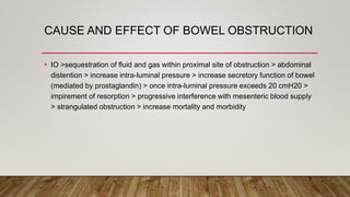
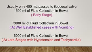
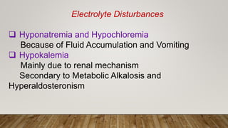
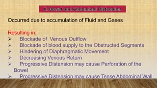
ANAESTHESIA FOR
INTESTINAL
OBSTRUCTION
DR YIN MIN THAN, DR. TENZIN YOEZER
KGUMSB
SIMPLE VS STRANGULATED BOWEL
OBSTRUCTION
• Simple obstruction – lumen is obstructed and blood supply is normal
• Mortality - <5 %
• Strangulation – obstruction of blood supply
• With stangulation – toxic fluid is discharged into lumen, bowel wall and peritoneal
cavity
• Carries high mortality – 8 – 25%
SMALL BOWEL AND LARGE BOWEL
• Small bowel – bowel distension with progressive accumulation of fluid and gas,
and quick systemic derangement.
• Large bowel – insidious onset( large capacity), less propensity to strangulate.
• Systemic derangement is less severe
SIGNS AND SYMPTOMS OF INTESTINAL
OBSTRUCTION
• Four cardinal symptoms:
• Crampy abdominal
• Nausea and vomiting
• Obstipation
• Abdominal distension
Vomiting is the earliest sign
Abdominal distension – late sign
Other signs:
Localized tenderness
Fever
Tachycardia
Leukocytosis
WBC – 15,000 – 25,000 –strong predictor
CAUSE AND EFFECT OF BOWEL OBSTRUCTION
• IO >sequestration of fluid and gas within proximal site of obstruction > abdominal
distention > increase intra-luminal pressure > increase secretory function of bowel
(mediated by prostaglandin) > once intra-luminal pressure exceeds 20 cmH20 >
impirement of resorption > progressive interference with mesenteric blood supply
> strangulated obstruction > increase mortality and morbidity
Perioperative Management
1.Fluid and Electrolyte Imbalance
2. Bowel and Abdominal Distension
3. Respiratory Problem
4. CVS Problem
5. Vomiting , Regurgitation and Aspiration
6. Investigation
7. Premedication
DEHYDRATION
 Normally 7-9 L of Fluids are secreted to the upper GI tract Daily
 In small intestinal obstruction , Fluid Loss occur due to;
 Accumulation of Fluid
 Increased Secretion
 Decreased Reabsorption
 This causes loss of Isotonic Salt Water resulting in Isotonic
Contraction of ECF volume
 So; Dehydration and Increased Hematocrit occur
Usually only 400 mL passes to ileocecal valve
1500 ml of Fluid Collection in Bowel
( Early Stage)
3000 ml of Fluid Collection in Bowel
( At Well Established cases with Vomiting)
6000 ml of Fluid Collection in Bowel
( At Late Stages with Hypotension and Tachycardia)
Degree of Dehydration is evaluated by
 Duration of illness
 Presence of Vomiting
 Skin Elasticity
 Sunken Eye
 Oliguria
The Degree of ECF loss can be monitored by serial
hematocrit determinations
 Rise in Hct is directly proportional to Fluid Loss
 If Hct increases to 55 % means 40% of Plasma and
ECF volume loss
 Can there be actual losses of red cell?
 Yes: long standing obstruction, esp with strangulation –
increased permeability of bowel walls with red cell loss
 Whole or PRC may be needed
Treatment
2-6 L of IV Fluids are needed according
to the degree of dehydration by RL or NS
Electrolyte Disturbances
 Hyponatremia and Hypochloremia
Because of Fluid Accumulation and Vomiting
 Hypokalemia
Mainly due to renal mechanism
Secondary to Metabolic Alkalosis and
Hyperaldosteronism
Acid Base Imbalance
Metabolic Acidosis is more common due to
 Dehydration and Loss of Alkaline Intestinal
Secretion
 Starvation Ketoacidosis
Occurred due to accumulation of Fluid and Gases
Resulting in;
 Blockade of Venous Outflow
 Blockade of blood supply to the Obstructed Segments
 Hindering of Diaphragmatic Movement
 Decreasing Venous Return
 Progressive Distension may cause Perforation of the
Bowel
 Progressive Distension may cause Tense Abdominal Wall
Nasogastric Tube for Abdominal Decompression As
Preoperative Management
1. Reverses peristalsis
2. Need Deep Anaesthesia and Muscle Relaxants to provide
adequate Operative Condition
3.Gradual Escape of Fluid - Monitor BP frequently during the
Surgical Procedure ( Sudden Escape esp opening of cavity
can cause Hypotension)
Implication of Tense abdominal
wall
Role of Nasogastric
Aspiration:
1.Reduce Bowel Distension
2.Improve Pulmonary
Ventilation
3.Reduce Risk of Subsequent
Aspiration during Induction
of Anaesthesia and Post
Extubation
Methods of abdominal decompression:
2 types of tube- short tube for stomach
and long tube for intestinal
Sump tube – for stomach, double lumen
tube ( one for aspiration and one for air
escape)
Miller-Abbott tube – double lumen long
intestinal tube with ballon
III. Respiratory Problems
Due to
1.Abdominal Distension which hinder the Diaphragm
resulting in inadequate Ventilation. This Decrease the Vt
and FRC which cause a decrease in PaO2 and Increase
in Pa CO2
2.Weakness of Intercostal Muscle due to Hypokalemia
 Hypotension and Tachycardia up to Shock
 Due to:
Hypovolemia
Decreased VR
Septic Shock
Hyponatremia
 Arrhythymia ( Ventricular), ST segement
changes
 Due to;
HypoKalemia
V. Vomiting, Regurgitation and Aspiration
Reversal of peristalsis and mechanical
obstruction pushes the intestinal juice in addition
to the gastric juice to produce a full stomach
with an increased intra abdominal pressure
Investigation
1. X ray: X-ray Abdomen in Supine and erect
position:
To ensure diagnosis of intestinal obstruction
2. Investigation to detect complications:
Hct
WBCs
Electrolytes
ABG for acid base balance, PaO2 and
Premedication
 Avoid all oral premedication
 Avoid drugs that may inhibit respiration
 Avoid anticholinergics if there is fever or
tachycardia
 Avoid antacids or H2 blockers although there is
a risk of aspiration
They may stimulate vomiting
They are of low value if a large volume of fluids
are already sequestrated in the bowel
Intra operative Management
1. Choice of Anaesthesia
2. Monitoring
3. Induction and Intubation
4. Maintenance
5. Extubation
 Choice of Anesthesia
 Regional Anesthesia
 General Anesthesia
 Monitoring
 Standard + UOP, CVP and PCWP
Intra operative Management
Intraoperative Management
 Induction and Intubation
There is a major risk of aspiration causing very high
mortality rates.
1.Awake Intubation: if difficult intubation
 Avoid loss of protective reflexes of the larynx
against vomiting or regurgitation(thus superior
laryngeal nerve block or transtracheal inject is
not warranted)
 Intubation followed by Induction
2. Rapid Sequence Crash Induction:
 Its done in supine or lateral position with head down tilt ( 10 degree) to avoid
aspiration if vomiting occurs.
 Preoxygenation : 8-10 L of 100% O2 for 2-5 mins
 Precurarization ( defasiculation) dose of NDMR to avoid Suxamithonium
Fasiculation.
 Naso Gastric Tube should be removed before intubation to:
 Allow effective cricoid pressure
 Avoid lower esophageal sphincter dysfunction.
Intraoperative Management
 IV Agents:
1.Thiopentone is a good choice if there is NO
Hypotension.
2.Ketamine or Etomidate are good choice if
there is Hypotension.
Intraoperative Management
MAINTENANCE
 O2 + Potent Inhalational Agent+ NDMR+ IPPV
 Careful Titration of Doses of Inhalational Agents is
Needed to avoid severe Hypotension
Intraoperative Management
N2O:
It should be avoided in Bowel Obstruction because it increases Gas
Distension which increases Intra Luminal Gas Volume and Pressure
This results in:
 More Increased Abdominal Distension(blood gas coefficient
=34 times N2, i.e N2O fills 34 times rapidly than N2)
 Increased Bowel Ischemia and Necrosis
 Difficulties with Abdominal closure at the end of Surgery
BENEFITS OF HIGH CONC. OXYGEN
• Reduces incidence of surgical wound infection ( 30% vs 80%)
• Factors that play for surgical wound infection:
• Tissue oxygen tension
• Presesnce of underlying illness
• Prophylactic antibiotic use
• Intravascular volume status,
• Pts body tepmerature,
• Post op pain control
GOALS OF FLUID MANAGEMENT
• Estimating fluid deficit is extremely difficult
• Poor preop nutrition and protein loss may lead to hypoabulminemia and further
fluid loss
• Pry goal – restoration of intravascular volume ( will increase tissue oxygenation)
• 2nd goal – correction of electrolyte, acid base
• Choice of fluid – balanced salt solution eg: RL
• Guide – CVP, UOP, BP, HR, skin turgor, electrolyte
Intraoperative
Management
Extubation:
 Awake Extubation in left lateral position.
 After returning of Upper Airway Reflexes
 After Good Suctioning
Post Operative Management
 Continue the Preoperative Management
 Fluid and Electrolyte Correction
 Respiratory and CVS Monitoring
 Postoperative ileus( functional failure of normal aboral intestinal transit, usually no
painful)
 Due to Hyponatremia and Hypokalemia, hypomagnesemia
 Small bowel recover within 24 h
 Gastric mortality – 24 – 48h
 Ileus may persist in Colon for3 – 5 days
 Postoperative Abdominal Decompression
 Continued for 5-6 days
Post Operative Management
 Post operative Respiratory Problem
 Abdominal Distension is present
 Abdominal Pain is present
 Residual Effects of Inhaled and IV
Anesthetics
 Reduction of 15-20% of FRC more than a
week after any upper abdominal Surgery
 Thereofre, have decide to keep endotracheal
tube and help in ventilation
Discussion

Anesthesia for Intestinal obstruction

  • 1.
    ANAESTHESIA FOR INTESTINAL OBSTRUCTION DR YINMIN THAN, DR. TENZIN YOEZER KGUMSB
  • 2.
    SIMPLE VS STRANGULATEDBOWEL OBSTRUCTION • Simple obstruction – lumen is obstructed and blood supply is normal • Mortality - <5 % • Strangulation – obstruction of blood supply • With stangulation – toxic fluid is discharged into lumen, bowel wall and peritoneal cavity • Carries high mortality – 8 – 25%
  • 3.
    SMALL BOWEL ANDLARGE BOWEL • Small bowel – bowel distension with progressive accumulation of fluid and gas, and quick systemic derangement. • Large bowel – insidious onset( large capacity), less propensity to strangulate. • Systemic derangement is less severe
  • 4.
    SIGNS AND SYMPTOMSOF INTESTINAL OBSTRUCTION • Four cardinal symptoms: • Crampy abdominal • Nausea and vomiting • Obstipation • Abdominal distension Vomiting is the earliest sign Abdominal distension – late sign Other signs: Localized tenderness Fever Tachycardia Leukocytosis WBC – 15,000 – 25,000 –strong predictor
  • 5.
    CAUSE AND EFFECTOF BOWEL OBSTRUCTION • IO >sequestration of fluid and gas within proximal site of obstruction > abdominal distention > increase intra-luminal pressure > increase secretory function of bowel (mediated by prostaglandin) > once intra-luminal pressure exceeds 20 cmH20 > impirement of resorption > progressive interference with mesenteric blood supply > strangulated obstruction > increase mortality and morbidity
  • 6.
    Perioperative Management 1.Fluid andElectrolyte Imbalance 2. Bowel and Abdominal Distension 3. Respiratory Problem 4. CVS Problem 5. Vomiting , Regurgitation and Aspiration 6. Investigation 7. Premedication
  • 7.
    DEHYDRATION  Normally 7-9L of Fluids are secreted to the upper GI tract Daily  In small intestinal obstruction , Fluid Loss occur due to;  Accumulation of Fluid  Increased Secretion  Decreased Reabsorption  This causes loss of Isotonic Salt Water resulting in Isotonic Contraction of ECF volume  So; Dehydration and Increased Hematocrit occur
  • 8.
    Usually only 400mL passes to ileocecal valve 1500 ml of Fluid Collection in Bowel ( Early Stage) 3000 ml of Fluid Collection in Bowel ( At Well Established cases with Vomiting) 6000 ml of Fluid Collection in Bowel ( At Late Stages with Hypotension and Tachycardia)
  • 9.
    Degree of Dehydrationis evaluated by  Duration of illness  Presence of Vomiting  Skin Elasticity  Sunken Eye  Oliguria
  • 10.
    The Degree ofECF loss can be monitored by serial hematocrit determinations  Rise in Hct is directly proportional to Fluid Loss  If Hct increases to 55 % means 40% of Plasma and ECF volume loss  Can there be actual losses of red cell?  Yes: long standing obstruction, esp with strangulation – increased permeability of bowel walls with red cell loss  Whole or PRC may be needed
  • 11.
    Treatment 2-6 L ofIV Fluids are needed according to the degree of dehydration by RL or NS
  • 12.
    Electrolyte Disturbances  Hyponatremiaand Hypochloremia Because of Fluid Accumulation and Vomiting  Hypokalemia Mainly due to renal mechanism Secondary to Metabolic Alkalosis and Hyperaldosteronism
  • 13.
    Acid Base Imbalance MetabolicAcidosis is more common due to  Dehydration and Loss of Alkaline Intestinal Secretion  Starvation Ketoacidosis
  • 14.
    Occurred due toaccumulation of Fluid and Gases Resulting in;  Blockade of Venous Outflow  Blockade of blood supply to the Obstructed Segments  Hindering of Diaphragmatic Movement  Decreasing Venous Return  Progressive Distension may cause Perforation of the Bowel  Progressive Distension may cause Tense Abdominal Wall
  • 15.
    Nasogastric Tube forAbdominal Decompression As Preoperative Management 1. Reverses peristalsis 2. Need Deep Anaesthesia and Muscle Relaxants to provide adequate Operative Condition 3.Gradual Escape of Fluid - Monitor BP frequently during the Surgical Procedure ( Sudden Escape esp opening of cavity can cause Hypotension) Implication of Tense abdominal wall
  • 16.
    Role of Nasogastric Aspiration: 1.ReduceBowel Distension 2.Improve Pulmonary Ventilation 3.Reduce Risk of Subsequent Aspiration during Induction of Anaesthesia and Post Extubation Methods of abdominal decompression: 2 types of tube- short tube for stomach and long tube for intestinal Sump tube – for stomach, double lumen tube ( one for aspiration and one for air escape) Miller-Abbott tube – double lumen long intestinal tube with ballon
  • 17.
    III. Respiratory Problems Dueto 1.Abdominal Distension which hinder the Diaphragm resulting in inadequate Ventilation. This Decrease the Vt and FRC which cause a decrease in PaO2 and Increase in Pa CO2 2.Weakness of Intercostal Muscle due to Hypokalemia
  • 18.
     Hypotension andTachycardia up to Shock  Due to: Hypovolemia Decreased VR Septic Shock Hyponatremia  Arrhythymia ( Ventricular), ST segement changes  Due to; HypoKalemia
  • 19.
    V. Vomiting, Regurgitationand Aspiration Reversal of peristalsis and mechanical obstruction pushes the intestinal juice in addition to the gastric juice to produce a full stomach with an increased intra abdominal pressure
  • 20.
    Investigation 1. X ray:X-ray Abdomen in Supine and erect position: To ensure diagnosis of intestinal obstruction 2. Investigation to detect complications: Hct WBCs Electrolytes ABG for acid base balance, PaO2 and
  • 21.
    Premedication  Avoid alloral premedication  Avoid drugs that may inhibit respiration  Avoid anticholinergics if there is fever or tachycardia  Avoid antacids or H2 blockers although there is a risk of aspiration They may stimulate vomiting They are of low value if a large volume of fluids are already sequestrated in the bowel
  • 22.
    Intra operative Management 1.Choice of Anaesthesia 2. Monitoring 3. Induction and Intubation 4. Maintenance 5. Extubation
  • 23.
     Choice ofAnesthesia  Regional Anesthesia  General Anesthesia  Monitoring  Standard + UOP, CVP and PCWP Intra operative Management
  • 24.
    Intraoperative Management  Inductionand Intubation There is a major risk of aspiration causing very high mortality rates. 1.Awake Intubation: if difficult intubation  Avoid loss of protective reflexes of the larynx against vomiting or regurgitation(thus superior laryngeal nerve block or transtracheal inject is not warranted)  Intubation followed by Induction
  • 25.
    2. Rapid SequenceCrash Induction:  Its done in supine or lateral position with head down tilt ( 10 degree) to avoid aspiration if vomiting occurs.  Preoxygenation : 8-10 L of 100% O2 for 2-5 mins  Precurarization ( defasiculation) dose of NDMR to avoid Suxamithonium Fasiculation.  Naso Gastric Tube should be removed before intubation to:  Allow effective cricoid pressure  Avoid lower esophageal sphincter dysfunction. Intraoperative Management
  • 26.
    Intraoperative Management  IVAgents: 1.Thiopentone is a good choice if there is NO Hypotension. 2.Ketamine or Etomidate are good choice if there is Hypotension.
  • 27.
    Intraoperative Management MAINTENANCE  O2+ Potent Inhalational Agent+ NDMR+ IPPV  Careful Titration of Doses of Inhalational Agents is Needed to avoid severe Hypotension
  • 28.
    Intraoperative Management N2O: It shouldbe avoided in Bowel Obstruction because it increases Gas Distension which increases Intra Luminal Gas Volume and Pressure This results in:  More Increased Abdominal Distension(blood gas coefficient =34 times N2, i.e N2O fills 34 times rapidly than N2)  Increased Bowel Ischemia and Necrosis  Difficulties with Abdominal closure at the end of Surgery
  • 29.
    BENEFITS OF HIGHCONC. OXYGEN • Reduces incidence of surgical wound infection ( 30% vs 80%) • Factors that play for surgical wound infection: • Tissue oxygen tension • Presesnce of underlying illness • Prophylactic antibiotic use • Intravascular volume status, • Pts body tepmerature, • Post op pain control
  • 30.
    GOALS OF FLUIDMANAGEMENT • Estimating fluid deficit is extremely difficult • Poor preop nutrition and protein loss may lead to hypoabulminemia and further fluid loss • Pry goal – restoration of intravascular volume ( will increase tissue oxygenation) • 2nd goal – correction of electrolyte, acid base • Choice of fluid – balanced salt solution eg: RL • Guide – CVP, UOP, BP, HR, skin turgor, electrolyte
  • 31.
    Intraoperative Management Extubation:  Awake Extubationin left lateral position.  After returning of Upper Airway Reflexes  After Good Suctioning
  • 32.
    Post Operative Management Continue the Preoperative Management  Fluid and Electrolyte Correction  Respiratory and CVS Monitoring  Postoperative ileus( functional failure of normal aboral intestinal transit, usually no painful)  Due to Hyponatremia and Hypokalemia, hypomagnesemia  Small bowel recover within 24 h  Gastric mortality – 24 – 48h  Ileus may persist in Colon for3 – 5 days  Postoperative Abdominal Decompression  Continued for 5-6 days
  • 33.
    Post Operative Management Post operative Respiratory Problem  Abdominal Distension is present  Abdominal Pain is present  Residual Effects of Inhaled and IV Anesthetics  Reduction of 15-20% of FRC more than a week after any upper abdominal Surgery  Thereofre, have decide to keep endotracheal tube and help in ventilation
  • 34.