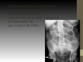
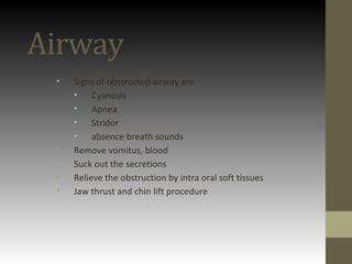
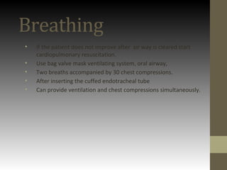
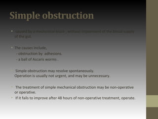
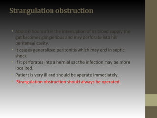
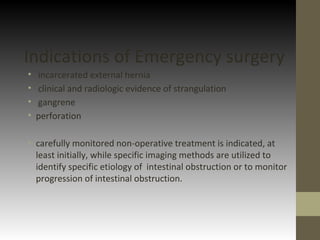
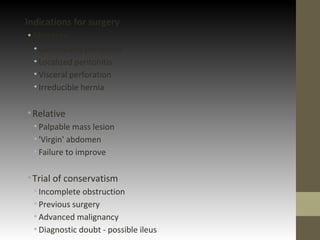
SlideShare a Scribd company logo
1 of 35
INTESTINAL
OBSTRUCTION
EmERgENCy
mANAgEmENT
Yanushka Herath
First Step
• Airway
• Breathing
• Circulation
Airway
• Signs of obstructed airway are
• Cyanosis
• Apnea
• Stridor
• absence breath sounds
• Remove vomitus, blood
• Suck out the secretions
• Relieve the obstruction by intra oral soft tissues
• Jaw thrust and chin lift procedure
Breathing
• If the patient does not improve after air way is cleared start
cardiopulmonary resuscitation.
• Use bag valve mask ventilating system, oral airway,
• Two breaths accompanied by 30 chest compressions.
• After inserting the cuffed endotracheal tube
• Can provide ventilation and chest compressions simultaneously.
Circulation
• Patient is lost large amount of fluid due to vomiting and
sequestered fluid into the gut. Two large wide bow cannulas
inserted.
• After assessing the blood pressure adequate amount of Normal
Saline or Hartman's solution is given as bolus dosage or rapid
infusion. If fails to achieve optimum BP adrenalin is given. Then
inotropes are given.
HISTORY
General
Abdominal pain –
first symptom
usually severe
colicky in nature
Abdominal distension
Vomiting
Constipation
absolute / relative
Specific
Age
•neonatal – imperforated anus, volvulus
•infants – intersusseption , strangulated hernia
•young adults and middle age – strangulated hernia ,
adhesions , IBD
•elderly – strangulated hernia , colonic CA
Previous history of surgery – post op adhesions
Complicated hernia – acute pain
Past medical history – inflammatory bowel disease ,
carcinoma of bowel or other structure
Examination
General Examination
Ill looking
Uncomfortably lying on the bed
Dyspnea
Febrile
Tachycardia
Dehydration
Abdominal Examination
Inspection
Asymmetrical abdomen
Abdominal distention
Surgical Scars
Palpation
Tenderness
Tense
Abdominal mass
Reduce or absent bowel sounds
Examine hernial orifices to exclude
strangulated hernia
Digital Rectal Examination(DRE)
• Obstructing Mass of pouch of Douglas
• Apex of an intersusseption
Investigations
• Imaging
• Haematological
• Biochemical
Imaging
Supine abdominal x-ray-
It may show abnormal bowel pattern (dilation of bowel
loops in case of obstruction or sentinel loop). It may also
show masses
Erect abdominal Xray-
Though It shows fluid levels in case of obstructed
bowel it is no longer routinely obtained.
Ultrasound-
It is less useful but may indicate presence of
intraperitoneal fluid or mass.
CT
It is performed with oral or Intravenous contrast. It
can localize the site of the obstruction. Lower
abdomen CT is useful in detection of acute
appendicitis, acute diverticulitis, intestinal
obstruction, aortic aneurysm & mesenteric
ischemia.
Radiological features of obstruction
■ The obstructed small bowel is
characterized by straight
segments that are generally central and
lie transversely. No
gas is seen in the colon
■ The jejunum is characterized by its valvulae
conniventes,
which completely pass across the width of the
bowel and
are regularly spaced, giving a ‘concertina’ or ladder
effe
■ Ileum – the distal ileum has been piquantly
described by
Wangensteen as featureless
■ Caecum – a distended caecum is shown by a
rounded gas
shadow in the right iliac fossa
■ Large bowel, except for the caecum, shows
haustral folds,
which, unlike valvulae conniventes, are spaced
irregularly,
do not cross the whole diameter of the bowel and
do not
have indentations placed opposite one another
A barium follow-through is contraindicated in
the presence of acute obstruction and may be life-
threatening.
Large bowel obstruction
Other investigation
• Full blood count
• Serum electrolytes
• Serum Creatinin
• Blood urea nitrogen
• Liver Function Test
• Serum Amylase
• Arterial blood gas
Full blood count
• Increased haemoglobin and haematocrit count.
• Due to heamoconcentration.
• Increased WBC
• Due to Infarction , Peritonitis
Serum electrolytes
• Hypokalaemia
• Due to severe emesis.
Blood urea nitrogen
• Due to vomiting and diarrhoea.
• Dehydration
Blood gas analysis.
• Metabolic acidosis
• Intestinal obstruction  ischemia  lactic acidosis.
Liver function tests.
• Increased serum bilirubin and alkaline phosphatase.
• Help to find the cause.
Serum amylase
• Increased levels due to irritation to pancreas by bowel loops. (Non
specific test.)
What is the next step ?
• NG tube placed to decompress the stomach.
• IV Fluids
• Catheterize the patient to monitor the urine out put.
• Maintain charts for pulse, BP, Temprature
• Repeated clinical assessment.
Decision Making
Simple obstruction
• caused by a mechanical block , without impairment of the blood supply
of the gut.
• The causes include,
- obstruction by adhesions.
- a ball of Ascaris worms .
• Simple obstruction may resolve spontaneously.
Operation is usually not urgent, and may be unnecessary.
• The treatment of simple mechanical obstruction may be non-operative
or operative.
• If it fails to improve after 48 hours of non-operative treatment, operate.
Strangulation obstruction
• About 6 hours after the interruption of its blood supply the
gut becomes gangrenous and may perforate into his
peritoneal cavity.
• It causes generalized peritonitis which may end in septic
shock.
• If it perforates into a hernial sac the infection may be more
localized.
• Patient is very ill and should be operate immediately.
• Strangulation obstruction should always be operated.
Indications of Emergency surgery
• incarcerated external hernia
• clinical and radiologic evidence of strangulation
• gangrene
• perforation
• carefully monitored non-operative treatment is indicated, at
least initially, while specific imaging methods are utilized to
identify specific etiology of intestinal obstruction or to monitor
progression of intestinal obstruction.
Indications for surgery
•Absolute
• Generalized peritonitis
• Localized peritonitis
• Visceral perforation
• Irreducible hernia
•Relative
• Palpable mass lesion
• 'Virgin' abdomen
• Failure to improve
•Trial of conservatism
• Incomplete obstruction
• Previous surgery
• Advanced malignancy
• Diagnostic doubt - possible ileus
Fluid Replacement
• Mechanical obstruction of small bowel causes accumulation of
fluid inside the distended bowel lumen. Therefore there is
depletion of intravascular volume & reduced perfusion of
other organs.
• Early recognition & restoration of intravascular volume to re-
establish the organ perfusion is important prior to operative
treatment, because induction of general anaesthesia in a
volume depleted patient may result in profound hypotension
& complications including renal failure.
Fluid entering small intestine per 24
hours
• Diet- 2-2.5 l
• Pancreatic juice- 0.75 – 1.5 l
• Saliva- 0.5 l
• Bile- 0.75 – 1.5 l
• Gastric juice- 1.5-3 l
• Total – 5.5-9 l
• Basic biochemical abnormality in intestinal obstruction is
sodium & water loss.
• Hartmann’s solution or normal saline can be used for fluid
replacement.
• The volume required is determined by clinical, hematological
& biochemical criteria.
• If renal functions are satisfactory & potassium is low,
potassium can be supplemented.
• If the patient is in shock, plasma expanders can be given.
• To calculate fluid losses & for accurate fluid replacement urine
output should be measured. Indwelling catheter is inserted.
• The patient should be kept nil by mouth.
Nasogastric Decompression
• This is achieved by the passage of a non-vented (Ryle) or
vented(Salem) tube.
• Tubes are normally placed on free drainage with 4 hourly
aspiration, but may be placed on continuous or intermitted
suction.
• As well as facilitating the decompression proximal to the
obstruction, they are essential to reduce the risk of
subsequent aspiration during induction of anaesthesia & post
extubation.
Thank you

More Related Content

What's hot

Management of enterocutaneous fistula
Management of enterocutaneous fistula Management of enterocutaneous fistula
Management of enterocutaneous fistula Zelalem Semegnew
 
Surgical treatment for peptic ulcer disease
Surgical treatment for peptic ulcer diseaseSurgical treatment for peptic ulcer disease
Surgical treatment for peptic ulcer diseaseBashir BnYunus
 
Management of intestinal obstruction
Management of intestinal obstructionManagement of intestinal obstruction
Management of intestinal obstructionyuyuricci
 
Gastro intestinal perforation
Gastro intestinal perforationGastro intestinal perforation
Gastro intestinal perforationsonali Harsh Raj
 
Acute and Chronic Pancreatitis
Acute and Chronic PancreatitisAcute and Chronic Pancreatitis
Acute and Chronic Pancreatitismarcosmachado
 
Intestinal Obstruction
Intestinal ObstructionIntestinal Obstruction
Intestinal ObstructionReynel Dan
 
Intestinal obstruction neo
Intestinal obstruction neoIntestinal obstruction neo
Intestinal obstruction neoNawin Kumar
 
Acute Intestinal obstruction by Dr. Daniel B. Yidana
Acute Intestinal obstruction by Dr. Daniel B. YidanaAcute Intestinal obstruction by Dr. Daniel B. Yidana
Acute Intestinal obstruction by Dr. Daniel B. YidanaDaniel Yidana
 
Large bowel obstruction
Large bowel obstructionLarge bowel obstruction
Large bowel obstructionairwave12
 
intestinal obstruction
intestinal obstructionintestinal obstruction
intestinal obstructionneha maurya
 
Cholecystectomy class
Cholecystectomy classCholecystectomy class
Cholecystectomy classsurgerymgmcri
 
SAFE LAPAROSCOPIC CHOLECYSTECTOMY
SAFE LAPAROSCOPIC CHOLECYSTECTOMYSAFE LAPAROSCOPIC CHOLECYSTECTOMY
SAFE LAPAROSCOPIC CHOLECYSTECTOMYDrAnandUjjwalSingh
 
GASTRIC OUTLET OBSTRUCTION
GASTRIC OUTLET OBSTRUCTIONGASTRIC OUTLET OBSTRUCTION
GASTRIC OUTLET OBSTRUCTIONRakesh Minocha
 
Gastric outlet obstruction
Gastric outlet obstructionGastric outlet obstruction
Gastric outlet obstructionikramdr01
 

What's hot (20)

Management of enterocutaneous fistula
Management of enterocutaneous fistula Management of enterocutaneous fistula
Management of enterocutaneous fistula
 
Surgical treatment for peptic ulcer disease
Surgical treatment for peptic ulcer diseaseSurgical treatment for peptic ulcer disease
Surgical treatment for peptic ulcer disease
 
Intestinal obstruction
Intestinal obstructionIntestinal obstruction
Intestinal obstruction
 
Management of intestinal obstruction
Management of intestinal obstructionManagement of intestinal obstruction
Management of intestinal obstruction
 
Gastro intestinal perforation
Gastro intestinal perforationGastro intestinal perforation
Gastro intestinal perforation
 
Acute and Chronic Pancreatitis
Acute and Chronic PancreatitisAcute and Chronic Pancreatitis
Acute and Chronic Pancreatitis
 
Intestinal Obstruction
Intestinal ObstructionIntestinal Obstruction
Intestinal Obstruction
 
Intestinal obstruction neo
Intestinal obstruction neoIntestinal obstruction neo
Intestinal obstruction neo
 
Acute Intestinal obstruction by Dr. Daniel B. Yidana
Acute Intestinal obstruction by Dr. Daniel B. YidanaAcute Intestinal obstruction by Dr. Daniel B. Yidana
Acute Intestinal obstruction by Dr. Daniel B. Yidana
 
Stoma
StomaStoma
Stoma
 
Large bowel obstruction
Large bowel obstructionLarge bowel obstruction
Large bowel obstruction
 
intestinal obstruction
intestinal obstructionintestinal obstruction
intestinal obstruction
 
Cholecystectomy class
Cholecystectomy classCholecystectomy class
Cholecystectomy class
 
SAFE LAPAROSCOPIC CHOLECYSTECTOMY
SAFE LAPAROSCOPIC CHOLECYSTECTOMYSAFE LAPAROSCOPIC CHOLECYSTECTOMY
SAFE LAPAROSCOPIC CHOLECYSTECTOMY
 
Paralytic ileus
Paralytic ileusParalytic ileus
Paralytic ileus
 
Splenic trauma
Splenic traumaSplenic trauma
Splenic trauma
 
GASTRIC OUTLET OBSTRUCTION
GASTRIC OUTLET OBSTRUCTIONGASTRIC OUTLET OBSTRUCTION
GASTRIC OUTLET OBSTRUCTION
 
Acute appendicitis
Acute appendicitisAcute appendicitis
Acute appendicitis
 
Gastric outlet obstruction
Gastric outlet obstructionGastric outlet obstruction
Gastric outlet obstruction
 
Colectomy
ColectomyColectomy
Colectomy
 

Viewers also liked

General management of intestinal obstruction Arindam Roy Medical College Ko...
General management  of intestinal  obstruction Arindam Roy Medical College Ko...General management  of intestinal  obstruction Arindam Roy Medical College Ko...
General management of intestinal obstruction Arindam Roy Medical College Ko...Chirantan MD
 
Intestinal obstruction
Intestinal obstructionIntestinal obstruction
Intestinal obstructionNote Noteenote
 
Medicine revision class Final year MBBS
Medicine revision class Final year MBBS Medicine revision class Final year MBBS
Medicine revision class Final year MBBS Pardeep Omani
 
Short cases in Respiration: in paediatrics-final MBBS
Short cases in Respiration: in paediatrics-final MBBSShort cases in Respiration: in paediatrics-final MBBS
Short cases in Respiration: in paediatrics-final MBBSYapa
 
Management Of Intestinal Obstruction
Management Of Intestinal ObstructionManagement Of Intestinal Obstruction
Management Of Intestinal ObstructionDr Harim Mohsin
 
Intestinal Obstruction Powerpoint Presentation
Intestinal Obstruction Powerpoint PresentationIntestinal Obstruction Powerpoint Presentation
Intestinal Obstruction Powerpoint PresentationKatherine 'Chingboo' Laud
 
Cholelithiasis final year mbbs lecture
Cholelithiasis   final year mbbs lectureCholelithiasis   final year mbbs lecture
Cholelithiasis final year mbbs lectureMr Adeel Abbas
 

Viewers also liked (9)

General management of intestinal obstruction Arindam Roy Medical College Ko...
General management  of intestinal  obstruction Arindam Roy Medical College Ko...General management  of intestinal  obstruction Arindam Roy Medical College Ko...
General management of intestinal obstruction Arindam Roy Medical College Ko...
 
Intestinal obstruction
Intestinal obstructionIntestinal obstruction
Intestinal obstruction
 
Intestinal obstruction
Intestinal obstructionIntestinal obstruction
Intestinal obstruction
 
Medicine revision class Final year MBBS
Medicine revision class Final year MBBS Medicine revision class Final year MBBS
Medicine revision class Final year MBBS
 
Short cases in Respiration: in paediatrics-final MBBS
Short cases in Respiration: in paediatrics-final MBBSShort cases in Respiration: in paediatrics-final MBBS
Short cases in Respiration: in paediatrics-final MBBS
 
intestinal obstruction
intestinal obstructionintestinal obstruction
intestinal obstruction
 
Management Of Intestinal Obstruction
Management Of Intestinal ObstructionManagement Of Intestinal Obstruction
Management Of Intestinal Obstruction
 
Intestinal Obstruction Powerpoint Presentation
Intestinal Obstruction Powerpoint PresentationIntestinal Obstruction Powerpoint Presentation
Intestinal Obstruction Powerpoint Presentation
 
Cholelithiasis final year mbbs lecture
Cholelithiasis   final year mbbs lectureCholelithiasis   final year mbbs lecture
Cholelithiasis final year mbbs lecture
 

Similar to Intestinal obstruction

Similar to Intestinal obstruction (20)

Urodynamic studies
Urodynamic studiesUrodynamic studies
Urodynamic studies
 
Acute abdomen
Acute abdomenAcute abdomen
Acute abdomen
 
Intestinal obstruction by Dr. Nitin Alapure
Intestinal obstruction by Dr. Nitin AlapureIntestinal obstruction by Dr. Nitin Alapure
Intestinal obstruction by Dr. Nitin Alapure
 
6 Intestinal Obstruction.pptx
6 Intestinal Obstruction.pptx6 Intestinal Obstruction.pptx
6 Intestinal Obstruction.pptx
 
Ude (urodynamic evaluation)
Ude (urodynamic evaluation)Ude (urodynamic evaluation)
Ude (urodynamic evaluation)
 
Cholelithiasis and cholecystitis
Cholelithiasis and cholecystitisCholelithiasis and cholecystitis
Cholelithiasis and cholecystitis
 
Chetan dialysis ppt
Chetan dialysis pptChetan dialysis ppt
Chetan dialysis ppt
 
Ascites ppts
Ascites pptsAscites ppts
Ascites ppts
 
Acute cholecystitis.pptx
Acute cholecystitis.pptxAcute cholecystitis.pptx
Acute cholecystitis.pptx
 
Cholelithiasis
Cholelithiasis Cholelithiasis
Cholelithiasis
 
Case study
Case studyCase study
Case study
 
La boob
La boobLa boob
La boob
 
Acute Pancreatitis
Acute PancreatitisAcute Pancreatitis
Acute Pancreatitis
 
intestinal obstruction in the Intestine.pptx
intestinal obstruction in the Intestine.pptxintestinal obstruction in the Intestine.pptx
intestinal obstruction in the Intestine.pptx
 
Pancreatitis.pptx
Pancreatitis.pptxPancreatitis.pptx
Pancreatitis.pptx
 
Abdominal Compartment Syndrome
Abdominal Compartment Syndrome Abdominal Compartment Syndrome
Abdominal Compartment Syndrome
 
Acute pancreatitis
Acute pancreatitisAcute pancreatitis
Acute pancreatitis
 
Pancreatitis
PancreatitisPancreatitis
Pancreatitis
 
ASCITIS IN CHILDREN BY DR VIJITHA
ASCITIS IN CHILDREN BY DR VIJITHAASCITIS IN CHILDREN BY DR VIJITHA
ASCITIS IN CHILDREN BY DR VIJITHA
 
liver cirrhosis.pptx
liver cirrhosis.pptxliver cirrhosis.pptx
liver cirrhosis.pptx
 

Intestinal obstruction

  • 2. First Step • Airway • Breathing • Circulation
  • 3. Airway • Signs of obstructed airway are • Cyanosis • Apnea • Stridor • absence breath sounds • Remove vomitus, blood • Suck out the secretions • Relieve the obstruction by intra oral soft tissues • Jaw thrust and chin lift procedure
  • 4. Breathing • If the patient does not improve after air way is cleared start cardiopulmonary resuscitation. • Use bag valve mask ventilating system, oral airway, • Two breaths accompanied by 30 chest compressions. • After inserting the cuffed endotracheal tube • Can provide ventilation and chest compressions simultaneously.
  • 5. Circulation • Patient is lost large amount of fluid due to vomiting and sequestered fluid into the gut. Two large wide bow cannulas inserted. • After assessing the blood pressure adequate amount of Normal Saline or Hartman's solution is given as bolus dosage or rapid infusion. If fails to achieve optimum BP adrenalin is given. Then inotropes are given.
  • 6. HISTORY General Abdominal pain – first symptom usually severe colicky in nature Abdominal distension Vomiting Constipation absolute / relative
  • 7. Specific Age •neonatal – imperforated anus, volvulus •infants – intersusseption , strangulated hernia •young adults and middle age – strangulated hernia , adhesions , IBD •elderly – strangulated hernia , colonic CA Previous history of surgery – post op adhesions Complicated hernia – acute pain Past medical history – inflammatory bowel disease , carcinoma of bowel or other structure
  • 8. Examination General Examination Ill looking Uncomfortably lying on the bed Dyspnea Febrile Tachycardia Dehydration
  • 9. Abdominal Examination Inspection Asymmetrical abdomen Abdominal distention Surgical Scars Palpation Tenderness Tense Abdominal mass Reduce or absent bowel sounds
  • 10. Examine hernial orifices to exclude strangulated hernia
  • 11. Digital Rectal Examination(DRE) • Obstructing Mass of pouch of Douglas • Apex of an intersusseption
  • 14. Supine abdominal x-ray- It may show abnormal bowel pattern (dilation of bowel loops in case of obstruction or sentinel loop). It may also show masses
  • 15. Erect abdominal Xray- Though It shows fluid levels in case of obstructed bowel it is no longer routinely obtained.
  • 16. Ultrasound- It is less useful but may indicate presence of intraperitoneal fluid or mass. CT It is performed with oral or Intravenous contrast. It can localize the site of the obstruction. Lower abdomen CT is useful in detection of acute appendicitis, acute diverticulitis, intestinal obstruction, aortic aneurysm & mesenteric ischemia.
  • 17. Radiological features of obstruction ■ The obstructed small bowel is characterized by straight segments that are generally central and lie transversely. No gas is seen in the colon
  • 18. ■ The jejunum is characterized by its valvulae conniventes, which completely pass across the width of the bowel and are regularly spaced, giving a ‘concertina’ or ladder effe ■ Ileum – the distal ileum has been piquantly described by Wangensteen as featureless ■ Caecum – a distended caecum is shown by a rounded gas shadow in the right iliac fossa
  • 19. ■ Large bowel, except for the caecum, shows haustral folds, which, unlike valvulae conniventes, are spaced irregularly, do not cross the whole diameter of the bowel and do not have indentations placed opposite one another A barium follow-through is contraindicated in the presence of acute obstruction and may be life- threatening.
  • 21. Other investigation • Full blood count • Serum electrolytes • Serum Creatinin • Blood urea nitrogen • Liver Function Test • Serum Amylase • Arterial blood gas
  • 22. Full blood count • Increased haemoglobin and haematocrit count. • Due to heamoconcentration. • Increased WBC • Due to Infarction , Peritonitis Serum electrolytes • Hypokalaemia • Due to severe emesis. Blood urea nitrogen • Due to vomiting and diarrhoea. • Dehydration
  • 23. Blood gas analysis. • Metabolic acidosis • Intestinal obstruction  ischemia  lactic acidosis. Liver function tests. • Increased serum bilirubin and alkaline phosphatase. • Help to find the cause. Serum amylase • Increased levels due to irritation to pancreas by bowel loops. (Non specific test.)
  • 24. What is the next step ? • NG tube placed to decompress the stomach. • IV Fluids • Catheterize the patient to monitor the urine out put. • Maintain charts for pulse, BP, Temprature • Repeated clinical assessment.
  • 26. Simple obstruction • caused by a mechanical block , without impairment of the blood supply of the gut. • The causes include, - obstruction by adhesions. - a ball of Ascaris worms . • Simple obstruction may resolve spontaneously. Operation is usually not urgent, and may be unnecessary. • The treatment of simple mechanical obstruction may be non-operative or operative. • If it fails to improve after 48 hours of non-operative treatment, operate.
  • 27. Strangulation obstruction • About 6 hours after the interruption of its blood supply the gut becomes gangrenous and may perforate into his peritoneal cavity. • It causes generalized peritonitis which may end in septic shock. • If it perforates into a hernial sac the infection may be more localized. • Patient is very ill and should be operate immediately. • Strangulation obstruction should always be operated.
  • 28. Indications of Emergency surgery • incarcerated external hernia • clinical and radiologic evidence of strangulation • gangrene • perforation • carefully monitored non-operative treatment is indicated, at least initially, while specific imaging methods are utilized to identify specific etiology of intestinal obstruction or to monitor progression of intestinal obstruction.
  • 29. Indications for surgery •Absolute • Generalized peritonitis • Localized peritonitis • Visceral perforation • Irreducible hernia •Relative • Palpable mass lesion • 'Virgin' abdomen • Failure to improve •Trial of conservatism • Incomplete obstruction • Previous surgery • Advanced malignancy • Diagnostic doubt - possible ileus
  • 30. Fluid Replacement • Mechanical obstruction of small bowel causes accumulation of fluid inside the distended bowel lumen. Therefore there is depletion of intravascular volume & reduced perfusion of other organs. • Early recognition & restoration of intravascular volume to re- establish the organ perfusion is important prior to operative treatment, because induction of general anaesthesia in a volume depleted patient may result in profound hypotension & complications including renal failure.
  • 31. Fluid entering small intestine per 24 hours • Diet- 2-2.5 l • Pancreatic juice- 0.75 – 1.5 l • Saliva- 0.5 l • Bile- 0.75 – 1.5 l • Gastric juice- 1.5-3 l • Total – 5.5-9 l
  • 32. • Basic biochemical abnormality in intestinal obstruction is sodium & water loss. • Hartmann’s solution or normal saline can be used for fluid replacement. • The volume required is determined by clinical, hematological & biochemical criteria. • If renal functions are satisfactory & potassium is low, potassium can be supplemented. • If the patient is in shock, plasma expanders can be given.
  • 33. • To calculate fluid losses & for accurate fluid replacement urine output should be measured. Indwelling catheter is inserted. • The patient should be kept nil by mouth.
  • 34. Nasogastric Decompression • This is achieved by the passage of a non-vented (Ryle) or vented(Salem) tube. • Tubes are normally placed on free drainage with 4 hourly aspiration, but may be placed on continuous or intermitted suction. • As well as facilitating the decompression proximal to the obstruction, they are essential to reduce the risk of subsequent aspiration during induction of anaesthesia & post extubation.

Editor's Notes

  1. <number>