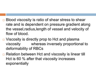
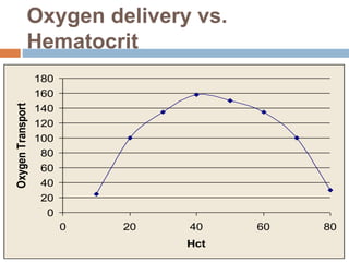
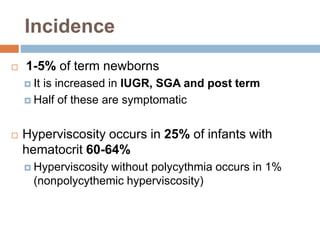
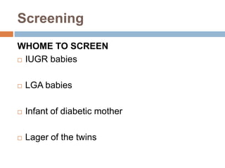
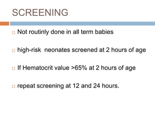
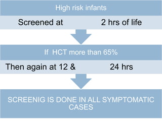
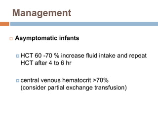
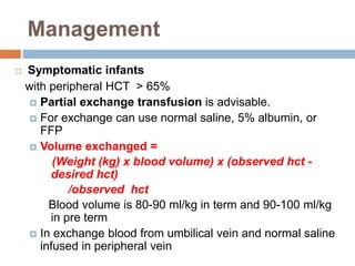
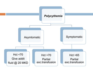
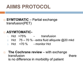
POLYCYTHEMIA IN NEWBORN
KANCHAN GAWADE
Definition
 Polycythemia ( polyglobulia)
 is increased total RBC mass
 Central venous hematocrit > 65%
 the hemoglobin is greater than 22 mg/dL
 Hct initially rises after birth due to placental transfer
of blood and decrease to baseline at 24 hrs
 Hyperviscosity is increased viscosity of the blood
more than 2 SD of mean resulting from
increased numbers of RBCs
 Blood viscosity is ratio of shear stress to shear
rate and is dependent on pressure gradient along
the vessel,radius,length of vessel and velocity of
flow of blood.
 Viscosity is directly prop to Hct and plasma
viscosity whereas inversely proportional to
deformability of RBCs
 Relation between Hct and viscosity is linear till
Hct is 60 % after that viscosity increases
exponentially
Oxygen delivery vs.
Hematocrit
0
20
40
60
80
100
120
140
160
180
0 20 40 60 80
Hct
OxygenTransport
Incidence
 1-5% of term newborns
 It is increased in IUGR, SGA and post term
 Half of these are symptomatic
 Hyperviscosity occurs in 25% of infants with
hematocrit 60-64%
 Hyperviscosity without polycythmia occurs in 1%
(nonpolycythemic hyperviscosity)
Pathophysiology
 Clinical signs result from regional effects of
hyperviscosity and from the formation of
microthrombi
 Tissue hypoxia
 Acidosis
 Hypoglycemia
 Organs affected – CNS, kidneys, adrenals,
cardiopulmonary system, GI tract
Pathophysiology of
Polycythemia
Causes
 Placental red cell transfusion --
-- Delayed cord clamping – at 1 min blood
volume of baby is 80 ml / kg.
-- At 2 min – blood vol of infant is 90 ml/kg
-- In newborn with polycythemia blood
volume
is inversly proportional to birth weight.
Causes
 Placental red cell transfusion --
--Holding baby below the mother at delivery
-- Maternal to fetal transfusion – diagnosed with
K-B test
--Twin to twin trasfusion
--forceful uterine contraction before cord
clamping
Causes
 Placental insufficiency
--Increased fetal erytropoesis d/t chronic
hypoxia
--SGA and IUGR
--Maternal hypertension like preeclampsia
--Post term
--Mothers with chronic hypoxia
(heart dis ,pulmonary dis)
--Pregnancy at hihg altitude ,maternal smoking
Other conditions
 Diabetic mother – raised erythropoiesis
 LGA
 CAH,
 patau syndrome,
 edward syndrome ,
 downs syndrome (PED)
 Maternal use of propranolol
 sepsis
Clinical features
 Mostly asymtomatic
 CNS – poor feeding ,lethargy,hypotonia, apnea,
tremers ,jitteriness,seizures, cerebral venous
thrombosis.
 CVS- cynosis, tachypnea, heart murmur, CHF,
cardiomegaly , increased pulmonary vascular
resisance,prominent vascular markings.
Clinical features
 RENAL- decreased GFR ,decrease sodium
excretion, renal vein thrombosis, Hematuria ,
proteinurea
 OTHERS – thrombosis ,thrombocytopenia,
poor feeding, decrease in calcium, NEC ,testicular
infracts, priapism, DIC
Screening
WHOME TO SCREEN
 IUGR babies
 LGA babies
 Infant of diabetic mother
 Lager of the twins
SCREENING
 Not routinly done in all term babies
 high-risk neonates screened at 2 hours of age
 If Hematocrit value >65% at 2 hours of age
 repeat screening at 12 and 24 hours.
SCREENIG IS DONE IN ALL SYMPTOMATIC
CASES
If HCT more than 65%
Then again at 12 & 24 hrs
High risk infants
Screened at 2 hrs of life
Diagnosis
 Central venous hematocrit > 65%
 ALWAYS draw a central venous sample if the
capillary hematocrit is > 65%
 Warmed capillary hematrocrit > 65% only suggestive
of polycythemia
 BLOOD VISCOSITY – may be measured where
facility available
Management
 Asymptomatic infants
 HCT 60 -70 % increase fluid intake and repeat
HCT after 4 to 6 hr
 central venous hematocrit >70%
(consider partial exchange transfusion)
Management
 Symptomatic infants
with peripheral HCT > 65%
 Partial exchange transfusion is advisable.
 For exchange can use normal saline, 5% albumin, or
FFP
 Volume exchanged =
(Weight (kg) x blood volume) x (observed hct -
desired hct)
/observed hct
Blood volume is 80-90 ml/kg in term and 90-100 ml/kg
in pre term
 In exchange blood from umbilical vein and normal saline
infused in peripheral vein
Polycythemia
Asymtomatic
Hct <70
Give addit
fluid @ 20 MKD
Hct >70
Partial
exc.transfusion
Symptomatic
Hct >65
Partial
exc.transfusion
AIIMS PROTOCOL
 SYMTOMATIC – Partial exchange
transfusion(PET)
 ASYMTOMATIC-
1. Hct >75% - transfusion
2. Hct 75 – 70 % - extra fluid alliquote @20 mkd
3. Hct <70 % - monitor Hct
 The Cochrane review – with exchange
transfusion there
is no difference in morbidity of patient
Other labs to check
 Serum glucose
 Hypoglycemia is common with polycythemia
 Serum bilirubin
 Increased bili due to increased RBC turnover
 Serum sodium, BUN, urine specific gravity
 Usually high if baby is deyhdrated
 Blood gas to rule-out inadequate oxygenation
as cause of symptoms
 Platelets, as thyrombocytopenia can be present
 Serum calcium b/c hypocalcemia can be seen
Prognosis
 Increased risk of GI disorders and NEC with
partial exchange transfusion (PET)
 PET is controversial!
 Infants with asymptomatic polycythemia have an
increased risk for neurologic sequelae.
Polycythemia   in  newborn

Polycythemia in newborn

  • 1.
  • 2.
    Definition  Polycythemia (polyglobulia)  is increased total RBC mass  Central venous hematocrit > 65%  the hemoglobin is greater than 22 mg/dL  Hct initially rises after birth due to placental transfer of blood and decrease to baseline at 24 hrs  Hyperviscosity is increased viscosity of the blood more than 2 SD of mean resulting from increased numbers of RBCs
  • 3.
     Blood viscosityis ratio of shear stress to shear rate and is dependent on pressure gradient along the vessel,radius,length of vessel and velocity of flow of blood.  Viscosity is directly prop to Hct and plasma viscosity whereas inversely proportional to deformability of RBCs  Relation between Hct and viscosity is linear till Hct is 60 % after that viscosity increases exponentially
  • 4.
  • 5.
    Incidence  1-5% ofterm newborns  It is increased in IUGR, SGA and post term  Half of these are symptomatic  Hyperviscosity occurs in 25% of infants with hematocrit 60-64%  Hyperviscosity without polycythmia occurs in 1% (nonpolycythemic hyperviscosity)
  • 6.
    Pathophysiology  Clinical signsresult from regional effects of hyperviscosity and from the formation of microthrombi  Tissue hypoxia  Acidosis  Hypoglycemia  Organs affected – CNS, kidneys, adrenals, cardiopulmonary system, GI tract
  • 7.
  • 8.
    Causes  Placental redcell transfusion -- -- Delayed cord clamping – at 1 min blood volume of baby is 80 ml / kg. -- At 2 min – blood vol of infant is 90 ml/kg -- In newborn with polycythemia blood volume is inversly proportional to birth weight.
  • 9.
    Causes  Placental redcell transfusion -- --Holding baby below the mother at delivery -- Maternal to fetal transfusion – diagnosed with K-B test --Twin to twin trasfusion --forceful uterine contraction before cord clamping
  • 10.
    Causes  Placental insufficiency --Increasedfetal erytropoesis d/t chronic hypoxia --SGA and IUGR --Maternal hypertension like preeclampsia --Post term --Mothers with chronic hypoxia (heart dis ,pulmonary dis) --Pregnancy at hihg altitude ,maternal smoking
  • 11.
    Other conditions  Diabeticmother – raised erythropoiesis  LGA  CAH,  patau syndrome,  edward syndrome ,  downs syndrome (PED)  Maternal use of propranolol  sepsis
  • 12.
    Clinical features  Mostlyasymtomatic  CNS – poor feeding ,lethargy,hypotonia, apnea, tremers ,jitteriness,seizures, cerebral venous thrombosis.  CVS- cynosis, tachypnea, heart murmur, CHF, cardiomegaly , increased pulmonary vascular resisance,prominent vascular markings.
  • 13.
    Clinical features  RENAL-decreased GFR ,decrease sodium excretion, renal vein thrombosis, Hematuria , proteinurea  OTHERS – thrombosis ,thrombocytopenia, poor feeding, decrease in calcium, NEC ,testicular infracts, priapism, DIC
  • 15.
    Screening WHOME TO SCREEN IUGR babies  LGA babies  Infant of diabetic mother  Lager of the twins
  • 16.
    SCREENING  Not routinlydone in all term babies  high-risk neonates screened at 2 hours of age  If Hematocrit value >65% at 2 hours of age  repeat screening at 12 and 24 hours.
  • 17.
    SCREENIG IS DONEIN ALL SYMPTOMATIC CASES If HCT more than 65% Then again at 12 & 24 hrs High risk infants Screened at 2 hrs of life
  • 18.
    Diagnosis  Central venoushematocrit > 65%  ALWAYS draw a central venous sample if the capillary hematocrit is > 65%  Warmed capillary hematrocrit > 65% only suggestive of polycythemia  BLOOD VISCOSITY – may be measured where facility available
  • 19.
    Management  Asymptomatic infants HCT 60 -70 % increase fluid intake and repeat HCT after 4 to 6 hr  central venous hematocrit >70% (consider partial exchange transfusion)
  • 20.
    Management  Symptomatic infants withperipheral HCT > 65%  Partial exchange transfusion is advisable.  For exchange can use normal saline, 5% albumin, or FFP  Volume exchanged = (Weight (kg) x blood volume) x (observed hct - desired hct) /observed hct Blood volume is 80-90 ml/kg in term and 90-100 ml/kg in pre term  In exchange blood from umbilical vein and normal saline infused in peripheral vein
  • 21.
    Polycythemia Asymtomatic Hct <70 Give addit fluid@ 20 MKD Hct >70 Partial exc.transfusion Symptomatic Hct >65 Partial exc.transfusion
  • 22.
    AIIMS PROTOCOL  SYMTOMATIC– Partial exchange transfusion(PET)  ASYMTOMATIC- 1. Hct >75% - transfusion 2. Hct 75 – 70 % - extra fluid alliquote @20 mkd 3. Hct <70 % - monitor Hct  The Cochrane review – with exchange transfusion there is no difference in morbidity of patient
  • 23.
    Other labs tocheck  Serum glucose  Hypoglycemia is common with polycythemia  Serum bilirubin  Increased bili due to increased RBC turnover  Serum sodium, BUN, urine specific gravity  Usually high if baby is deyhdrated  Blood gas to rule-out inadequate oxygenation as cause of symptoms  Platelets, as thyrombocytopenia can be present  Serum calcium b/c hypocalcemia can be seen
  • 24.
    Prognosis  Increased riskof GI disorders and NEC with partial exchange transfusion (PET)  PET is controversial!  Infants with asymptomatic polycythemia have an increased risk for neurologic sequelae.