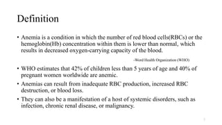
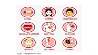
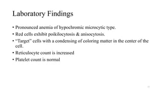
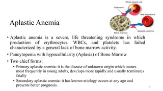
ANEMIA
Presented By:
Nishant Shrestha
M Pharm (2nd Sem)
Definition
• Anemia is a condition in which the number of red blood cells(RBCs) or the
hemoglobin(Hb) concentration within them is lower than normal, which
results in decreased oxygen-carrying capacity of the blood.
-Word Health Organization (WHO)
• WHO estimates that 42% of children less than 5 years of age and 40% of
pregnant women worldwide are anemic.
• Anemias can result from inadequate RBC production, increased RBC
destruction, or blood loss.
• They can also be a manifestation of a host of systemic disorders, such as
infection, chronic renal disease, or malignancy.
2
Fig No. 1: Regulation of Erythropoiesis
Clinical Manifestation of Anemia
• Tiredness
• Generalized muscular weakness
• Headache in older patients
• Angina pectoris
• Confusion
• Visual disturbance
• Fatigue
• Dizziness
• Irritability
• Weakness
• Lethargy
• Palpitations
• Vertigo
• Shortness of breath
• Chest pain
4
Fig No. 2: Symptoms of Anemia
5
Signs of Anemia
• Tachycardia
• Pale appearance (most prominent in conjunctiva)
• Decreased mental sharpness
• Increased intensity of some cardiac valvular murmurs
• Gait abnormality in vitamin B12 deficiency
6
Diagnosis of Anemia
History
• Diet history-vegeterian or non-vegeterian
• h/o-chronic blood loss (menorrhagia, hemorrhoids)
• h/o-drugs like anticancerous agents, chloramphenicol, penicillamine
(aplastic anemia,) Dapsone, quinine (hemolytic anemia)
• Family history of anemia (thalassemia, sickle cell anemia)
• h/o alcohol addiction
• h/o renal disease
• h/o systemic symptoms like fever, weight loss, night sweats
• Obstetric and menstrual history
7
Examination
General Examination
• Pallor
• Icterus
• Edema
• Lymphadenopathy
• Petechiae
Systemic Examination
• CVS-flow murmur, loud S1
• Chest –crepts
• P/A-splenomegaly
8
Pallor
Lymphadenopathy
Icterus
Petechiae
edema
Tests for Anemia
Complete Blood Count Tests
Test What Does This Test Do? What Clues Does It Reveal About Possible Anemia?
Red blood cell
(RBC), white blood
cell (WBC), and
platelet count
Counts all the RBCs, WBCs, and platelets
in a sample of blood. Gives information
about the size, shape, and physical
characteristics of the blood cells.
A lower than normal number of RBCs suggests anemia. Specific
changes in number, size, or shape point to whether the anemia is
caused by less production, more destruction, or loss of RBCs.
WBC differential Identifies the five types of WBCs in a
blood sample and the relative percentage
of each in the sample.
Specific types of WBCs increase in response to certain diseases and
conditions. A WBC differential can help identify a condition that may
be causing anemia.
Hemoglobin Measures the amount of oxygen-carrying
protein in the blood.
A low level of hemoglobin points to diseases (such as iron-deficiency
anemia) that usually cause the body to make too few RBCs.
Hematocrit Measures how much space RBCs take up
in your blood.
A low hematocrit level points to anemia. An abnormal hematocrit
level also may be a sign of a blood or bone marrow disorder.
Mean corpuscular
volume (MCV) Measures the average size of RBCs.
RBC size gives a clue to the type of anemia. Larger than normal
RBCs may suggest pernicious anemia caused by vitamin B12 or
folate deficiency. Smaller than normal RBCs suggest iron-deficiency
anemia or thalassemia (a rare, inherited anemia).
9
Tests for Anemia
Complete Blood Count Tests
10
Test What Does This Test Do? What Clues Does It Reveal About Possible Anemia?
Mean corpuscular
hemoglobin (MCH)
Calculates the average amount of
oxygen-carrying hemoglobin inside
an RBC
Mirrors MCV results: Larger than normal RBCs have more
oxygen-carrying hemoglobin; smaller than normal RBCs
often have less.
Mean corpuscular
hemoglobin
concentration
(MCHC)
Calculates average concentration of
hemoglobin inside an RBC.
Abnormal values may offer clues to the type of anemia or
other possible conditions.
Red cell distribution
width
Calculates the difference in size of
RBCs.
Amount of difference in size may suggest the body is trying to
make new RBCs to correct the anemia or may suggest a cause
for the anemia.
Blood smear Shows size, shape, and number of
RBCs, WBCs, and platelets. Used
when complete blood count results
are abnormal.
The presence of abnormal or immature blood cells can point
to possible causes for the anemia.
Tests for Anemia
Complete Blood Count Tests
11
Test What Does This Test Do? What Clues Does It Reveal About Possible Anemia?
Reticulocyte count Measures the number of young RBCs
in the blood. Shows whether the bone
marrow is making enough RBCs at
the correct rate or at a higher rate in
an appropriate response to the
anemia.
A markedly higher reticulocyte count may point to hemolytic
anemia. A lower reticulocyte count can point to iron-
deficiency anemia, pernicious anemia, aplastic anemia, or
other anemias caused by reduced RBC production.
Serum iron Measures the total amount of iron in the
blood.
Iron is a part of hemoglobin. Nearly all iron in the blood is
bound to a protein called transferrin. Transferrin transports
iron to the bone marrow, where hemoglobin and RBCs are
made, or to body tissues for storage. Abnormal results on
these tests can point to iron-deficiency anemia.
Total iron-binding
capacity (TIBC)
Measures the total amount of iron that
can be bound by transferrin.
Unsaturated iron-
binding capacity
(UIBC)
Determines the portion of transferrin that
is not yet saturated with iron.
Tests for Anemia
Complete Blood Count Tests
12
Test What Does This Test Do? What Clues Does It Reveal About Possible Anemia?
Transferrin
saturation
Shows the percentage of transferrin
that is saturated with iron. It is
calculated using results of serum iron,
TIBC, and UIBC tests.
Serum ferritin Reflects the amount of stored iron in your
whole body.
Coombs test Looks for antibodies directed against
RBCs.
A positive result points to the presence of antibodies (proteins
made by the immune system). These results can point to
hemolytic anemia.
G6PD test Measures the amount of glucose-6-
phosphate dehydrogenase (G6PD) in
RBCs.
If RBCs lack the enzyme G6PD, they become weak and can break
apart. (Enzymes are proteins that drive chemical reactions in the
body.) Abnormal results can point to a G6PD deficiency, an inherited
condition that can lead to hemolytic anemia.
Bone Marrow Tests
13
Test What Does This Test Do? What Clues Does It Reveal About Possible
Anemia?
Bone marrow
aspiration and
bone marrow
biopsy
Gives information about the
marrow’s ability to make
enough healthy blood cells.
(The doctor collects and
evaluates a sample of bone
marrow.)
Abnormal results showing only a few cells
in the bone marrow can point to aplastic
anemia.
14
Tab No. 1: Normal Hematologic Values
Source: Pharmacotherapy Handbook 7th edition
15
Fig No. 3: General algorithm for diagnosis of anemias
Classification of Anemia
I. Morphological classification
II. Etiological classification
III. Pathophysiology
16
I. Morphological Classification
• Macrocytic anemias
• Megaloblastic anemias
• Vitamin B12 deficiency
• Folic acid deficiency anemias
• Microcytic hypochromic anemias
• Iron-deficiency anemia
• Genetic anomaly
• Sickle cell anemia
• Thalassemia
• Other hemoglobinopathies (abnormal hemoglobins)
• Normocytic anemias
• Recent blood loss
• Hemolysis
• Bone marrow failure
• Anemia of chronic disease
• Renal failure
• Endocrine disorders
• Myelodysplastic anemias
17
II. Etiologic Classification
• Deficiency
• Iron
• Vitamin B12
• Folic acid
• Pyridoxine
• Central, caused by impaired bone marrow function
• Anemia of chronic disease
• Anemia of the elderly
• Malignant bone marrow disorders
• Peripheral
• Bleeding (hemorrhage)
• Hemolysis (hemolytic anemias)
18
III. Pathophysiology
• Excessive blood loss
• Recent hemorrhage
• Trauma
• Peptic ulcer
• Gastritis
• Hemorrhoids
• Chronic hemorrhage
• Vaginal bleeding
• Peptic ulcer
• Intestinal parasites
• Aspirin and other nonsteroidal anti-inflammatory agents
• Excessive RBC destruction
• Extracorpuscular (outside the cell) factors
• RBC antibodies
• Drugs
• Physical trauma to RBC (artificial valves)
• Excessive sequestration in the spleen
• Intracorpuscular factors
• Heredity
• Disorders of hemoglobin synthesis 19
III. Pathophysiology
• Inadequate production of mature RBCs
• Deficiency of nutrients (B12, folic acid, iron, protein)
• Deficiency of erythroblasts
• Aplastic anemia
• Isolated (often transient) erythroblastopenia
• Folic acid antagonists
• Antibodies
• Conditions with infiltration of bone marrow
• Lymphoma
• Leukemia
• Myelofibrosis
• Carcinoma
• Endocrine abnormalities
• Hypothyroidism
• Adrenal insufficiency
• Pituitary insufficiency
• Chronic renal disease
• Chronic inflammatory disease
• Granulomatous diseases
• Collagen vascular diseases
• Hepatic disease
20
REDISPOSING FACTORS:
Age: 13-30 y.o.
Gender: Female
Conditions: Pregnancy, excessive menstruation, alcoholics
Underlying disease conditions: Cancer, Kidney Diseases, Diabetes
Mellitus
PRECIPITATING FACTORS:
Dietary intake (Iron, Vitamin B12 and Folic Acid), erythropoietin
Malabsorption: L ack of intrinsic factor, Inappropriate interventions
upon intake of Supplements, Gastrectomy, Inflammatory
Bowel Disease
Folic Acid
Deficiency
Proliferation of large immature dysfunctional
RBC
Iron Deficiency
Vitamin B 12 Deficiency
Lack of hemoglobin in the RBC
Manifestations:
• Smooth sour tongue
• Diarrhea
• Paresthesias of
hands and feet
• Impaired
coordination and
position sense
Alterations of Normal production of RBCs
Manifestations:
• Pale skin
• shortness of breath
• rapid heartbeat
• low vitality
• dizziness
Decrease in oxygen Carrying Capability
Hypoxia
Hypoxemia
Organ Dysfunction
Organ Failure
DEATH
Fig No. 4: Algorithm of Pathophysiology of Anemia 21
Iron Deficiency Anemia
• Iron deficiency anemia is a condition in which the body lacks enough
red blood cell because of deficiency and inadequate bioavailability of
dietary iron.
• Iron is an essential mineral that is needed to form hemoglobin, an
oxygen carrying protein inside red blood cells.
• Defective synthesis of hemoglobin, resulting in red cells that are
smaller than normal (microcytic) and contain reduced amounts of
hemoglobin (hypochromic).
• Iron deficiency is usually easily diagnosed from the red cell indices.
22
Causes of Iron Deficiency Anemia
• Inadequate intake of Iron.
• Malabsorption of iron due to hypochlorhydria & diarrhea.
• Increased requirement of iron in a growing child & in pregnancy.
• Increased loss of iron due to injury and peptic ulcer.
• Chronic blood loss such as menstrual & menopausal bleeding
parturition.
• Gastrotomy.
23
Role of Iron
• Iron is an essential element for blood production.
• About 70 percent of your body's iron is found in the red blood cells of
blood called hemoglobin and in muscle cells called myoglobin.
• Hemoglobin is essential for transferring oxygen in blood from the
lungs to the tissues.
• About 25 percent of the iron in the body is stored as ferritin, found in
cells and circulates in the blood.
• Iron helps form and oxygenate our blood cells and hemoglobin.
• Iron helps maintain a normal immune system
24
25
Fig No. 5: Body iron distribution and storage
Iron Requirements
Age Male Female Pregnancy Lactation
Birth to 6 months 0.27 mg* 0.27 mg*
7–12 months 11 mg 11 mg
1–3 years 7 mg 7 mg
4–8 years 10 mg 10 mg
9–13 years 8 mg 8 mg
14–18 years 11 mg 15 mg 27 mg 10 mg
19–50 years 8 mg 18 mg 27 mg 9 mg
51+ years 8 mg 8 mg
Tab No. 2: Recommended Dietary Allowances (RDAs) for
Iron
Source: National Institute of Health
• Total serum iron: 26–170 mcg/dL in women and 76–198 mcg/dL in men
Clinical Features
• Pallor
• Koilonychia
• Fatigue and weakness
• Anxiety
• Irritability
• Sleepiness
• Palpitations
• Hair loss
• Fainting
• Depression
• Breathlessness
27
Koilonychia
Hematological Findings
• Microcytic & hypochromic & the peripheral smear shows abnormal
forms of RBCs.
• Reduced hemoglobin level, as low as 13.5 g/dL
• Normal or slightly reduced RBC count.
• MCV, MCH & MCHC are all reduced.
28
Bone Marrow Findings
• Cellularity – Normal
• Erythropoiesis – normoblastic erythropoiesis with small polychromatic
normoblast.
• Marrow iron – decreases and absence of siderotic granules from
developing normoblast.
Biochemical findings:
• Low serum iron level
• Total iron binding capacity is high
• Serum ferritin is low
29
Treatment
• Diet: Green leafy vegetables (Spinach, Turnip, Peas, Asparagus), nuts,
dates, meat.
• Iron therapy
• Oral therapy Ferrous sulphate 200mg/day, Ferrous fumarate 200mg/day,
Ferrous gluconate 300mg/day
• Parenteral therapy Iron dextran, Iron sorbitol citrate, Iron carbohydrate
complex
• Blood transfusion: Transfusion of packed red cells can be given if
there is excessive blood loss, congestive cardiac failure or immediate
replenishment is required.
30
Megaloblastic Anemia
• Megaloblastic anemia is a condition in which the bone marrow
produces unusually large, structurally abnormal, immature red blood
cells (megaloblasts).
• It is hematological disorder caused due to impaired DNA synthesis.
• Deficiency of Vitamin B12 & Folate or both, resulting in disordered
cell proliferation leading to Megaloblastic anemia.
31
Vitamin B12
• Important for pyrimidine synthesis in the production of DNA.
• Only Dietary source are foods of animal origin (fish, egg, cheese,
milk)
• Deficiency - delay in nuclear maturation and cell division.
• Vit B12 is synthesised in the human large bowel by bacteria but is not
absorbed, thus, humans are entirely dependent on dietary source.
• Vit B12 is stored in liver
• The stores may last for 3 years
• Daily requirement is 1-2 mcg/day
32
Vitamin B12 Deficiency
Etiology:
A. Inadequate dietary intake
B. Malabsorption
• Gastric causes - Pernicious anemia, Gastrectomy
• Intestinal causes - tropical sprue, Crohn’s disease
• Removal of B12 from intestines – Fish tape worm infestation
33
Folate (Vit B9)
Function of folate
• To synthesize DNA, RNA and to repair
• Aiding rapid cell division and growth
• To produce healthy red blood cells
• It is important for pregnant women to have enough folic acid to prevent major
birth defects of her baby's brain or spine
34
Causes of Folate Deficiency
• Inadequate dietary intake
• Alcoholism
• Malnutrition
• Excessive cooking
• Inadequate absorption
• Tropical sprue
• Non-tropical sprue
• Drugs : Phenytoin, barbiturates, methotrexate
• Increased requirement – infancy, pregnancy, lactation
• Others : Hemodialysis, hemolytic anemia
35
Clinical Features
• Fatigue
• Weakness
• Body ache
• Vertigo
• Beefy tongue
• Diarrhea
• Anorexia
• Weight loss
36
Beefy tongue
Anorexia
Laboratory Findings
• A) General laboratory findings:
• 1) Blood picture and red cell indices:
a. Hemoglobin – reduced
b. Red cell – macrocytic
c. Reticulocyte count – low
d. Red cell indices– MCV increased MCH increased, MCHC normal or reduced
e. Leucocytes – hyper segmented neutrophils
f. Platelets – moderately reduced
• 2) Bone marrow findings:
a. Cellularity – hypercellular with decreased myloid- erythroid ratio
b. Erythropoiesis – erythroid hyperplasia, megaloblasts with fenestrated chromatin
network
c. Gaint metamyelocytes may be present
37
Laboratory Findings (Cont)
• 3) Biochemical findings:
• Serum Vit B12 is reduced in Vit B12 deficiency
• Serum folate is reduced in folate deficiency
• Rise in the serum unconjugated bilirubin and LDH
• Serum iron and ferritin may be normal or elevated
• Urinary formiminoglutamate (FIGLU) levels increased
• Red cell folate is reduced in folate deficiency
38
Treatment
• Treatment of B12 deficiency
a. Diet: Non vegetarian food, diary products
b. Replacement therapy: 1000mcg B12 I.M once a day for 2 weeks. Continue
1000mcg B12 I.M per week until blood count normalizes.
• Treatment of folic acid deficiency
a. Diet: green leafy vegetables, nuts, meat
b. Replacement therapy: 5mg/day for 2-4 months
c. Folinic acid 15 mg/day used in patients on treatment with methotrexate or
trimethoprim
39
Pernicious Anemia (Addison’s Anemia,
Biermer’s Anemia, Hunter –Addison Anemia)
• Pernicious anemia is a deficiency in red blood cells caused by lack of
vitamin B12 in the blood.
• Pernicious anemia is defined as a type of vitamin B12 deficiency that
results from impaired uptake of vitamin B-12 due to the lack of a
substance known as intrinsic factor (IF) produced by the stomach
lining.
• It is autoimmune disorder, because autoantibodies to gastric parietal
cells are often found in patients.
40
Clinical Features
• Slightly more women than men are affected by pernicious anemia.
• Triad of symptoms present are:
• Generalized weakness.
• Sore painful tongue.
• Numbness / Tingling of extremities
• Exhibit yellow color on the skin & sclera.
• Paresthetic sensations of the extremities.
• Weakness, stiffness, difficulty in walking, drowsiness,
• In-coordination or loss of vibratory sensation.
• Degeneration of myelin sheaths, loss of nerve fibers.
41
Laboratory Findings
• RBC count is decreased
• Macrocytosis is present – a chief characteristic blood.
• Poikilocytosis, a variation in shape.
• WBC count & MCH decreased
• Bone marrow shows great number of immature red cells
• Achlorhydria or lack of gastric hydrochloric acid secretion is a
constant feature
• Schilling test detects the absence of intrinsic factor
42
Treatment
• Vitamin B-12 injections that are followed closely over time
• Following the blood level of vitamin B-12 over the course of therapy
• Making adjustments accordingly in vitamin B-12 dosing
43
Hemolytic Anemia
• Hemolytic anemia is defined as anemia resulting from an increase in the rate of red cell
destruction.
• When blood cells die, the body's bone marrow makes more blood cells to replace them.
However, in hemolytic anemia, the bone marrow can't make red blood cells fast enough to
meet the body's needs.
Classification:
I. ACQUIRED (EXTRA CORPUSCULAR)
A. Antibody: Immunohemolytic anemias
1. Autoimmune hemolytic anemia (AIHA)
• Warm antibody AIHA
• Cold antibody AIHA
2. Drug-induced immunohemolytic anemia
3. Iso-immune hemolytic anemia
44
Hemolytic Anemia
B. Mechanical trauma: Microangiopathic hemolytic anemia
C. Direct toxic effects: Malaria, bacterial, infection and other agents
D. Acquired red cell membrane abnormalities: paroxysmal nocturnal
hemoglobinuria (PNH)
E. Splenomegaly
II. HEREDITARY (INTRACORPUSCULAR)
A. Abnormalities of red cell membrane
1. Hereditary spherocytosis
2. Hereditary elliptocytosis (hereditary ovalocytosis)
3. Hereditary stomatocytosis
45
Hemolytic Anemia
B. Disorders of red cell interior
1. Red cell enzyme defects (Enzymopathies)
i. Defects in the hexose monophosphate shunt: G6PD deficiency
ii. Defects in the Embden-Meyerhof (or glycolytic) pathway: pyruvate kinase
deficiency
2. Disorders of hemoglobin (Hemoglobinopathies)
i. Structurally abnormal hemoglobins: sickle syndromes other
hemoglobinopathies
ii. Reduced globin chain synthesis: thalassaemias
46
Sickle Cell Anemia
• Sickle Cell Anemia is a genetic disorder that affects erythrocytes
(RBC) causing them to become sickle or crescent shaped.
• Sickle cells increase blood viscosity and tend to reduce blood flow
leading to thrombosis and tissue infarction.
• In sickle cell anemia, the red blood cells become rigid and sticky and
are shaped like sickles or crescent moons
• The effects of this condition due to an abnormality of the hemoglobin
molecules found in erythrocytes.
47
Clinical Features
• Common in females
• Clinical manifestations begin only after several months as fetal Hb
protects against sickling phenomenon.
• Fatigue, weakness and shortness of breath.
• Severe abdominal pain, muscle and joint pain, at high temperature
which may result in circulatory collapse also occur.
• Painless hematuria.
• Most of people may expire before the age of 40 years.
48
Laboratory Findings
• RBC count decrease up to 10,00,000/mm3.
• Decrease Hb level concentration 6-9 g/dl
• Blood smear shows typical sickle-shaped red cell.
• Presence of howell-jolly bodies
• Hb electrophoresis – no normal HbA, more of HbS.
• Reticulocyte count is raised.
49
Treatment
• Prevention of episode by avoiding the precipitating factors
• Folic acid supplement (5mg/daily)
• Prophylactic antibiotics are given
• Blood transfusion should be given during crisis only
50
Thalassemia 'Cooleys Anemia', 'Mediterranean
Anemia' and ‘Erythroblastic Anemia'.
• Thalassemia is an inherited blood disorder in which the body produces
an abnormal form of hemoglobin which results in excessive
destruction of red blood cells and further leads to anemia.
• Genetic defect, which could be either mutation or deletion, results in
reduced rate of synthesis or no synthesis of one of the globin chains
that make up hemoglobin.
• This can cause the formation of abnormal
hemoglobin molecules, thus causing anemia.
51
Clinical Features
• Pallor of skin , fever, chills, prominent cheek bone and mild
hepatosplenomegaly.
• Deposition of iron in various organs (due to multiple transfusions)
leads to signs and symptoms of organ failure.
• Most patients die in childhood due to anemia and cardiac failure.
• Mongoloid appearance
52
Laboratory Findings
• Pronounced anemia of hypochromic microcytic type.
• Red cells exhibit poikilocytosis & anisocytosis.
• “Target” cells with a condensing of coloring matter in the center of the
cell.
• Reticulocyte count is increased
• Platelet count is normal
53
Laboratory Findings (cont.)
• MCV, MCH, MCHC are significantly reduced
• WBC’S count increased to 10,000-25,000/mm3.
• Bone marrow cells show cellular hyperplasia
• Serum bilirubin & fecal & urinary urobilinogen are elevated because
of severe hemolysis.
54
Treatment
• Blood transfusion
• Splenectomy indicated when there is severity of anemia
• Chelating agent- iron overload can be reduced by regular use of
chelating agents like Desferrioxamine therapy.
• Folic acid owing to increase erythropoiesis.
55
Aplastic Anemia
• Aplastic anemia is a severe, life threatening syndrome in which
production of erythrocytes, WBCs, and platelets has failed
characterized by a general lack of bone marrow activity.
• Pancytopenia with hypocellularity (Aplasia) of Bone Marrow
• Two chief forms:
• Primary aplastic anemia: it is the disease of unknown origin which occurs
most frequently in young adults, develops more rapidly and usually terminates
fatally
• Secondary aplastic anemia: it has known etiology occurs at any age and
presents better prognosis. 56
Etiology
• Drugs: Methotrexate, Chloramphenicol, Sulpha drugs
• Toxic chemicals: Benzene derivatives, insecticides, arsenicals
• Infections: viral hepatitis, AIDS
• Miscellaneous: Irradiation, Pregnancy.
57
Clinical Features
• Severe weakness with dyspnea
• Exhibit skin Pallor
• Numbness & tingling of extremities
• Edema is present.
• Petechiae in the skin and mucous membrane occur, due to platelet
deficiency.
• Neutropenia leads to a decreased resistance to infection.
58
Laboratory Findings
• RBC count is remarkably reduced to 10,00,000cells/mm3.
• The absolute granulocyte count is particularly low with relative
lymphocytosis.
• Thrombocytopenia results in a prolonged BT
• CT remains normal.
• Clot retraction is poor; tourniquet test (capillary fragility test) is
positive.
• The classical finding is that of pancytopenia along with reduction of
absolute reticulocyte count
59
Treatment
• Withdrawal of the etiological agent
• Supportive therapy—it is in the form of antibiotics and transfusion.
• Hemopoiesis—stimulation of hemopoiesis by androgens.
• Bone marrow transplantation—it can be useful in some cases.
• Antifibrinolytic agents—gingival bleeding can be reduced by using
systemic antifibrinolytic agents such as aminocaproic acid or
transexamic acid and local treatment.
60
Anemia Due to Abnormalities of RBC
Membrane
• Hereditary spherocytosis:
• Red blood cells are excessively permeable to sodium ion. The osmostic
fragility of red cells is abnormal in these conditions. It will lead to loss of cell
membrane and gradually the cell becomes spherical in shape and are destroyed
by spleen, giving rise to hemolytic anemia.
61
Clinical Features
• Age—symptoms are usually present in childhood.
• Symptoms—there is mild to moderate hepatosplenomegaly, jaundice
and anemia.
• A hemolytic and aplastic crisis may be precipitated by infection or
may occur with any cause. Such patients will have shivering, fever,
marked weakness, vomiting, abdominal pain, dyspnea and palpitation.
62
Hematological Findings
• The peripheral smear shows presence of spherocytes.
• Reticulocyte count is increased.
• Hb ranges from 5 to 12 gm/dl.
Management
• Splenectomy is the treatment of choice but should be done after the child is 6
years old.
• Folic acid 5 mg daily.
63
Isoimmune Hemolytic Anemia
Erythroblastosis fetalis:
• It occurs due to isoimmune antibodies.
• Erythroblastosis fetalis is hemolytic anemia in the fetus (or neonate, as
erythroblastosis neonatorum) caused by transplacental transmission of
maternal antibodies to fetal red blood cells.
• It is also called as Hemolytic Disease of Newborn (HDN).
• It is congenital hemolytic anemia due to Rh incompatibility results from
destruction of fetal blood brought about by a reaction between maternal and
fetal blood factors.
64
Clinical Features
• Some infants are stillborn.
• Symptoms: anemia with pallor, jaundice, edema resulting in fetal
hydrops.
65
Laboratory Findings
• RBCs count may vary less than 1,000,000 cells /cubic mm
• Large number of normoblasts and nucleated RBC
• The icterus index is very high and may reach a level of 100 units
66
Treatment
• Antenatal—maternal sensitization is prevented by the administration
of 300 mg of Rh antiglobulin after amniocentesis has been done.
• Management of affected fetus can be done by intensive plasmapheresis
of mother during pregnancy in order to remove maternal antibodies or
by intravenous transfusion of blood into the fetus to prevent fetal
anemia.
• Postnatal—in infants exhibiting only anemia, fresh packed RBCs are
given to correct anemia.
67
Autoimmune Hemolytic Anemia
• Antibodies are formed the against the red cells antigens and there is
immune mediated destruction of RBCs.
• Anemia caused by antibodies produced by the patient's own immune
system that destroy red blood cells.
• These antibodies, depending on thermal characteristics, are divided
into
1. Warm antibodies
2. Cold antibodies
68
Warm Autoimmune Hemolytic Anemia
• Occur at any age and in either sex.
• It is defined by the presence of autoantibodies that attach to and
destroy red blood cells at temperatures equal to or greater than normal
body temperature.
• Disease may occur without any apparent cause (idiopathic), but 20-
25% patients develop this disorder as a complication of underlying
disease i.e. chronic lymphatic leukaemia, lymphoma, systemic lupus
erythematosis (SLE).
• It may occur in association with drugs (alpha methyldopa).
69
Clinical Features
• Symptoms vary with severity of the disease and its cause.
• Vomiting
• Fever
• Hepatosplenomegaly
70
Investigations
• Mild to moderate chronic anemia
• Reticulocytosis
• Spherocytosis and polychromasia on blood film examination
• Positive Direct Coomb's (antiglobin) test for warm antibodies on red
cells
• Positive Indirect Coomb's (antiglobin) test for warm antibodies in the
serum
71
Management
• Drug therapy - Oral prednisolone 40-60 mg daily may be given to
suppress antibody formation and continued for 4- 6 weeks.
• Blood transfusion is not necessary except in life threatening situations.
• Splenectomy is considered if response to steroids fails.
72
Cold Autoimmune Hemolytic Anemia
• Cold agglutinins induce hemolysis at low temperature by binding the
complement on the red cells.
• It occurs in two conditions.
1. Idiopathic cold agglutinin disease
2. Cold agglutinin type disease
73
Idiopathic Cold Agglutinin Disease
• It occurs mainly in the Elderly.
• Raynaud's phenomenon (intense vasospasm of peripheral arteries on
exposure to cold leading to tricolor response - paleness, cyanosis and
redness) and acrocyanosis occurs when a person is exposed to cold.
• Characteristic findings demonstrate increased red cells turnover and
agglutination on the blood film which is reversed on warming.
• Cold agglutinins are present in high titres.
• The antiglobin test is always positive and demonstrates complement
binding.
74
Treatment
• Treatment consists of keeping the extremities warm.
• Transfusion should he avoided as far as possible.
• Steroids (oral prednisolone 40-60 mg daily may be given to suppress
antibody formation and continued for 4- 6 weeks) and splenectomy
have no role.
• Immunosuppressive therapy may help to decrease levels of antibody in
severe cases.
75
Cold Agglutinin Type Disease
• It occurs on 2 conditions:
• Infection with mycoplasma pneumonia or infectious mononucleosis.
• This is self limiting hemolysis.
• Paraxysmal cold hemoglobinuria.
• Seen in association with syphilis or certain viral infection such as mumps &
measles.
76
Treatment
• Avoidance of cold temperature
• Folic acid supplementation
• Cytotoxic drugs
• α-interferon
• Plasmapheresis (process in which the liquid part of the blood, or plasma, is
separated from the blood cells)
• Immunosuppressant E.g., Rituximab
• Efforts should be made to limit transfusion
• Difficult to match
• Transfusion worsens hemolysis
• Blood warmer may reduce the risk of further hemolysis
77

Anemia

  • 1.
  • 2.
    Definition • Anemia isa condition in which the number of red blood cells(RBCs) or the hemoglobin(Hb) concentration within them is lower than normal, which results in decreased oxygen-carrying capacity of the blood. -Word Health Organization (WHO) • WHO estimates that 42% of children less than 5 years of age and 40% of pregnant women worldwide are anemic. • Anemias can result from inadequate RBC production, increased RBC destruction, or blood loss. • They can also be a manifestation of a host of systemic disorders, such as infection, chronic renal disease, or malignancy. 2
  • 3.
    Fig No. 1:Regulation of Erythropoiesis
  • 4.
    Clinical Manifestation ofAnemia • Tiredness • Generalized muscular weakness • Headache in older patients • Angina pectoris • Confusion • Visual disturbance • Fatigue • Dizziness • Irritability • Weakness • Lethargy • Palpitations • Vertigo • Shortness of breath • Chest pain 4
  • 5.
    Fig No. 2:Symptoms of Anemia 5
  • 6.
    Signs of Anemia •Tachycardia • Pale appearance (most prominent in conjunctiva) • Decreased mental sharpness • Increased intensity of some cardiac valvular murmurs • Gait abnormality in vitamin B12 deficiency 6
  • 7.
    Diagnosis of Anemia History •Diet history-vegeterian or non-vegeterian • h/o-chronic blood loss (menorrhagia, hemorrhoids) • h/o-drugs like anticancerous agents, chloramphenicol, penicillamine (aplastic anemia,) Dapsone, quinine (hemolytic anemia) • Family history of anemia (thalassemia, sickle cell anemia) • h/o alcohol addiction • h/o renal disease • h/o systemic symptoms like fever, weight loss, night sweats • Obstetric and menstrual history 7
  • 8.
    Examination General Examination • Pallor •Icterus • Edema • Lymphadenopathy • Petechiae Systemic Examination • CVS-flow murmur, loud S1 • Chest –crepts • P/A-splenomegaly 8 Pallor Lymphadenopathy Icterus Petechiae edema
  • 9.
    Tests for Anemia CompleteBlood Count Tests Test What Does This Test Do? What Clues Does It Reveal About Possible Anemia? Red blood cell (RBC), white blood cell (WBC), and platelet count Counts all the RBCs, WBCs, and platelets in a sample of blood. Gives information about the size, shape, and physical characteristics of the blood cells. A lower than normal number of RBCs suggests anemia. Specific changes in number, size, or shape point to whether the anemia is caused by less production, more destruction, or loss of RBCs. WBC differential Identifies the five types of WBCs in a blood sample and the relative percentage of each in the sample. Specific types of WBCs increase in response to certain diseases and conditions. A WBC differential can help identify a condition that may be causing anemia. Hemoglobin Measures the amount of oxygen-carrying protein in the blood. A low level of hemoglobin points to diseases (such as iron-deficiency anemia) that usually cause the body to make too few RBCs. Hematocrit Measures how much space RBCs take up in your blood. A low hematocrit level points to anemia. An abnormal hematocrit level also may be a sign of a blood or bone marrow disorder. Mean corpuscular volume (MCV) Measures the average size of RBCs. RBC size gives a clue to the type of anemia. Larger than normal RBCs may suggest pernicious anemia caused by vitamin B12 or folate deficiency. Smaller than normal RBCs suggest iron-deficiency anemia or thalassemia (a rare, inherited anemia). 9
  • 10.
    Tests for Anemia CompleteBlood Count Tests 10 Test What Does This Test Do? What Clues Does It Reveal About Possible Anemia? Mean corpuscular hemoglobin (MCH) Calculates the average amount of oxygen-carrying hemoglobin inside an RBC Mirrors MCV results: Larger than normal RBCs have more oxygen-carrying hemoglobin; smaller than normal RBCs often have less. Mean corpuscular hemoglobin concentration (MCHC) Calculates average concentration of hemoglobin inside an RBC. Abnormal values may offer clues to the type of anemia or other possible conditions. Red cell distribution width Calculates the difference in size of RBCs. Amount of difference in size may suggest the body is trying to make new RBCs to correct the anemia or may suggest a cause for the anemia. Blood smear Shows size, shape, and number of RBCs, WBCs, and platelets. Used when complete blood count results are abnormal. The presence of abnormal or immature blood cells can point to possible causes for the anemia.
  • 11.
    Tests for Anemia CompleteBlood Count Tests 11 Test What Does This Test Do? What Clues Does It Reveal About Possible Anemia? Reticulocyte count Measures the number of young RBCs in the blood. Shows whether the bone marrow is making enough RBCs at the correct rate or at a higher rate in an appropriate response to the anemia. A markedly higher reticulocyte count may point to hemolytic anemia. A lower reticulocyte count can point to iron- deficiency anemia, pernicious anemia, aplastic anemia, or other anemias caused by reduced RBC production. Serum iron Measures the total amount of iron in the blood. Iron is a part of hemoglobin. Nearly all iron in the blood is bound to a protein called transferrin. Transferrin transports iron to the bone marrow, where hemoglobin and RBCs are made, or to body tissues for storage. Abnormal results on these tests can point to iron-deficiency anemia. Total iron-binding capacity (TIBC) Measures the total amount of iron that can be bound by transferrin. Unsaturated iron- binding capacity (UIBC) Determines the portion of transferrin that is not yet saturated with iron.
  • 12.
    Tests for Anemia CompleteBlood Count Tests 12 Test What Does This Test Do? What Clues Does It Reveal About Possible Anemia? Transferrin saturation Shows the percentage of transferrin that is saturated with iron. It is calculated using results of serum iron, TIBC, and UIBC tests. Serum ferritin Reflects the amount of stored iron in your whole body. Coombs test Looks for antibodies directed against RBCs. A positive result points to the presence of antibodies (proteins made by the immune system). These results can point to hemolytic anemia. G6PD test Measures the amount of glucose-6- phosphate dehydrogenase (G6PD) in RBCs. If RBCs lack the enzyme G6PD, they become weak and can break apart. (Enzymes are proteins that drive chemical reactions in the body.) Abnormal results can point to a G6PD deficiency, an inherited condition that can lead to hemolytic anemia.
  • 13.
    Bone Marrow Tests 13 TestWhat Does This Test Do? What Clues Does It Reveal About Possible Anemia? Bone marrow aspiration and bone marrow biopsy Gives information about the marrow’s ability to make enough healthy blood cells. (The doctor collects and evaluates a sample of bone marrow.) Abnormal results showing only a few cells in the bone marrow can point to aplastic anemia.
  • 14.
    14 Tab No. 1:Normal Hematologic Values Source: Pharmacotherapy Handbook 7th edition
  • 15.
    15 Fig No. 3:General algorithm for diagnosis of anemias
  • 16.
    Classification of Anemia I.Morphological classification II. Etiological classification III. Pathophysiology 16
  • 17.
    I. Morphological Classification •Macrocytic anemias • Megaloblastic anemias • Vitamin B12 deficiency • Folic acid deficiency anemias • Microcytic hypochromic anemias • Iron-deficiency anemia • Genetic anomaly • Sickle cell anemia • Thalassemia • Other hemoglobinopathies (abnormal hemoglobins) • Normocytic anemias • Recent blood loss • Hemolysis • Bone marrow failure • Anemia of chronic disease • Renal failure • Endocrine disorders • Myelodysplastic anemias 17
  • 18.
    II. Etiologic Classification •Deficiency • Iron • Vitamin B12 • Folic acid • Pyridoxine • Central, caused by impaired bone marrow function • Anemia of chronic disease • Anemia of the elderly • Malignant bone marrow disorders • Peripheral • Bleeding (hemorrhage) • Hemolysis (hemolytic anemias) 18
  • 19.
    III. Pathophysiology • Excessiveblood loss • Recent hemorrhage • Trauma • Peptic ulcer • Gastritis • Hemorrhoids • Chronic hemorrhage • Vaginal bleeding • Peptic ulcer • Intestinal parasites • Aspirin and other nonsteroidal anti-inflammatory agents • Excessive RBC destruction • Extracorpuscular (outside the cell) factors • RBC antibodies • Drugs • Physical trauma to RBC (artificial valves) • Excessive sequestration in the spleen • Intracorpuscular factors • Heredity • Disorders of hemoglobin synthesis 19
  • 20.
    III. Pathophysiology • Inadequateproduction of mature RBCs • Deficiency of nutrients (B12, folic acid, iron, protein) • Deficiency of erythroblasts • Aplastic anemia • Isolated (often transient) erythroblastopenia • Folic acid antagonists • Antibodies • Conditions with infiltration of bone marrow • Lymphoma • Leukemia • Myelofibrosis • Carcinoma • Endocrine abnormalities • Hypothyroidism • Adrenal insufficiency • Pituitary insufficiency • Chronic renal disease • Chronic inflammatory disease • Granulomatous diseases • Collagen vascular diseases • Hepatic disease 20
  • 21.
    REDISPOSING FACTORS: Age: 13-30y.o. Gender: Female Conditions: Pregnancy, excessive menstruation, alcoholics Underlying disease conditions: Cancer, Kidney Diseases, Diabetes Mellitus PRECIPITATING FACTORS: Dietary intake (Iron, Vitamin B12 and Folic Acid), erythropoietin Malabsorption: L ack of intrinsic factor, Inappropriate interventions upon intake of Supplements, Gastrectomy, Inflammatory Bowel Disease Folic Acid Deficiency Proliferation of large immature dysfunctional RBC Iron Deficiency Vitamin B 12 Deficiency Lack of hemoglobin in the RBC Manifestations: • Smooth sour tongue • Diarrhea • Paresthesias of hands and feet • Impaired coordination and position sense Alterations of Normal production of RBCs Manifestations: • Pale skin • shortness of breath • rapid heartbeat • low vitality • dizziness Decrease in oxygen Carrying Capability Hypoxia Hypoxemia Organ Dysfunction Organ Failure DEATH Fig No. 4: Algorithm of Pathophysiology of Anemia 21
  • 22.
    Iron Deficiency Anemia •Iron deficiency anemia is a condition in which the body lacks enough red blood cell because of deficiency and inadequate bioavailability of dietary iron. • Iron is an essential mineral that is needed to form hemoglobin, an oxygen carrying protein inside red blood cells. • Defective synthesis of hemoglobin, resulting in red cells that are smaller than normal (microcytic) and contain reduced amounts of hemoglobin (hypochromic). • Iron deficiency is usually easily diagnosed from the red cell indices. 22
  • 23.
    Causes of IronDeficiency Anemia • Inadequate intake of Iron. • Malabsorption of iron due to hypochlorhydria & diarrhea. • Increased requirement of iron in a growing child & in pregnancy. • Increased loss of iron due to injury and peptic ulcer. • Chronic blood loss such as menstrual & menopausal bleeding parturition. • Gastrotomy. 23
  • 24.
    Role of Iron •Iron is an essential element for blood production. • About 70 percent of your body's iron is found in the red blood cells of blood called hemoglobin and in muscle cells called myoglobin. • Hemoglobin is essential for transferring oxygen in blood from the lungs to the tissues. • About 25 percent of the iron in the body is stored as ferritin, found in cells and circulates in the blood. • Iron helps form and oxygenate our blood cells and hemoglobin. • Iron helps maintain a normal immune system 24
  • 25.
    25 Fig No. 5:Body iron distribution and storage
  • 26.
    Iron Requirements Age MaleFemale Pregnancy Lactation Birth to 6 months 0.27 mg* 0.27 mg* 7–12 months 11 mg 11 mg 1–3 years 7 mg 7 mg 4–8 years 10 mg 10 mg 9–13 years 8 mg 8 mg 14–18 years 11 mg 15 mg 27 mg 10 mg 19–50 years 8 mg 18 mg 27 mg 9 mg 51+ years 8 mg 8 mg Tab No. 2: Recommended Dietary Allowances (RDAs) for Iron Source: National Institute of Health • Total serum iron: 26–170 mcg/dL in women and 76–198 mcg/dL in men
  • 27.
    Clinical Features • Pallor •Koilonychia • Fatigue and weakness • Anxiety • Irritability • Sleepiness • Palpitations • Hair loss • Fainting • Depression • Breathlessness 27 Koilonychia
  • 28.
    Hematological Findings • Microcytic& hypochromic & the peripheral smear shows abnormal forms of RBCs. • Reduced hemoglobin level, as low as 13.5 g/dL • Normal or slightly reduced RBC count. • MCV, MCH & MCHC are all reduced. 28
  • 29.
    Bone Marrow Findings •Cellularity – Normal • Erythropoiesis – normoblastic erythropoiesis with small polychromatic normoblast. • Marrow iron – decreases and absence of siderotic granules from developing normoblast. Biochemical findings: • Low serum iron level • Total iron binding capacity is high • Serum ferritin is low 29
  • 30.
    Treatment • Diet: Greenleafy vegetables (Spinach, Turnip, Peas, Asparagus), nuts, dates, meat. • Iron therapy • Oral therapy Ferrous sulphate 200mg/day, Ferrous fumarate 200mg/day, Ferrous gluconate 300mg/day • Parenteral therapy Iron dextran, Iron sorbitol citrate, Iron carbohydrate complex • Blood transfusion: Transfusion of packed red cells can be given if there is excessive blood loss, congestive cardiac failure or immediate replenishment is required. 30
  • 31.
    Megaloblastic Anemia • Megaloblasticanemia is a condition in which the bone marrow produces unusually large, structurally abnormal, immature red blood cells (megaloblasts). • It is hematological disorder caused due to impaired DNA synthesis. • Deficiency of Vitamin B12 & Folate or both, resulting in disordered cell proliferation leading to Megaloblastic anemia. 31
  • 32.
    Vitamin B12 • Importantfor pyrimidine synthesis in the production of DNA. • Only Dietary source are foods of animal origin (fish, egg, cheese, milk) • Deficiency - delay in nuclear maturation and cell division. • Vit B12 is synthesised in the human large bowel by bacteria but is not absorbed, thus, humans are entirely dependent on dietary source. • Vit B12 is stored in liver • The stores may last for 3 years • Daily requirement is 1-2 mcg/day 32
  • 33.
    Vitamin B12 Deficiency Etiology: A.Inadequate dietary intake B. Malabsorption • Gastric causes - Pernicious anemia, Gastrectomy • Intestinal causes - tropical sprue, Crohn’s disease • Removal of B12 from intestines – Fish tape worm infestation 33
  • 34.
    Folate (Vit B9) Functionof folate • To synthesize DNA, RNA and to repair • Aiding rapid cell division and growth • To produce healthy red blood cells • It is important for pregnant women to have enough folic acid to prevent major birth defects of her baby's brain or spine 34
  • 35.
    Causes of FolateDeficiency • Inadequate dietary intake • Alcoholism • Malnutrition • Excessive cooking • Inadequate absorption • Tropical sprue • Non-tropical sprue • Drugs : Phenytoin, barbiturates, methotrexate • Increased requirement – infancy, pregnancy, lactation • Others : Hemodialysis, hemolytic anemia 35
  • 36.
    Clinical Features • Fatigue •Weakness • Body ache • Vertigo • Beefy tongue • Diarrhea • Anorexia • Weight loss 36 Beefy tongue Anorexia
  • 37.
    Laboratory Findings • A)General laboratory findings: • 1) Blood picture and red cell indices: a. Hemoglobin – reduced b. Red cell – macrocytic c. Reticulocyte count – low d. Red cell indices– MCV increased MCH increased, MCHC normal or reduced e. Leucocytes – hyper segmented neutrophils f. Platelets – moderately reduced • 2) Bone marrow findings: a. Cellularity – hypercellular with decreased myloid- erythroid ratio b. Erythropoiesis – erythroid hyperplasia, megaloblasts with fenestrated chromatin network c. Gaint metamyelocytes may be present 37
  • 38.
    Laboratory Findings (Cont) •3) Biochemical findings: • Serum Vit B12 is reduced in Vit B12 deficiency • Serum folate is reduced in folate deficiency • Rise in the serum unconjugated bilirubin and LDH • Serum iron and ferritin may be normal or elevated • Urinary formiminoglutamate (FIGLU) levels increased • Red cell folate is reduced in folate deficiency 38
  • 39.
    Treatment • Treatment ofB12 deficiency a. Diet: Non vegetarian food, diary products b. Replacement therapy: 1000mcg B12 I.M once a day for 2 weeks. Continue 1000mcg B12 I.M per week until blood count normalizes. • Treatment of folic acid deficiency a. Diet: green leafy vegetables, nuts, meat b. Replacement therapy: 5mg/day for 2-4 months c. Folinic acid 15 mg/day used in patients on treatment with methotrexate or trimethoprim 39
  • 40.
    Pernicious Anemia (Addison’sAnemia, Biermer’s Anemia, Hunter –Addison Anemia) • Pernicious anemia is a deficiency in red blood cells caused by lack of vitamin B12 in the blood. • Pernicious anemia is defined as a type of vitamin B12 deficiency that results from impaired uptake of vitamin B-12 due to the lack of a substance known as intrinsic factor (IF) produced by the stomach lining. • It is autoimmune disorder, because autoantibodies to gastric parietal cells are often found in patients. 40
  • 41.
    Clinical Features • Slightlymore women than men are affected by pernicious anemia. • Triad of symptoms present are: • Generalized weakness. • Sore painful tongue. • Numbness / Tingling of extremities • Exhibit yellow color on the skin & sclera. • Paresthetic sensations of the extremities. • Weakness, stiffness, difficulty in walking, drowsiness, • In-coordination or loss of vibratory sensation. • Degeneration of myelin sheaths, loss of nerve fibers. 41
  • 42.
    Laboratory Findings • RBCcount is decreased • Macrocytosis is present – a chief characteristic blood. • Poikilocytosis, a variation in shape. • WBC count & MCH decreased • Bone marrow shows great number of immature red cells • Achlorhydria or lack of gastric hydrochloric acid secretion is a constant feature • Schilling test detects the absence of intrinsic factor 42
  • 43.
    Treatment • Vitamin B-12injections that are followed closely over time • Following the blood level of vitamin B-12 over the course of therapy • Making adjustments accordingly in vitamin B-12 dosing 43
  • 44.
    Hemolytic Anemia • Hemolyticanemia is defined as anemia resulting from an increase in the rate of red cell destruction. • When blood cells die, the body's bone marrow makes more blood cells to replace them. However, in hemolytic anemia, the bone marrow can't make red blood cells fast enough to meet the body's needs. Classification: I. ACQUIRED (EXTRA CORPUSCULAR) A. Antibody: Immunohemolytic anemias 1. Autoimmune hemolytic anemia (AIHA) • Warm antibody AIHA • Cold antibody AIHA 2. Drug-induced immunohemolytic anemia 3. Iso-immune hemolytic anemia 44
  • 45.
    Hemolytic Anemia B. Mechanicaltrauma: Microangiopathic hemolytic anemia C. Direct toxic effects: Malaria, bacterial, infection and other agents D. Acquired red cell membrane abnormalities: paroxysmal nocturnal hemoglobinuria (PNH) E. Splenomegaly II. HEREDITARY (INTRACORPUSCULAR) A. Abnormalities of red cell membrane 1. Hereditary spherocytosis 2. Hereditary elliptocytosis (hereditary ovalocytosis) 3. Hereditary stomatocytosis 45
  • 46.
    Hemolytic Anemia B. Disordersof red cell interior 1. Red cell enzyme defects (Enzymopathies) i. Defects in the hexose monophosphate shunt: G6PD deficiency ii. Defects in the Embden-Meyerhof (or glycolytic) pathway: pyruvate kinase deficiency 2. Disorders of hemoglobin (Hemoglobinopathies) i. Structurally abnormal hemoglobins: sickle syndromes other hemoglobinopathies ii. Reduced globin chain synthesis: thalassaemias 46
  • 47.
    Sickle Cell Anemia •Sickle Cell Anemia is a genetic disorder that affects erythrocytes (RBC) causing them to become sickle or crescent shaped. • Sickle cells increase blood viscosity and tend to reduce blood flow leading to thrombosis and tissue infarction. • In sickle cell anemia, the red blood cells become rigid and sticky and are shaped like sickles or crescent moons • The effects of this condition due to an abnormality of the hemoglobin molecules found in erythrocytes. 47
  • 48.
    Clinical Features • Commonin females • Clinical manifestations begin only after several months as fetal Hb protects against sickling phenomenon. • Fatigue, weakness and shortness of breath. • Severe abdominal pain, muscle and joint pain, at high temperature which may result in circulatory collapse also occur. • Painless hematuria. • Most of people may expire before the age of 40 years. 48
  • 49.
    Laboratory Findings • RBCcount decrease up to 10,00,000/mm3. • Decrease Hb level concentration 6-9 g/dl • Blood smear shows typical sickle-shaped red cell. • Presence of howell-jolly bodies • Hb electrophoresis – no normal HbA, more of HbS. • Reticulocyte count is raised. 49
  • 50.
    Treatment • Prevention ofepisode by avoiding the precipitating factors • Folic acid supplement (5mg/daily) • Prophylactic antibiotics are given • Blood transfusion should be given during crisis only 50
  • 51.
    Thalassemia 'Cooleys Anemia','Mediterranean Anemia' and ‘Erythroblastic Anemia'. • Thalassemia is an inherited blood disorder in which the body produces an abnormal form of hemoglobin which results in excessive destruction of red blood cells and further leads to anemia. • Genetic defect, which could be either mutation or deletion, results in reduced rate of synthesis or no synthesis of one of the globin chains that make up hemoglobin. • This can cause the formation of abnormal hemoglobin molecules, thus causing anemia. 51
  • 52.
    Clinical Features • Pallorof skin , fever, chills, prominent cheek bone and mild hepatosplenomegaly. • Deposition of iron in various organs (due to multiple transfusions) leads to signs and symptoms of organ failure. • Most patients die in childhood due to anemia and cardiac failure. • Mongoloid appearance 52
  • 53.
    Laboratory Findings • Pronouncedanemia of hypochromic microcytic type. • Red cells exhibit poikilocytosis & anisocytosis. • “Target” cells with a condensing of coloring matter in the center of the cell. • Reticulocyte count is increased • Platelet count is normal 53
  • 54.
    Laboratory Findings (cont.) •MCV, MCH, MCHC are significantly reduced • WBC’S count increased to 10,000-25,000/mm3. • Bone marrow cells show cellular hyperplasia • Serum bilirubin & fecal & urinary urobilinogen are elevated because of severe hemolysis. 54
  • 55.
    Treatment • Blood transfusion •Splenectomy indicated when there is severity of anemia • Chelating agent- iron overload can be reduced by regular use of chelating agents like Desferrioxamine therapy. • Folic acid owing to increase erythropoiesis. 55
  • 56.
    Aplastic Anemia • Aplasticanemia is a severe, life threatening syndrome in which production of erythrocytes, WBCs, and platelets has failed characterized by a general lack of bone marrow activity. • Pancytopenia with hypocellularity (Aplasia) of Bone Marrow • Two chief forms: • Primary aplastic anemia: it is the disease of unknown origin which occurs most frequently in young adults, develops more rapidly and usually terminates fatally • Secondary aplastic anemia: it has known etiology occurs at any age and presents better prognosis. 56
  • 57.
    Etiology • Drugs: Methotrexate,Chloramphenicol, Sulpha drugs • Toxic chemicals: Benzene derivatives, insecticides, arsenicals • Infections: viral hepatitis, AIDS • Miscellaneous: Irradiation, Pregnancy. 57
  • 58.
    Clinical Features • Severeweakness with dyspnea • Exhibit skin Pallor • Numbness & tingling of extremities • Edema is present. • Petechiae in the skin and mucous membrane occur, due to platelet deficiency. • Neutropenia leads to a decreased resistance to infection. 58
  • 59.
    Laboratory Findings • RBCcount is remarkably reduced to 10,00,000cells/mm3. • The absolute granulocyte count is particularly low with relative lymphocytosis. • Thrombocytopenia results in a prolonged BT • CT remains normal. • Clot retraction is poor; tourniquet test (capillary fragility test) is positive. • The classical finding is that of pancytopenia along with reduction of absolute reticulocyte count 59
  • 60.
    Treatment • Withdrawal ofthe etiological agent • Supportive therapy—it is in the form of antibiotics and transfusion. • Hemopoiesis—stimulation of hemopoiesis by androgens. • Bone marrow transplantation—it can be useful in some cases. • Antifibrinolytic agents—gingival bleeding can be reduced by using systemic antifibrinolytic agents such as aminocaproic acid or transexamic acid and local treatment. 60
  • 61.
    Anemia Due toAbnormalities of RBC Membrane • Hereditary spherocytosis: • Red blood cells are excessively permeable to sodium ion. The osmostic fragility of red cells is abnormal in these conditions. It will lead to loss of cell membrane and gradually the cell becomes spherical in shape and are destroyed by spleen, giving rise to hemolytic anemia. 61
  • 62.
    Clinical Features • Age—symptomsare usually present in childhood. • Symptoms—there is mild to moderate hepatosplenomegaly, jaundice and anemia. • A hemolytic and aplastic crisis may be precipitated by infection or may occur with any cause. Such patients will have shivering, fever, marked weakness, vomiting, abdominal pain, dyspnea and palpitation. 62
  • 63.
    Hematological Findings • Theperipheral smear shows presence of spherocytes. • Reticulocyte count is increased. • Hb ranges from 5 to 12 gm/dl. Management • Splenectomy is the treatment of choice but should be done after the child is 6 years old. • Folic acid 5 mg daily. 63
  • 64.
    Isoimmune Hemolytic Anemia Erythroblastosisfetalis: • It occurs due to isoimmune antibodies. • Erythroblastosis fetalis is hemolytic anemia in the fetus (or neonate, as erythroblastosis neonatorum) caused by transplacental transmission of maternal antibodies to fetal red blood cells. • It is also called as Hemolytic Disease of Newborn (HDN). • It is congenital hemolytic anemia due to Rh incompatibility results from destruction of fetal blood brought about by a reaction between maternal and fetal blood factors. 64
  • 65.
    Clinical Features • Someinfants are stillborn. • Symptoms: anemia with pallor, jaundice, edema resulting in fetal hydrops. 65
  • 66.
    Laboratory Findings • RBCscount may vary less than 1,000,000 cells /cubic mm • Large number of normoblasts and nucleated RBC • The icterus index is very high and may reach a level of 100 units 66
  • 67.
    Treatment • Antenatal—maternal sensitizationis prevented by the administration of 300 mg of Rh antiglobulin after amniocentesis has been done. • Management of affected fetus can be done by intensive plasmapheresis of mother during pregnancy in order to remove maternal antibodies or by intravenous transfusion of blood into the fetus to prevent fetal anemia. • Postnatal—in infants exhibiting only anemia, fresh packed RBCs are given to correct anemia. 67
  • 68.
    Autoimmune Hemolytic Anemia •Antibodies are formed the against the red cells antigens and there is immune mediated destruction of RBCs. • Anemia caused by antibodies produced by the patient's own immune system that destroy red blood cells. • These antibodies, depending on thermal characteristics, are divided into 1. Warm antibodies 2. Cold antibodies 68
  • 69.
    Warm Autoimmune HemolyticAnemia • Occur at any age and in either sex. • It is defined by the presence of autoantibodies that attach to and destroy red blood cells at temperatures equal to or greater than normal body temperature. • Disease may occur without any apparent cause (idiopathic), but 20- 25% patients develop this disorder as a complication of underlying disease i.e. chronic lymphatic leukaemia, lymphoma, systemic lupus erythematosis (SLE). • It may occur in association with drugs (alpha methyldopa). 69
  • 70.
    Clinical Features • Symptomsvary with severity of the disease and its cause. • Vomiting • Fever • Hepatosplenomegaly 70
  • 71.
    Investigations • Mild tomoderate chronic anemia • Reticulocytosis • Spherocytosis and polychromasia on blood film examination • Positive Direct Coomb's (antiglobin) test for warm antibodies on red cells • Positive Indirect Coomb's (antiglobin) test for warm antibodies in the serum 71
  • 72.
    Management • Drug therapy- Oral prednisolone 40-60 mg daily may be given to suppress antibody formation and continued for 4- 6 weeks. • Blood transfusion is not necessary except in life threatening situations. • Splenectomy is considered if response to steroids fails. 72
  • 73.
    Cold Autoimmune HemolyticAnemia • Cold agglutinins induce hemolysis at low temperature by binding the complement on the red cells. • It occurs in two conditions. 1. Idiopathic cold agglutinin disease 2. Cold agglutinin type disease 73
  • 74.
    Idiopathic Cold AgglutininDisease • It occurs mainly in the Elderly. • Raynaud's phenomenon (intense vasospasm of peripheral arteries on exposure to cold leading to tricolor response - paleness, cyanosis and redness) and acrocyanosis occurs when a person is exposed to cold. • Characteristic findings demonstrate increased red cells turnover and agglutination on the blood film which is reversed on warming. • Cold agglutinins are present in high titres. • The antiglobin test is always positive and demonstrates complement binding. 74
  • 75.
    Treatment • Treatment consistsof keeping the extremities warm. • Transfusion should he avoided as far as possible. • Steroids (oral prednisolone 40-60 mg daily may be given to suppress antibody formation and continued for 4- 6 weeks) and splenectomy have no role. • Immunosuppressive therapy may help to decrease levels of antibody in severe cases. 75
  • 76.
    Cold Agglutinin TypeDisease • It occurs on 2 conditions: • Infection with mycoplasma pneumonia or infectious mononucleosis. • This is self limiting hemolysis. • Paraxysmal cold hemoglobinuria. • Seen in association with syphilis or certain viral infection such as mumps & measles. 76
  • 77.
    Treatment • Avoidance ofcold temperature • Folic acid supplementation • Cytotoxic drugs • α-interferon • Plasmapheresis (process in which the liquid part of the blood, or plasma, is separated from the blood cells) • Immunosuppressant E.g., Rituximab • Efforts should be made to limit transfusion • Difficult to match • Transfusion worsens hemolysis • Blood warmer may reduce the risk of further hemolysis 77

Editor's Notes

  • #16 TIBC: total iron binding capacity WBC: White blood cells MCV: mean Corpuscular volume IF: intrinsic factor A2,F: Hemoglobin
  • #29 A blood smear reveals information about the number and shape of blood cells in the body.
  • #43 The Schilling test is a medical procedure used to determine whether you're absorbing vitamin B-12 properly. Your doctor may choose to order this test if you have vitamin B-12 deficiency, or pernicious anemia. 
  • #65 Alloimmunity is an immune response to nonself antigens from members of the same species, which are called alloantigens or isoantigens.
  • #72 Coombs Test: Detects presence of either antibody on RBC or of antibody in serum, Helpful in determining if a hemolytic anemia is immune-mediated